
Abstract
Xenogeneic scaffolds derived from the decellularized pancreas are plausible biomedical materials for pancreatic tissue engineering applications. During the decellularized process, the ultrastructure of extracellular matrices, including collagen fibers, was destructed, which leads to the decrease of mechanical strength and the immune-inflammatory response after transplantation in vivo. The cross-linking method plays an important role in increasing mechanical strength and reducing the inflammatory potential of decellularized scaffolds. However, no ideal cross-linking agent has been identified for decellularized pancreatic scaffolds yet. In this study, a cyclic perfusion system was used to cross-link decellularized pancreatic scaffolds from Sprague Dawley rat with silver nanoparticles (AgNPs). The optimum concentration of AgNPs was selected according to the scanning electron microscope observation and mechanical evaluation, as well as cytotoxicity to human umbilical vein endothelial cells and MIN-6 cell lines in vitro. The inflammation after transplantation in vivo was evaluated by hematoxylin and eosin staining; M1/M2 polarization phenotype of macrophages was further evaluated. Our results showed that after cross-linking, the scaffold possessed better mechanical property and biocompatibility, with the polarization of M2 macrophages increased. Thus, AgNP-cross-linked pancreatic acellular scaffold can provide an ideal scaffold source for pancreatic tissue engineering.
Introduction
Pancreas transplantation is an ideal method for the treatment of type 1 diabetes, but the clinical application was limited due to the shortage of donor resources. Scaffold derived from decellularized pancreas (DP) was biocompatible, which could be recellularized for pancreas transplantation. Decellularized pancreatic scaffold has three-dimensional ultra-microstructure and retains the original extracellular matrix (ECM) components and active factors to a large extent, which can be an ideal material for pancreatic tissue engineering (Baptista et al., 2009). Decellularized process reduces immunogenicity by removing cellular components, membrane antigens, and soluble proteins. However, a significant inflammatory response still occurs after transplantation in vivo.
Also, collagen and elastin fibers constitute the architectural framework of decellularized scaffold, which degrades quickly with endogenous enzymes at the implantation site and the mechanical strength is decreased. Cross-linking has been successfully applied to reduce the immunogenicity and enhance the mechanical strength after decellularization (Mikhailov et al., 2016) However, although there are several protocols for decellularization, sterilization, and cell reseeding (Moreira et al., 2017), few studies associated with the cross-linking of the decellularized pancreatic scaffolds have been published (Somers et al., 2008).
The most widely used cross-linking agent is glutaraldehyde (Gulbins et al., 2003). However, several limitations, such as cytocompatibility and cytotoxicity, have been reported (Filová et al., 2020). The use of proanthocyanidins and genipin, resulting in an overt change in surface color, is also undesirable (Jinlin et al., 2020; Mina et al., 2014). Recently, as people become more interested in nanotechnology, several nanomaterials have been applied to modify acellular tissues of different shapes and sizes to suit various purposes (Alcor et al., 2009; Nair et al., 2017; Ostdiek et al., 2015; Shevach et al., 2014). Among these nanomaterials, the application of metal nanoparticles, including silver nanoparticles (AgNPs), has increased due to their unique properties (Beck et al., 2015). AgNPs have the capability to bind with collagen fibers in a highly ordered manner through various noncovalent binding mechanisms (Kwan et al. 2011). In addition, AgNPs provide multiple sites of attachment, while most cross-linking agents only provide two-point connections between the collagen molecules.
Previous studies have demonstrated that AgNPs can increase the stability of collagen, modulating their alignment and assisting in the development of newly formed collagen fibers (Srivatsan et al., 2015). Moreover, AgNPs improve the resistance of different polymers to calcification and provide them with anti-inflammatory and antibacterial properties, further reducing their immunogenicity (Manikandan et al., 2015; Saleh et al., 2018).
This study aimed to evaluate the ability of AgNPs (100 nm) in improving the structural stability and biocompatibility of decellularized rat pancreas. Our results showed that the AgNPs cross-linked not only improved the mechanical properties according to the resistance to degradation and tensile strength test but also induced M2 polarization of macrophages and reduced inflammatory response. The decellularized pancreatic scaffold cross-linked with AgNPs can provide an ideal scaffold source for pancreatic tissue engineering.
Materials and Methods
Pancreas harvesting and decellularization
Pancreas were harvested from Sprague Dawley rats (250 g body weight; 8 weeks old) from the Experimental Animal Center of Nantong University. The pancreas was decellularized as follows: a cannula was placed into the isolated pancreas, then connected to a peristaltic pump, and the pancreatic tissue filled with fluid through the vascular system to remove the cells. The decellularization process was performed by perfusing the tissue with phosphate-buffered saline (PBS) for 1 hour, followed by deionized water for 30 minutes at 4 mL/min, and then 1% (w/v) Triton X-100 (Amresco, Solon, OH)/0.1% ammonium hydroxide (Xilong Chemical Reagent Co. Ltd., Nanjing, China) in distilled water for 20 hours, until the pancreatic tissue had become translucent.
Subsequently, lysed cells and debris were rinsed out by perfusion with distilled water for a further 2 hours. Finally, the pancreas was perfused with PBS for a further 4 hours to remove any remaining cellular debris and to maintain isotonicity. Decellularized pancreatic scaffolds with an intact ECM were preserved in PBS at 4°C (Kwan et al., 2011). All animal procedures were performed according to institutional and national guidelines and approved by the Animal Care Ethics Committee of Nantong University.
Gross and morphological examination, and scanning electron microscope imaging
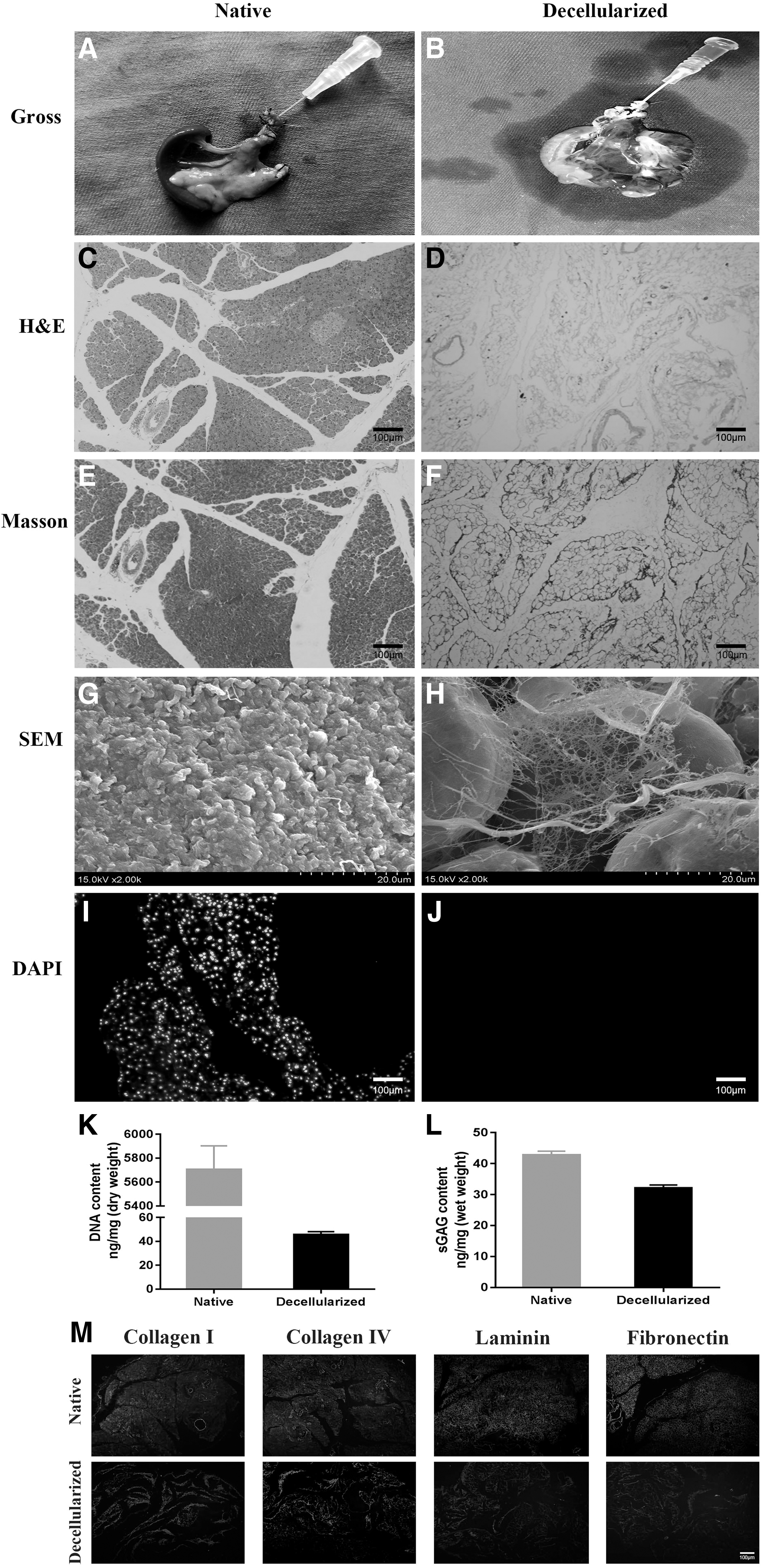
The transparency of the DP scaffold was evaluated by gross examination with the naked eye. The native pancreatic tissue and decellularized pancreatic tissue were fixed in 30% sucrose for 3 days, then the tissue pieces were taken out, soaked in the OCT embedding agent (Sakura Finetek Japan Co., Tokyo, Japan) for 2 hours, and then the samples were taken out and embedded in the embedding agent and placed in a −80° refrigerator frozen into pieces. Then the tissues were cut into 10 μm thick slices at the level of Bregma on a cryostat (LM3050S; Leica Microsystems, Bannockburn, III, Germany) and mounted on poly-L-lysine-coated glass slides.
Histological examination was performed using hematoxylin and eosin (H&E; Solarbio, Beijing, China) staining on frozen sections from DP scaffold to evaluate cellular removal and to examine the structural integrity of decellularized scaffolds in comparison with the native ones. In addition, 4,6-diamidino-2-phenylindole (DAPI; Sigma-Aldrich, St. Louis, MO) staining was performed to confirm the complete removal of cells. Imaging under a scanning electron microscope (SEM, SU-8010; Hitachi, Tokyo, Japan) was also performed to confirm the complete removal of cells and the preservation of the ECM ultrastructure.
Quantification of DNA and sulfated glycosaminoglycans
Total DNA was extracted from the pancreas using G-spinTM Total DNA Extraction kit (D6943–02; Promega, MA) in accordance with the manufacturer's instructions. Total DNA was then evaluated with a Nanodrop One spectrophotometer (Thermo Fisher Scientific, Waltham, MA). Sulfated glycosaminoglycans (sGAG) were measured by using Blyscan sGAG Assay, (110 assays), [Standard Kit Size] (Biocolor Ltd., Carrickfergus, UK), quantitative detection of sulfated proteoglycan and glycosaminoglycan (sGAG) content by dyeing combined release method.
Draw a standard curve
Processing of samples to be tested: Take 50 μL sample to be tested for experiment. If the initial absorption peak of the reactant at 656 nm is greater than 1.5, the sample is diluted. When the concentration of the sample to be tested falls into the middle section of the standard curve, the accuracy is higher.
Obtaining of sulfated aminoglycan-dye complex: Add 1.0 mL of Blyscan dye solution to the sample to be tested, mix well, and when the insoluble matter appears, centrifuge, discard the supernatant, and then obtain a purple aminoglycan sulfate-dye complex
The release of dye in the aminoglycan sulfate-dye complex: Add 1.0 mL of dye release agent and mix well. Dissolve for more than 5 minutes to fully release the dye
Determination: Use a spectrophotometer to measure at a wavelength of 656 nm, or use a colorimeter red filter to measure
In vitro immunofluorescent staining
The presence of four matrix proteins (collagens I and IV, fibronectin, and laminin) was ascertained by immunofluorescence (IF) staining. Polyclonal rabbit anti-collagen I (ab34710, 1:500; Abcam, Cambridge, MA), polyclonal rabbit anti-collagen IV (ab19808, 1:500; Cambridge, MA), rabbit anti-fibronectin (ab45688, 1:500; Abcam), and polyclonal rabbit anti-laminin (ab11575, 1:500; Abcam) antibodies were used as primary antibodies and incubated overnight at 4°C, followed by incubation with secondary antibody, Goat anti-rabbit IgG (Alexa flour 594, ab150080, 1:1000; Abcam), for 1 hour at room temperature. Their expressions were visualized using a fluorescence microscope (Olympus, Tokyo, Japan).
Cross-linking study
Pancreas scaffolds were immersed in different cross-linking solutions for 4 hours at 37°C with cyclic perfusion, as follows: (1) AgNP 100 nm (730777; Sigma-Aldrich) (at four concentrations: 0.5, 2.5, 5, and 7.5 μg/mL) (AgNP group) and (2) ultrapure water as a control (decellularized group, DP). All scaffolds were then rinsed in PBS, after which, the effects of cross-linking were evaluated. SEM imaging was conducted to evaluate structural changes in the pancreas ECM following cross-linking. We choose the lowest concentration for testing, Sections were imaged by SEM and analyzed by energy-dispersive X-ray spectroscopy (EDS) to assess retention of nanoparticles. Transmission electron microscope (TEM) was also performed on sections of the AgNP group to qualitatively evaluate the retention of nanoparticles on the sections.
Maximum load
We used the CT3 texture analyzer (Brookfield Engineering Laboratories, Middleboro, MA) to do the indentation test from the different groups (Cheung et al., 2014; Kim et al., 2012; Tse Justin and Long Jennifer 2014). Then we calculated the values of different groups divided by the value of the DP group, and assessed the results of the maximum loads.
In vitro remained collagen content ratio
Immersed dry DP scaffolds (n ¼ 6 for each group) in 0.1 M Tris-HCL containing 50 mM calcium chloride and incubated at 37°C for 30 minutes, then treated with 0.1 M Tris-HCL containing 50 U of collagenase type I (Worthington Biochemical Co., Lakewood, NJ), and incubated with shaking at 37°C for 12 hours. The samples were treated with 0.25 M of ethylenediaminetetraacetic acid (EDTA; MediaTech, Inc., Manassas, VA) on ice to stop digestion. Lysates of degraded segments from the different groups were centrifuged, after the hydrolyzed supernatants (6 N HCl, 110°C). Ninhydrin solution (Sigma-Aldrich) was mixed and the mixtures were heated for 30 minutes at 100°C. The optical densities (ODs) of the mixtures from the different groups were measured using a UV-vis spectrophotometer at a wavelength of 570 nm and evaluated semiquantitatively by dividing the values of ODs of the mixtures from the different treated groups.
Swelling ratio
Air-dried different groups of specimens, weighed to obtain data (W0), then soaked for 2 hours in PBS at room temperature, and weighed to obtain data (W1) again, and calculated the wet weight increase to the original dry weight (W1–W0/W0).
In vitro cytotoxicity test
Human umbilical vein endothelial cells (HUVECs) and MIN6 cells were cultured in a 96-well plate with the supernatant of four cross-linked scaffold groups, soaked for 5 days in a 37°C incubator containing 5% CO2. A CCK-8 assay (CK04–500, Dojindo Laboratories, Kumamoto, Japan) and a PowerWave XS microplate reader (Bio-Tek) were used to quantitatively assess cytotoxicity from measurements of the OD at 450 nm to calculate cell survival rate (Cheung et al., 2014; Tse Justin et al., 2014) after incubation with different concentrations of cross-linked groups.
Based on the previous tensile strength, water absorption, and remaining collagen content combined with the CCK-8 experiment, 0.5 μg/mL was selected as the follow-up experimental group under the premise of the lowest cytotoxic side effects.
In vivo implantation
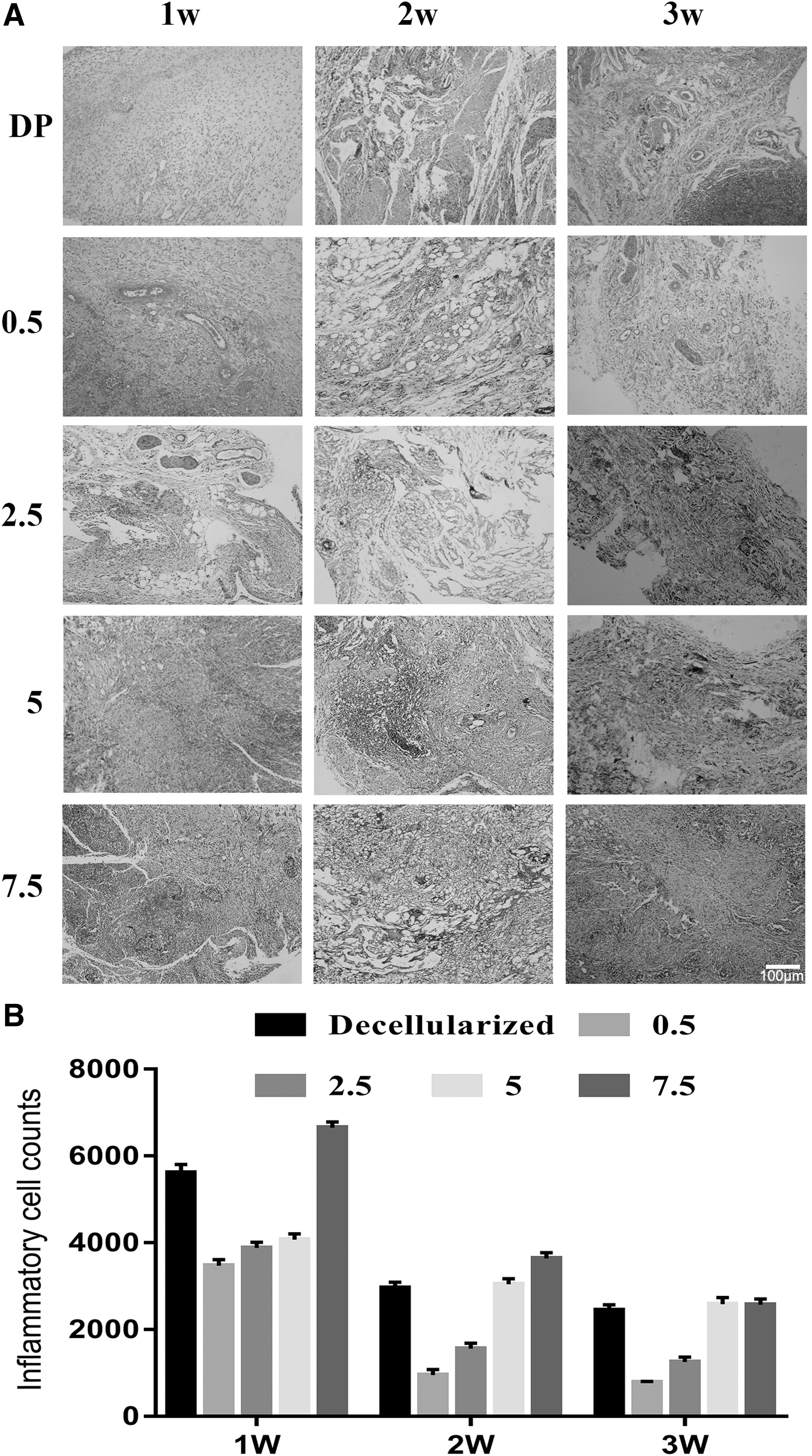
Native decellularized and four concentrations of AgNP groups were sterilized by immersing in 0.1% peracetic acid (PAA; Sigma-Aldrich) for 2 hours, followed by washing with PBS for 48 hours. Sprague Dawley rats (250 g body weight; 8 weeks old) were anesthetized; a small incision was made down the back of the rats, and then a dorsal subcutaneous pouch was created by blunt dissection. Implanted decellularized rat pancreas scaffolds went into this pouch, oriented away from the suture site, and the wound was sutured and sterilized. Animals were euthanized at 7, 14, and 21 days postimplantation (PI). The implanted tissues were cut into 10 μm thick slices at the level of Bregma on a freezing microtome (LM3050S; Leica Microsystems, Bannockburn, III) and loaded on poly-L-lysine-coated glass slides. H&E staining was conducted to assess the xenografts for signs of immunological rejection.
To quantify immune cell infiltration, the total number of neutrophils, lymphocytes, and macrophages was expressed as the number of inflammatory cells. Infiltrating immune cells were quantified in three fields at the border between the implanted material and surrounding tissue on each slide at a 100 × magnification. For assessment of immune cell phenotype, IF was conducted with anti-CCR7 (Alexa FluorVR 555) (ab207018, 1:1000; Abcam) and anti-CD206 (Alexa FluorVR 594) (#141726, 1:1000; Biolegend, San Diego, CA) antibodies as M1 and M2 markers; DAPI was used as a nuclear counterstain, respectively. The M1/M2 ratio was calculated using Image J software (Srivatsan et al., 2015). All results were evaluated histologically by the same researcher in a blinded manner.
Statistical analysis
All data were analyzed using SPSS v19.0 (Chicago, IL) and displayed using GraphPad Prism software (La Jolla, CA). A Student's t-test was used to determine significance between two groups and by analysis of variance for multiple comparisons. Data are presented as mean ± standard error of the mean. Sample size and statistical details can be found in the figures and legends. A level of p < 0.05 was considered significant.
Results
DP matrix
During the decellularization process, the original organs gradually become transparent, while the tissue morphology and natural vascular structure are preserved (Fig. 1A, B). Histological analysis using H&E and Masson's trichrome staining demonstrated the removal of nuclear and cytoplasmic material and the presence of an intact ECM (Fig. 1C–F). SEM analysis confirmed that the decellularized scaffolds had an intact ECM structure, without any cellular material (Fig. 1G, H). DAPI staining confirmed that no nuclear content remained after decellularization (Fig. 1I, J). Data analysis indicated that the DNA content was reduced by more than half and that more than 70% of sGAG content was retained (Fig. 1K, L). IF demonstrated a large degree of collagen retention before and after decellularization (Fig. 1M).
Characterization of DP scaffolds.
In vitro examination
SEM observations indicated that the microstructure of the scaffold changed after cross-linking. The ultrastructure of the pancreatic decellularized scaffold was improved by the nanosilver overlay connection between the matrix fibers (Fig. 2A). TEM and EDS analysis of the AgNPs group sections demonstrated that the nanoparticles remained bound and were retained on the sections, indicating that the nanosilver has been successfully loaded onto the scaffold after cross-linking (Fig. 2B, C).

Effects of different modifications to DP scaffold.
The UV-visible spectrum of the supernatant collected by extraction with ultrapure water was used to soak the sections of the lowest concentration AgNP group and was recorded, demonstrating no characteristic AgNP peaks over a period of 5 days (Fig. 2D). As the cross-linking concentration increased, maximum load capacity of the tissue increased (Fig. 2E). AgNP concentration increased and swelling rate of scaffolds increased (Fig. 2F). In addition, as concentration of AgNPs increased, the proportion of collagen increased (Fig. 2G).
Evaluation of in vitro cytotoxicity test by using HUVECs and MIN-6 cells
CCK-8 assays of HUVECs and MIN6 cell lines were performed in 96-well plates with scaffolds loaded with different concentrations of AgNPs. The results showed that as the concentration of AgNPs increased, cell viability decreased at the concentration of 2.5 μg/mL. A concentration of 0.5 μg/mL was the concentration that resulted in a decrease in HUVEC and MIN-6 cell viability that was not significant (p > 0.05). So we choose the concentration of 0.5 μg/mL as the maximum nontoxic concentration (Fig. 3).

CCK-8 assay. (*p < 0.05, **p < 0.01, ***p < 0.001).
Histological characterization of implanted pancreas scaffolds in vivo
In the control group, implants were surrounded by the tissue of the subcutaneous back and a thick capsule has formed 1 week PI. In comparison, the 0.5 μg/mL group was covered with a considerably thinner capsule. Histological analysis showed that a less intense immune cell infiltration had occurred into the cross-linked materials, indicating a weaker immunological response.
Two weeks after implantation, most of the neovascularization was detected in the 0.5 μg/mL concentration group, and the inflammatory infiltration was at a low level. At the same time point, the decellularized group had continuous infiltration and accumulation of inflammatory cells with no signs of newly formed blood vessels within the implant. After 3 weeks, the inflammatory response in the experimental group was stable with a greater number of vascular lumen-like structures observed in the 0.5 μg/mL group. According to the numbers of lymphocytes and granulocytes visible in H&E-stained sections, the acute inflammatory reaction was significantly reduced in the 0.5 μg/mL group compared with the control group 3 weeks PI (p < 0.01) (Fig. 4).
Host immune reaction against scaffolds from the different groups:
IF staining of macrophages (M1 & M2) was conducted at 7, 14, and 21 days after surgery. The cross-linked group exhibited an intense expression of M2 phenotype macrophages compared with the control group. The M1/M2 ratio at 7, 14, and 21 days PI significantly decreased in the 0.5 μg/mL group compared with that of the DP group (p < 0.05). M1 macrophages were marked by CCR7+ staining, and M2 macrophages by CD206+ staining.
There were clear differences in the distribution of the M1 and M2 macrophages in the different implant groups. One week after implantation, the DP group had a greater number of M1 macrophages than the cross-linked scaffolds. The M2 macrophage distribution was the converse of the M1 distribution. In the second week, a greater number of M2 macrophages were present in the 0.5 μg/mL AgNP group. In the third week, the situation of all experimental groups basically tended to be the same (Fig. 5).

Polarization of macrophages in the different groups:
The M2-dominant macrophage polarization within the implants indicated that tissue formation processes were occurring due to the host response induced by the cross-linked matrices (Manikandan et al., 2015; Saleh et al., 2018).
Discussion
Reducing the immunogenicity associated with DP scaffolds is vital for increasing the clinical utility of pancreas transplantation. We hypothesized that cross-linking decellularized rat pancreas scaffolds with AgNPs may upgrade their biocompatibility and structural stability, mainly based on the ability of AgNPs to bind to collagen fibers, and the combination with their anti-inflammatory properties, which would reduce host immune response and improve collagen fiber stability against decellularized natural tissue-derived scaffolds (Saleh et al., 2018; Saleh et al., 2019).
The AgNPs cross-linking also avoided the previously reported limitations against with other cross-linking agents, such as calcification, toxicity, weak bonding, abnormal coloration, and difficult extraction (Baptista et al., 2009; Somers et al., 2008). As far as we know, this is the first study to use nanoparticles to modify the structural properties and biocompatibility of decellularized pancreatic scaffolds. In this study, we evaluated the effect of cross-linking after using AgNPs (size 100 nm).
Characterization of DP scaffolds revealed that complete decellularization was achieved without massive damage to the native-mimic structure of the decellularized tissues before commencing the cross-linking process. After histological and morphological evaluation, there were obvious changes in the scaffold before and after AgNP cross-linking, the broken scaffold fibers were reconnected together to form a dense network structure, and samples from the AgNP group displayed more regular coiled collagen fibers. This might be caused by the unique properties and very large surface area of the AgNPs (Srivatsan et al., 2015). AgNP-related structural improvement of collagen fibers has also been reported in other studies of collagen hydrogels, collagen nanofibers, and wound healing in vivo.
The maximum load data indicated that, as AgNP concentration increased, the mechanical strength increased, and also indicated that the mechanical properties of the scaffold improve (Kwan et al., 2011; Qimeng et al., 2020; Saleh et al., 2018; Saleh et al., 2019). With increased cross-linking, the swelling rate of scaffolds was correspondingly improved; it may be due to the ability of AgNPs to bind with water, and electrostatic interactions in watery environments may be to increase the scaffold surface network area, furthermore, increases the hydrophilic properties of the scaffolds (Saleh et al. 2019).
We found that the proportion of collagen remaining, as indicated by the results of a ninhydrin assay, increased as AgNP concentration increased, probably due to the broken collagen structure repaired after nanosilver cross-linking; this finding may indicate that the cross-linked nanosilver is linked at the cleavage site of collagen to prevent enzymes from penetrating into the treated scaffold. An increase in cross-linking concentration affected collagen stability, consistent with the observed mechanical properties.
Large AgNPs (100 nm) were chosen in this study to avoid the known cytotoxicity of smaller particles (Beck et al., 2015; Kwan et al., 2011; Xu et al., 2018). As one of the most important characteristics of any modifying agent is that it should be noncytotoxic, cytocompatibility of the different modified scaffolds was evaluated by CCK-8 assay. The CCK-8 assay indicated that an AgNP concentration of 0.5 μg/mL was superior nontoxicity for cross-linking in terms of their effect on HUVECs and MIN-6 cells. These cell types will be used for future studies. In the next step, HUVECs and MIN-6 cells will be seeded onto scaffolds and then implanted subcutaneously into rats. After a certain period of time, neovascularization and insulin secretion will be evaluated by observation of HUVECs and MIN-6 in the decellularized scaffolds after recellularization.
Through previous experiments, comparing the maximum tensile strength test, water absorption test, residual collagen content test experiment, and cytotoxicity test, we found that the 0.5 μg/mL concentration brings about a strong mechanical tensile force and good level water absorption capacity on the basis of maximum nontoxicity, while retaining relatively much collagen content. Based on these conditions, we chose 0.5 μg/mL concentration as the ideal concentration for in vitro experiments.
Based on previous tests, we screened out the excellent concentration of the in vitro cross-linking. To prove whether it is suitable for in vivo transplantation, we initially selected all concentration groups and blank groups of stent, all transplanted to the rat's back skin to observe the inflammation and infiltration. Our focus was that of the host immune response that affects the regeneration process of a recellularized transplantable material. Microscopic examination of different modified implanted scaffolds revealed that samples from the AgNP groups exhibited the most promising results in terms of biocompatibility. Compared with the control group, the AgNP groups exhibited the lowest number of inflammatory cells on days 7, 14, and 21 by H&E staining.
This finding attested that, in the AgNP group, the biocompatibility of the scaffolds may be improved, possibly owing to the ability of the AgNPs to restrain the production of inflammatory cytokines such as the different proinflammatory interleukins, tumor necrosis factor, and interferon gamma (Manikandan et al., 2015; Saleh et al., 2018; Saleh et al., 2019). Subsequently, the host immune response decreased, and the immunogenicity of AgNP-loaded pancreas scaffolds was restrained.
Polarization of macrophages was detected in this study by differentiating their phenotypes (M1 and M2) using IF staining; we selected the control group to compare against the 0.5 μg/mL AgNP group for M1/M2 IF staining, which indicated that the 0.5 μg/mL group caused macrophages to enter the M2 phase earlier than the simple decellularized group. This finding indicates that the biocompatibility of the AgNP group scaffold is superior due to inhibition of inflammatory cytokine production by AgNPs (such as various proinflammatory interleukins, tumor necrosis factor, and interferon-gamma) (Manikandan et al., 2015).
Consequently, the host immune response was suppressed, and the immunogenicity of the pancreatic scaffolds with AgNPs was inhibited (Alcor et al., 2009; Manikandan et al., 2015). And AgNPs improve biocompatibility of the DP scaffold by increasing the polarization of the macrophage M2 phenotype. In addition, the resembled blood vessels surrounding M2 cells were observed after the second week, suggesting that M2 macrophages had a proangiogenic effect, which further verified the excellent performance of the scaffold cross-linked with AgNPs (Brown et al., 2009; Spiller et al., 2014). Thus, the durability of the transplanted pancreatic scaffold may increase in a low inflammatory environment, allowing it to retain its integrity until recellularized, before cells construct new tissue (Spiller et al., 2014).
In summary, the results indicated that the decellularized pancreatic scaffolds cross-linked using AgNPs exhibited the enhanced mechanical strength, invoked a reduced inflammatory response, and induced M2 polarization of macrophages in vivo. In the future, additional in vitro and in vivo investigation are required to evaluate the ability of the cross-linked acellular pancreatic scaffolds to support cellular activity, function, and tissue regeneration. It is predicted that AgNP cross-linked pancreatic decellularized scaffolds will provide a new direction in allogeneic transplantation in tissue engineering and that the decellularized scaffolds prepared using this method have broad applicability for the construction of future organoids and human organ transplantation.
Footnotes
Author Disclosure Statement
The authors declare they have no potential conflicts of interest with respect to the research, authorship, and/or publication of this article.
Funding Information
This research was supported by the National Key Research and Development Program of China (Grant No. 2018YFC1105603) and the National Natural Science Foundation of China (Grant No. 31830028, 81471801) and the Science and Technology Project of Nantong City (MS12018077, MS12018058) and the Postgraduate Research and Practice Innovation Program of Jiang su Province (KYCX18_2408).