




Abstract
The decrease of endometrial receptivity leads to repeated implantation failure (RIF) during in vitro fertilization and embryo transfer. To explore the therapeutic potential of menstrual blood-derived mesenchymal stem cells (MenSCs) in addressing RIF, we established a murine model of embryonic implantation dysfunction using mifepristone. Subsequently, we administered MenSCs to these mice via tail vein injection and assessed their impact on the implantation and pregnancy rates of the affected mice. Furthermore, we conducted immunohistochemical staining on uterine tissues from these mice to examine the expression of endometrial receptivity markers, specifically vascular endothelial growth factor (VEGF)-A, HAND2, and HOXA10 following MenSCs transplantation. In parallel, we conducted in vitro studies to elucidate the molecular mechanisms of cell therapy by measuring the expression levels of VEGF-A, HAND2, and HOXA10 in endometrial stromal cells using real-time PCR and western blotting. In our mifepristone-induced mouse models, we observed a reduction in both pregnancy rates and implantation sites; however, these parameters were significantly improved after MenSCs transplantation. Similarly, the expression levels of VEGF-A, HAND2, and HOXA10 in the uterine tissues of the mifepristone group were diminished, but these levels were restored following MenSCs therapy. In vitro, after mifepristone treating, the expression of VEGF-A, HAND2, and HOXA10 decreased in endometrial stromal cells, but their expression increased after MenSCs coculture supernatant. In conclusion, these results demonstrated that MenSCs transplantation could increase endometrial receptivity by upregulating VEGF-A, HAND2, and HOXA10 expression. This study suggests MenSCs as a novel stem cell candidate in the treatment of RIF.
Introduction
With the rapid development of assisted reproductive technology (ART), in vitro fertilization–embryo transfer (IVF-ET) and related derivative technologies makes the clinical pregnancy rate increase continuously. However, there are still some infertile patients who fail to achieve a clinical pregnancy after multiple ETs, which can be defined as repeated implantation failure (RIF). Currently, there is no consensus on the definition of RIF. In 2005, the European Society of Human Reproduction and Embryology (PGD) defined RIF as the failure to obtain clinical pregnancy after 3 or more high-quality ETs or multiple ETs with more than 10 embryos (Thornhill et al., 2005). The techniques, including endometrium scratching (van Hoogenhuijze et al., 2023), aspirin (Sung et al., 2021), sildenafil (Moini et al., 2020), hormone regulation (Vera-Montoya et al., 2023), and intrauterine infusion of Granulocyte colony stimulating factor, are currently used to treat patients with RIF to improve pregnancy rate (Wu et al., 2022). However, despite a fundamental understanding of RIF’s causes and the effectiveness of certain interventions, RIF remains one of the most significant challenges to improving clinical pregnancy rates in ART.
Endometrial receptivity refers to the receptivity of maternal endometrium to blastocysts, which is an important factor affecting the success rate of ART (Hiraoka et al., 2023). Endometrial receptivity varies with the menstrual cycle and shows maximum tolerance at a particular time known as the “window of implantation” which spans between days 20 and 24 of the menstrual cycle. Several studies have shown that two thirds of patients with RIF are due to insufficient endometrial receptivity, while embryos account for one-third of implantation failures (Achache and Revel, 2006). Enhancing endometrial receptivity is pivotal in the quest for RIF treatments.
Stem cell therapy has been a new research direction for repairing endometrium and improving endometrium receptivity in recent years. Menstrual blood-derived mesenchymal stem cells (MenSCs) were chosen in this study, which have unique advantages in the treatment of RIF. First, MenSCs are widely available and can be obtained noninvasively, which are superior to bone marrow-derived mesenchymal stem cells (MSCs) and adipose tissue-derived stem cells. Second, autologous MenSCs have no ethical controversy and immune rejection, which are better than other allogeneic stem cells. Moreover, MenSCs have low tumor-forming, noninvasive, and stable biological characteristics (Zhang et al., 2023). So they are favored by researchers. In recent clinical studies that are based in small sample sizes, it has been confirmed that there were safety and beneficial effects of MenSCs in multiple sclerosis (Zhong et al., 2009), muscular dystrophy (Ichim et al., 2010a), congestive heart failure (Ichim et al., 2010b), and Asherman syndrome (Wu et al., 2023), which indicates the application value of MenSCs in clinical treatment. Most notably, MenSCs, being easily derived from the endometrium, are particularly advantageous for endometrial repair and receptivity enhancement compared to other stem cell types. Currently, reproductive research on MenSCs primarily concentrates on Asherman syndrome, where their therapeutic effectiveness has been established.
However, there are no reports on the use of MenSCs for RIF treatment. The object of this study is to: (1) assess the impact of tail vein MenSCs administration on enhancing endometrial receptivity in mice with failed embryo implantation, (2) calculate embryo implantation rate and pregnancy rate, and (3) investigate the underling mechanisms of their therapeutic effects.
Materials and Methods
Cell isolation and culture
Three healthy volunteers aged from 28 to 39 were selected to collect menstrual blood by using menstrual cups on the second day of menstruation (all volunteers were provided with written informed consent before menstrual blood collection, and ethical approval was obtained from Ethics Committee of Liaoning Province Research Institute of Family Planning). The menstrual blood samples were mixed with equal volume of phosphate-buffered saline (PBS) (Corning) containing 100 U/mL penicillin, and 100 mg/mL streptomycin (Solarbio) at 4°C for 24 hours. Then, the samples were transferred onto the equal volume of Ficoll (TBD Science, China) and was centrifuged at 500 g for 20 minutes. Mononuclear cells were fractionated in Ficoll and washed with PBS twice. Then, cell precipitation was resuspended and cultured in Dulbecco’s modified Eagle medium/Ham’s F-12 (DMEM/F12) (Corning) with 10% fetal bovine serum (CellSeraTM) and 100 U/mL penicillin, and 100 mg/mL streptomycin, cultured at 37°C in a 5% CO2 humidified incubator. The culture medium was changed every 3–5 days and the cells were passaged by trypsin digestion after reaching 80%–90% confluence. In the following experiment, we used these cells that have been passaged 4–6 times.
Characterization of the MenSCs
The surface markers of the MenSCs were evaluated by using Flow cytometric analysis. 1 × 10⁶ MenSCs (passaged 4 times) were collected and resuspended in PBS (containing with 5% Bovine Serum Albumin). FITC-conjugated anti-human antibodies for CD90, CD73, CD105, CD45, CD34 as well as phycoerythrin-conjugated anti-human antibodies for CD90, CD73, CD105, CD45, CD34 (Biolengend) were used to characterize MenSCs. Then, the stained cells were washed twice with stain buffer and were analyzed by using a FACScan flow cytometer (BD Biosciences). MenSCs were characterized for their capability to differentiate into adipocytes, osteoblasts, and chondroblasts. MenSCs were cultured at 80%–90% confluence and were subsequently differentiated with differentiation medium at 37°C in 5% CO2.
In brief, for differentiation assays, the growth medium was subsequently replaced with adipogenic, osteogenic or chondroblasts inducing medium. The induced differentiation experiment was conducted according to the instructions of the kit (Cyagen). After induction, the cells were washed with PBS, fixed with 4% paraformaldehyde for 10 minutes, and then washed with PBS twice. Adipogenic, osteogenic, and chondroblasts differentiation was confirmed via Oil Red, Alizarin Red, and Alysin Blue staining, respectively.
Supernatant of MenSCs cocultured with endometrial stromal cells
MenSCs were cultured at 90% confluence. Subsequently, 3 mL of DMEM/F12 medium (Corning) was replaced, and the cells were continued to be cultured for an additional 48 hours. The culture supernatant was collected and centrifuged at 3000 rpm for 5 minutes to remove cells and cell debris. The supernatant after centrifugation was stored at −70°C in a refrigerator for subsequent experiments. Endometrial stromal cells (CP-H233, Pricena, China) were seeded in 60 mm culture dishes at a density of 1 × 10⁵ for 24 hours. The cells were treated with (1) control group: regular cell culture solution; (2) mifepristone group: regular cell culture solution with 25 ng/mL mifepristone (Yuanye Bio-Technology); and (3) MenSCs supernatant group: supernatant of MenSCs with 25 ng/mL mifepristone. After 48 hours, endometrial stromal cells were collected for the experiments of quantitative real-time polymerase chain reaction (PCR) and western blotting.
Quantitative Real-Time PCR
Total RNA was extracted with Total RNA Kit I (Omega) from endometrial stromal cells according to the manufacturer’s instructions. Reverse transcription was performed by using a PrimeScriptTM RT reagent kit (Takara), and quantitative real-time PCR was performed by using a TB GreenR)Premix Ex TaqTM II (Tli RNaseH Plus) (Takara) on an ABI 7500 real-time PCR system. The primers used in the experiments are listed in Table 1. All target gene mRNAs were normalized to the housekeeping gene glyceraldehyde-3-phosphate dehydrogenase (GAPDH), and the results were calculated by means of relative quantification using the 2 -△△CT method.
Sequences of Primers Used for Real-Time Reverse Transcription Polymerase Chain Reaction
GAPDH, glyceraldehyde-3-phosphate dehydrogenase; VEGF, Vascular endothelial growth factor.
Western blotting
The cells were prepared by using RIPA (KeyGEN). Protein concentration was quantified by using the bicinchoninic acid (BCA) protein assay kit (Bio-platform). A total of 25 µg protein/lane was separated by SDS-PAGE (8%–12%) and transferred onto polyvinylidene difluoride membranes. The membranes were blocked with 5% nonfat milk for 1.5 hours at room temperature. Then the membranes were incubated with primary antibodies against HAND2 (1:1000; ab200040, Abcam), HOXA10 (1:1000; ab191470, Abcam), vascular endothelial growth factor (VEGF)-A (1:1000; ab46154, Abcam), and GAPDH (1:3000; BA002, Bio-platform) at 4°C overnight. Following the primary incubation, membranes were incubated with HRP-conjugated second anti-mouse/goat/rabbit antibodies (1:5000; UT2001, Utibody/1:1000; KGAA38, Keygen/1:5000; UT2003, Utibody) for 1 hour. Protein bands were visualized by using enhanced chemiluminescence (ECL; Thermo). The densitometric analysis for the quantification of the bands was performed by using Gel-Pro Analyzer 4 (Media Cybernetics, USA). GAPDH was used as an endogenous control.
Tube formation assay
Matrigel (Corning, Bedford, USA) was diluted with serum-free Dulbecco’s Modified Eagle Medium (DMEM) medium at a ratio of 1:1 and distributed in a 96-well plate for gel formation at 37°C for 30 minutes. Human Umbilical Vein Endothelial Cells (HUVECs) (CP-H082, Pricena, China) were divided into two groups and were labeled with CellTracker Green (Invitrogen, California, USA). (1) Control group: a total of 1 × 10⁴ HUVECs were seeded per well with regular cell culture solution; (2) MenSCs group: a total of 1 × 10⁴ HUVECs were seeded per well with supernatant of MenSCs. Following a 5-hour incubation period, tubular network structures were imaged in three randomly selected fields per well using a Nikon instrument (Japan). The acquired images from both experimental groups were quantitatively analyzed using the angiogenesis module of Image J software. Key parameters including the number of nodes and total tube length, as automatically calculated by the software, were selected for statistical analysis.
Animal models and treatment
Animals and grouping
Kunming mice of SPF grade (6–8 weeks old, weighing 25–28 g) were purchased from Changsheng Biotechnology Company. The females were not mated and the males were proved to be fertile. The animals were housed in our laboratory animal center on a 12-hour light, and 12-hour dark regimen. After a 1-week acclimation period, female and male mice (2:1 ratio) were mated at 6:00 PM, and the presence of a vaginal plug was checked 15 hours later. The presence of vaginal plug was considered as evidence of mating, and this day was designated as Day 0.5 post-coitum (Day0.5). A total of 34 pregnant mice were randomly divided into three groups: control group (n = 10), mifepristone group (n = 12), and mifepristone with MenSCs group (n = 12). All animal experiments were planned and performed in accordance with national legislation on laboratory animals’ protection and were approved by Liaoning Province Research Institute of Family Planning (JHSY-2019-05).
Animal models
On Day 3.5 post-coitum (Day 3.5) at 9:00 am, the mifepristone group and MenSCs group were treated with mifepristone solution (Yuanye Bio-Technology) at a dose of 5.5 mg/kg via intraperitoneal injection, while the control group received an equivalent volume of PBS.
Administration of MenSCs
MenSCs were cultured in DMEM/F12 medium as described above. After 48 hours of culture, MenSCs (1 × 10⁶ cells, passaged 4 to 6 times) were collected and resuspended in 0.2 mL of PBS. On Day 3.5 post-coitum (Day 3.5), 0.2 mL of MenSCs suspension was administered to the MenSCs group via tail vein injection, while the mifepristone group and control group received an equivalent volume of PBS instead.
Collection of tissue
At 9:00 am on Day 7.5 post-coitum (Day 7.5), all 34 female mice from each group were sacrificed. The uterus horns were excised to determine the number of implantation sites and pregnancy rate. The number of pregnant mice and implanted embryos in each uterus was recorded. All uterus tissues were fixed in 4% paraformaldehyde for subsequent immunohistochemistry experiments.
Immunohistochemistry
Tissues were fixed in 4% paraformaldehyde, dehydrated and embedded in paraffin, and cut into 5.0 µm sections for immunohistochemistry. Sections were fixed in 3% H2O2 for 30 minutes to block endogenous peroxidase reactivity and were blocked with normal goat serum (Boster, China) for 15 minutes. Then, sections were immunolabeled with anti-HAND2 antibody (1:1000; ab200040, Abcam), anti-HOXA10 antibody (1:500; ab191470, Abcam), and anti-VEGF-A antibody (1:800; ab46154, Abcam) overnight at 4°C. After washed with PBS, the sections were incubated with the corresponding secondary antibody and the results were stopped with 3,3-diaminobenzidine (DAB; Boster, China). %Area was calculated by dividing the area with absorbance over a minimum threshold by the total area analyzed for normalization and data comparison. The %Area of VEGF-A, HAND2, and HOXA10 was counted with a 200-fold horizon for 4 fields per sample.
Statistical analysis
Values are presented as the mean ± scanning electron microscope. Significance within groups was compared by using one-way Analysis of Variance. The numbers of pregnant mice were analyzed by chi-square test. Differences were considered significant at p < 0.05. All statistical analyses were performed by using SPSS17.0 software (SPSS Inc.)
Results
Characterization of MenSCs
MenSCs showed a colony-like morphology was clearly observed in the primary cultures. With the increase of the number of passages, the cell morphology tends to be consistent, showing typical fibroblast-like, spindle, or compact vortex shape (Fig. 1A). Flow cytometry analysis revealed that the MenSCs were positive for CD73, CD105, and CD90, which were typical MSC markers, and the MenSCs were negative for CD34, CD45, and HLA-DR which were absent in MSCs (Fig. 1B).

Morphology and immunophenotypes of MenSCs.
To evaluate the differentiation capacity of MenSCs, the cells were induced to differentiate to adipocytes, osteoblasts, and chondroblasts. The chemical staining results indicate that MenSCs have the ability to differentiate into three lineages (Fig. 1C).
Supernatant of MenSCs coculture promotes HUVECs tube formation
Tube formation assay on Matrigel was used to evaluate the angiogenic effect of MenSCs. Compared with the control group, the number of nodes (608 ± 56.51 vs. 1016 ± 128.29, p < 0.05) and the total length of tubular network (17,907 ± 1707.93 vs. 25,234.67 ± 2325.82, p < 0.05) were both significantly increased in the MenSCs supernatant incubated group (Fig. 2).

Supernatant of MenSCs positively regulates tube formation in HUVECs.
Supernatant of MenSCs coculture promotes the expression of VEGF-A, HOXA10, and HAND2 in endometrial stromal cells
VEGF-A, HOXA10, and HAND2 are important genes related to endometrial receptivity. The expression of VEGF-A, HOXA10, and HAND2 in endometrial stromal cells was downregulated after 48 hours of treatment with 25 ng/mL mifepristone. After adding the supernatant of MenSCs, the expression level of VEGF-A, HOXA10, and HAND2 genes in damaged endometrial stromal cells was upregulated and the level returned to normal level (Fig. 3).
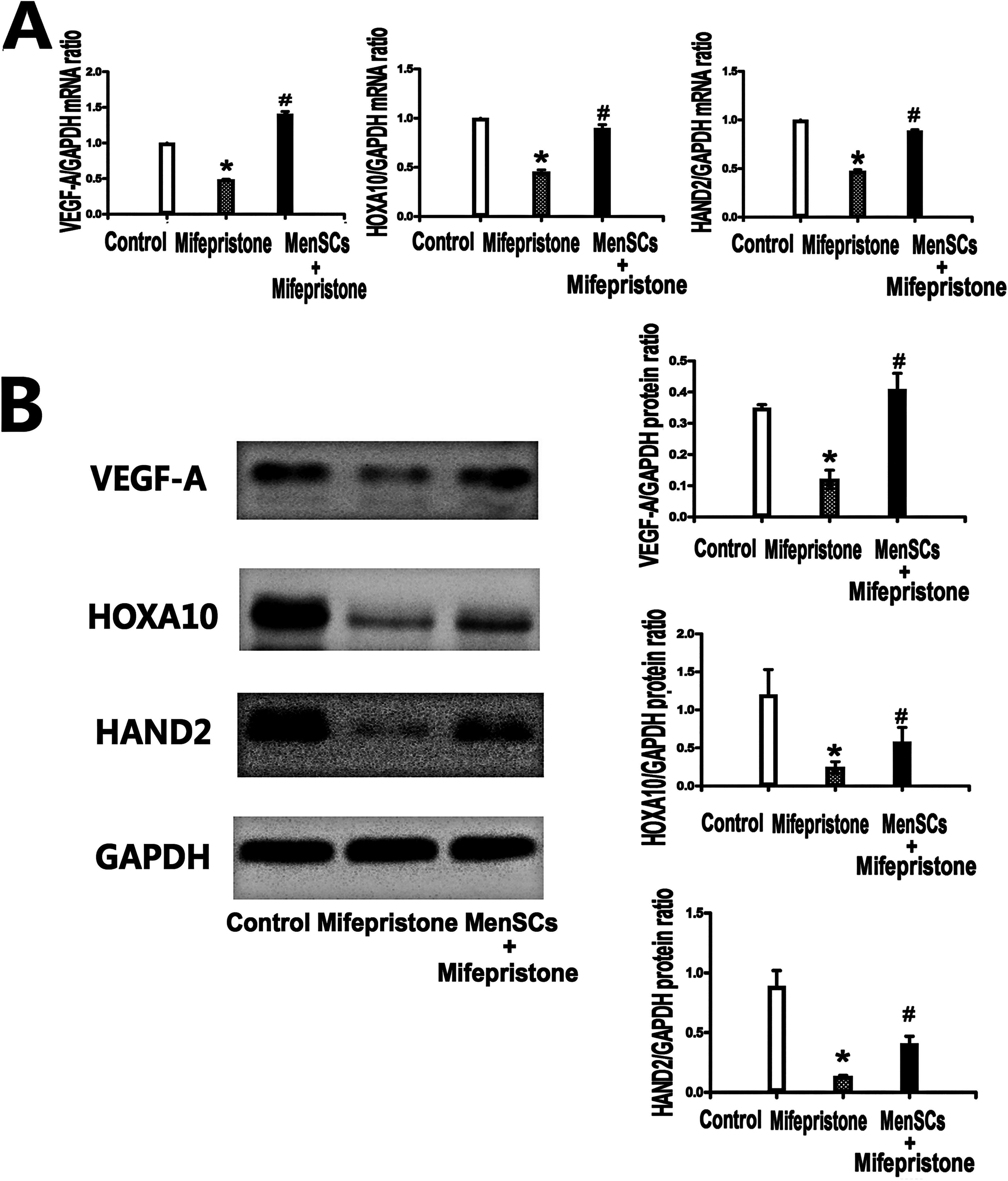
Supernatant of MenSCs stimulates VEGF-A, HAND2, and HOXA10 expression after treatment of mifepristone.
Effect of MenSCs on pregnancy rate and the implantation sites of pregnancy mice
The pregnancy rate in mifepristone group significantly decreased, compared with control group (33.3% vs. 90%, p < 0.05). As compared with mifepristone group, the pregnancy rate in MenSCs group with mifepristone significantly increased (66.7% vs. 33.3%, p < 0.05). In mifepristone group, the number of implanted embryos was lower than that in control group (1.17 ± 2.37 vs. 14.60 ± 5.36, p < 0.05). After the MenSCs were injected into the tail vein, the number of implanted embryos increased, compared with that of mifepristone group (6.67 ± 6.66 vs. 1.17 ± 2.37, p < 0.05) (Fig. 4A and B, Table 2).

MenSCs increased the implantation rate of mice embryos.
Comparison of Number of Implanted Embryos and Pregnancy Rate Among Different Groups
MenSCs, menstrual blood-derived mesenchymal stem cells.
#p < 0.05 compare with control group; ##p < 0.05 compare with mifepristone group.
*p < 0.05 compare with control group; **p < 0.05 compare with mifepristone group.
Effect of MenSCs on endometrial receptivity
In order to illustrate the role played by MenSCs in endometrial receptivity by regulating VEGF-A, HAND2, and HOXA10, we used uterine tissue of paraffin-embedded mice, which were stained with VEGF-A, HAND2, and HOXA10 antibodies. The expression of VEGF-A in uterine tissue of mifepristone group (4.50 ± 0.98 vs. 19.22 ± 2.00, p < 0.05) dramatically decreased, compared with the control group. By contrast, the expression of VEGF-A significantly increased after MenSCs therapy was compared with that in the mifepristone group (17.20 ± 1.60 vs. 4.50 ± 0.98, p < 0.05). Mifepristone group also showed lower staining intensities of HAND2 than those of control group (3.69 ± 1.47 vs. 12.81 ± 1.48, p < 0.05). MenSCs group showed higher staining intensities (13.75 ± 2.21 vs. 3.69 ± 1.47, p < 0.05) than those of mifepristone group. Meanwhile, the expression of HOXA10 of mifepristone group (5.18 ± 1.69 vs. 15.89 ± 2.30, p < 0.05) decreased, compared with control group. After using MenSCs therapy, the expression of HOXA10 significantly increased, compared with mifepristone group (11.72 ± 1.68 vs. 5.18 ± 1.69, p < 0.05) (Fig. 5).

Discussion
The investigation and management of RIF usually focus on the quality of the embryo and endometrial receptivity (Ma et al., 2022). Multiple embryos fail to implant, and a relevant percentage of IVF/ICSI treatment failures is due to endometrial receptivity disorders (Zhang et al., 2022). In this study, we created an embryo implantation dysfunction model in female Kunming rats using intraperitoneal injection of mifepristone to mimic the pathological mechanisms observed in RIF patients. Utilizing the mifepristone model, we demonstrated that the pregnancy rate was lower and the number of embryonic implantation sites was reduced in the mifepristone group compared to the control group. Concurrently, transplanting MenSCs into mifepristone-treated mice resulted in an increase in both pregnancy rate and embryonic implantation sites. MenSCs treatment proved to be highly effective in embryo implantation dysfunction models. The results of the present study show that (1) transplantation of MenSCs to the embryo implantation dysfunction models has a good effect on the pregnancy rate and embryonic implantation; (2) MenSCs treatment facilitates endometrial angiogenesis and promotes the expression of VEGF-A; and (3) MenSCs treatment enhances endometrial receptivity and the expression of HAND2 and HOXA10. These findings could guide more effective clinical trials and treatment in the future.
There are numerous factors that could lead to RIF, and abnormal angiogenesis is one of them (Wang and Chen, 2024). VEGF-A (also called VEGF), has been proved to be the most important and potent factor in angiogenesis (Eguchi et al., 2022). The VEGF family was key mediators of vascular growth and remodeling in a variety of tissues, including the human endometrium (Lei et al., 2021). Elevated VEGF-A expression caused a significantly increased implantation rate and endometrial receptivity (Guo et al., 2021; You et al., 2022). Our study demonstrated that MenSCs administration improved the pregnancy rate in mice with embryonic implantation dysfunction. Furthermore, VEGF-A expression was diminished in the mifepristone group and upregulated following MenSCs therapy in the MenSCs group. In vitro, we showed that MenSCs supernatant stimulated tube formation in HUVECs. Using real-time PCR and western blotting, we found that VEGF-A mRNA and protein expression levels in endometrial stromal cells were downregulated by mifepristone treatment, and upregulated after the addition of MenSCs supernatant. These results suggest that MenSCs treatment may potentially enhance uterine angiogenesis by regulating VEGF-A expression, thereby improving uterine receptivity and increasing the pregnancy rate in mice with embryonic implantation dysfunction.
Heart and neural crest derivatives protein 2 (HAND2) is considered as a progesterone-induced transcription factor expressed in the stroma. HAND2 actively contributes to the receptivity, implantation, and decidualization of the uterus in mice (Niknafs et al., 2021). In uterine tissue-specific HAND2 knockout mice, continued induction of paracrine mitogenic mediators in the stroma maintains epithelial proliferation and stimulates E-induced pathways, resulting in impaired implantation (Cho et al., 2013). In vitro, we determined that HAND2 expression levels in endometrial stromal cells decreased after 48 hours of mifepristone treatment, but were upregulated following MenSCs supernatant treatment. Similarly, HAND2 expression was reduced in the mifepristone group, and significantly increased in the MenSCs group compared to the mifepristone group. These results indicate that MenSCs can regulate HAND2 expression in injured.
HOXA10 gene belongs to homeobox (HOX) gene, which is named after a 183 bp HOX is contained in this gene family (Cho et al., 2013). In the embryo implantation window stage, high levels of estrogen and progesterone were bound with corresponding receptors on the endometrium to activate the transcription of HOXA10 gene, then resulted in decidualization of endometrial stromal cells, a peak state of endometrial differentiation, and good receptivity of the endometrium, which provide conditions for embryo implantation. Lim et al. (1999) found that HOXA10 gene is involved in mediating stromal cell proliferation, and its mutation may lead to implantation failure by inhibiting stromal cell proliferation and decreasing decidua response. HOXA10 expression decreased in endometrium of patients with RIF or recurrent abortion (Xue et al., 2021). HOXA10 is abnormally hyperacylated in the endometrium at the secretary stage in patients with RIF, and the acylation of HOXA10 can lead to the reduction of its transcriptional activity and the destruction of protein structure stability (Jiang et al., 2017). During the implantation window period for IVF-ET patients, especially RIF patients, the specific regulation of HOXA10 expression is of great significance for improving the implantation rate of embryos and ultimately increasing the pregnancy rate. Our study confirmed that HOXA10 expression decreased in the mifepristone group, while it was upregulated in the MenSCs group in both in vivo and in vitro experiments.
In the human IVF clinical treatment, some couples suffered from RIF even if they transferred high-quality embryos. Adequate uterine vascularity and the regulating factors are needed at the time of implantation, otherwise recurrent implantation failure may occur (Günther et al., 2023). Decreased endometrial receptivity has been considered to be responsible for implantation failure. This study explored the potential of MenSCs therapy in RIF and preliminarily investigated its therapeutic mechanism. MenSCs play a powerful role in angiogenesis and paracrine regulation, offering a definitive therapeutic advantage in the field of infertility.
In conclusion, our results indicate that MenSCs can improve endometrial receptivity, by significantly increasing endometrial angiogenesis and upregulating the expression of VEGF-A, HAND2, and HOXA10. In addition, MenSCs therapy could increase the pregnancy rate and embryonic implantation sites in cases of embryonic implantation dysfunction. This study provides a new therapeutic option for improving treatment outcomes in RIF.
Footnotes
Authors’ Contributions
M.C. designed the study and wrote the article. M.C. and Y.Y. performed the experiments. M.C. contributed to data analysis and interpreted data. C.G. contributed to the correction of the article and gave final approval to publish this article. All authors confirm the authenticity of all the raw data and agree to be accountable for all aspects of the study. All authors read and approved the final version of the article.
Author Disclosure Statement
The authors declare they have no conflicting financial interests.
Funding Information
This study was supported by the Natural Science Foundation Program of Liaoning Province (No. 2019-ZD-1093).