
Abstract


The principle underlying molecular diagnostics is simple—devise a methodology that quickly detects an altered state through the analysis of a biological marker. With that philosophy in mind, companies go to great lengths to develop sophisticated equipment that will accurately measure either a single or collection of biomarkers that are known indicators of disease. Yet, with advances continually making technology smaller, faster, and cheaper, a molecular diagnostic device is only as good as the biological entity that it seeks to identify.
In many ways, nucleic acids seem like ideal candidates for researchers to use as markers of disease pathology. Indeed, there are a fair number of molecular diagnostic tests that exploit the subtle genetic variations (SNPs, copy number variations, etc.,) that exist between abnormal and unaffected genomes. Yet, protein samples offer a significantly greater degree of differentiated information content than their nucleic acid counterparts. Moreover, there are a considerable number of protein molecules that are secreted by cells which are often sought after as diagnostic markers due to their relative ease of harvesting. In contrast, the methodology required to isolated nucleic acid markers can be elaborate and time-consuming.
Adding to their versatility, many protein biomarkers have the additional advantage of not being just static molecules that are merely sought after for their presence in biological samples. For instance, in the case of enzymatic diagnostics, the marker of clinical disease is also an active component of the test—such is the case for the lactic acid dehydrogenase test, a classic example of an active biomarker for cell damage that is utilized for a variety of pathological states.
selvanegra / Getty Images
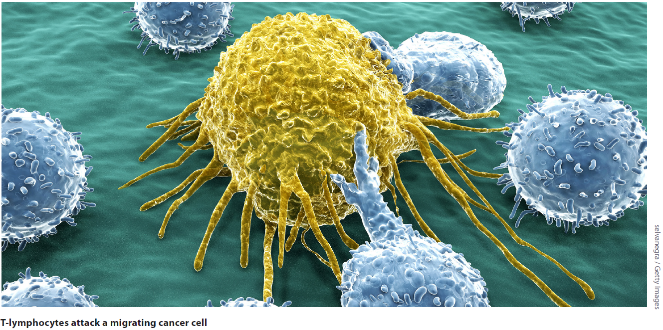
Immunoglobulins are another example of protein biomarkers that can be an active part of the testing procedure. Antibodies have been an essential part of lab diagnostics for decades. Their use in enzyme-linked immunosorbent assays (ELISA) or other such immunoassays are integral to “wet-lab” style of bioanalytical methods. Antibodies are typically produced by the immune system in response to infections, yet in some instances of cancer, the immune system produces immunoglobulin molecules to control the growth of various tumors. This innate immunotherapy did not go unnoticed by research scientists and over the years many of them have intently tried to stimulate the immune system, in a variety of manners, to keep cancerous growths in check—effectively spawning the burgeoning field of Immuno-oncology therapeutics.
Keeping It All in Check
Since immunotherapy continues to revolutionize the cancer treatment landscape, the American Society of Clinical Oncology has bestowed upon it the moniker of top cancer advance for 2016. Currently, checkpoint inhibitors represent some of the most exciting therapies in the last 20 years, demonstrating extraordinary rates of long-lasting responses for a variety of the most difficult-to-treat cancers. Though for all their success, many patients don't respond to immunotherapies and in some cases can have serious, life-threating side-effects—owing to the growing demand for biomarkers that can discern which patients will positively respond to treatment.
Two of the most relevant protein biomarkers in recent years are the programmed cell death protein 1 (PD-1), a surface receptor belonging to the immunoglobulin superfamily, and its ligand (PD-L1) a small transmembrane protein that has been implicated in playing a major role in immune suppression. PD-1 is expressed in most tumor-infiltrating T cells, including antigen-specific CD8+ T cells. Moreover, PD-1 expression occurs after T-cell activation and has been used as a marker of T-cell exhaustion.
The PD-1 receptor has the capacity to bind two ligands, PD-L1 and PD-L2. Typically, when PD-1 is bound to its ligands the result is the elimination of activated effector T-cells once they have completed their tasks—thus making PD-1 an immune checkpoint molecule. When the interaction of PD-1 and PD-L1 is disrupted, for instance with checkpoint inhibitor antibodies such as nivolumab (Opdivo), T cells remain activated, seeking out cancer cells to eliminate. PD-L1 expression has been detected in a number of tumor types, including melanoma, non-small cell lung cancer (NSCLC) renal, and ovarian cancers—underscoring the importance of checkpoint inhibitor drugs.
Because of its role in immune suppression and expression pattern in cancerous tissues, PD-L1 has begun to emerge as a potential prognostic marker. Reporting in the April issue of Oncotarget, Chowdhury et al., studied the effectiveness of using PD-L1 as a predictive biomarker for papillary thyroid carcinoma (PTC) and its variants. Using standard immunohistochemical (IHC) staining techniques, the researchers analyzed “251 archived formalin fixed and paraffin embedded (FFPE) surgical tissue samples (66 benign thyroid nodules and 185 PTCs).” The investigators found that “PD-L1 positive expression in PTC correlates with a greater risk of recurrence and shortened disease-free survival supporting its potential application as a prognostic marker for PTC.”
Scheel et al., reporting ahead of print in August for Pathologe, found similar results with the use of IHC methods when surveying for PD-L1 biomarker expression in NSCLC tissue. “The use of PD-L1 IHC in NSCLC is suitable for identification of patients with an increased probability of a clinical benefit from immunotherapy,” the authors wrote. “The various proportional cut-offs used to interpret the staining results can be summarized in a total score, which can be reproducibly assessed. The staining patterns of the four assays investigated were, however, not congruent in all situations.”
Different Direction, Similar Endpoint
The PD-1/PD-L1 molecules aren't the only high-profile protein biomarkers associated with immunotherapy. The cytotoxic T-lymphocyte-associated protein 4 (CTLA-4) is a receptor that also functions as an immune checkpoint, downregulating immune responses. CTLA-4 is constitutively expressed in regulatory T cells (Tregs) after their activation, serving as an off switch when bound to its ligands CD80 or CD86 present on the surface of antigen-presenting cells (APC). CTLA-4 transmits an inhibitory signal to Tregs, causing them to deactivate—the disruption of which is the basis for the development of antagonist drugs such as ipilimumab (Yervoy). Conversely, researchers have developed CTLA-4 agonist drugs such as abatacept (Orencia) as potential therapies for autoimmune disorders.
Previous work done to track the expression patterns of CTLA-4 have shown that the receptor is constitutively expressed in a variety of cancers—in particular, melanoma, for which ipilimumab has been FDA approved for treatment of the disease. Due to the remarkable success of treating melanoma patients with checkpoint inhibitors that target the CTLA-4 pathway, investigators are now attempting to validate CTLA-4 as a prognostic marker for other tumor types.
Reporting recently in the Journal of Thoracic Oncology, Deng et al., looked at the roles of CTLA-4 and PDCD1 (gene responsible for PD-1 production) expression in lung cancer patient samples. “We used a lung cancer database of 1715 patients measured by Affymetrix microarrays to analyze the association of gene expression with clinicopathological factors and survival,” the authors penned.
The researchers found that in 909 patients with histology information, a significantly higher level of PDCD1 and CTLA-4 expression were observed for squamous carcinoma than adenocarcinoma. Moreover, the authors found that in 848 patients with known smoking history, current and former smokers were found to have significantly elevated gene expression when compared to nonsmokers. “In this study with large number of patients, PDCD1 and CTLA-4 expression was significantly higher in squamous carcinoma and current/former smokers,” the authors wrote. This led the researchers to conclude that “higher expression of CTLA-4, but not PDCD1 predicts worse survival.”
A New Day Dawns
The era of immunotherapy has descended upon clinical medicine, offering scientists and physicians the opportunity to dramatically improve disease outcomes. However, the path toward better prognosis and treatment options is not unfettered, and translational biomarker research is one way to overcome many of the limitations of immunotherapy. Increased validation of novel biomarkers that provide insight into whether immunotherapies will be successful for individual patients, especially when the timing of treatment is a factor, as it most often is for cancer, is essential.
While putative examples of critical immunotherapy biomarkers for cancer have been presented here with proteins from the immune checkpoint pathways (PD-1, PDL-1, and CTL-4), the search continues for the next soluble factor or cell-surface receptor that is wholly indicative of disease and open the door to even better immunotherapy compounds.