




Abstract

Advances in precision medicine are determined, in large part, by the ability to more carefully and completely analyze and understand the genome. This requires new developments in how we look at DNA that go way beyond sequencing data.
The new developments in this space promise limitless creativity and versatility, from methods that detect DNA in such small amounts that rare variants can be found, to tools that uncover the 3D architecture of the genome to facilitate further insights into structural variation. Such advances could dramatically improve patient outcomes by allowing clinicians to follow a tumor’s progression through a simple blood draw, or discover a bacterial infection before the patient even feels sick. The work at the forefront of precision medicine is original, exciting, transformative, and moving at breakneck speed.
Knowing the Locations of Loci Could Transform Genomics
The structural variation of the genome is a hot topic, with many companies developing novel ways of making strides in this area. One such company, Phase Genomics, located in Seattle, WA, has developed a library preparation kit that has the potential to transform genome science.
The approach used by Phase Genomics to study the 3D architecture of genomes comes from work done in the laboratory of Jay Shendure, M.D., Ph.D., professor of genome sciences at the University of Washington. The method is based on Hi-C, which was first developed about a decade ago in the laboratories of Job Dekker, Ph.D., professor of biochemistry and molecular pharmacology at the University of Massachusetts Medical School, and Eric Lander, Ph.D., professor of biology at the Massachusetts Institute of Technology.
“Although Hi-C has been around for a while, the way that Phase Genomics is applying it is novel,” said Ivan Liachko, Ph.D., and CEO of Phase Genomics. “Hi-C data is somewhat esoteric, but extremely powerful. It provides an orthogonal datatype that can supplement all sorts of genomic efforts.”
Ivan Liachko, Ph.D., CEO, Phase Genomics

Hi-C starts by crosslinking the DNA within intact cells, causing interacting loci to bind to one another before sequencing. In sequencing the junctions that are formed, Hi-C allows the capture of the sequences that are physically close to each other in the cell.
There are multiple applications for this type of sequence analysis, one of which is assembling a genome with ultralong genomic contiguity without the requirement of high molecular weight DNA. Indeed, Phase Genomics, in collaboration with PacBio, published the goat genome last year—the highest contiguity de novo genome ever assembled. And during the second week of October this year, PacBio announced that it has produced the most contiguous diploid human genome assembly of a single individual to date, representing the nearly complete DNA sequence from all 46 chromosomes inherited from both parents. This was done, again, using technology from Phase Genomics.
An additional application is in microbiome research, enabling the identification of which specific sequences start in the same cell from a large, heterogeneous sample of bacteria and other microbes. This is especially important when dealing with mobile elements such as plasmids or transposons—pieces of DNA that can move antibiotic resistance genes from species to species. “Because the crosslinks occur inside intact cells, any two loci that interact by Hi-C must have originated in the same cell, and this data can be used to deconvolute high-quality genomes directly from mixed populations,” explained Liachko.
Researchers at Karius are leveraging NGS for infectious disease diagnosis through the detection of cell-free DNA (cfDNA).
seksan Mongkhonkhamsao / Getty Images

Liachko said that sometimes, “you need more than a sequence. Sometimes you need to know the location of a piece of DNA.” Proximity ligation tools such as Hi-C allow the capture of sequences that are physically close to each other in the nucleus, enabling haplotype phasing—or determinations of which two sequences are on the same chromo-some, and of which mutations are on the same haplotype. By allowing genomic structural and phase information to be obtained in just a day from a library preparation kit, the Phase Genomics technology gives genomic science a power it previously lacked and sorely needed.
Identifying ‘Fevers of Known Origins’
Although next-generation sequencing (NGS) may be best known for its use in oncology, the researchers at Karius are using it to transform infectious disease diagnosis through the detection of cell-free DNA (cfDNA). The test that Karius has developed is currently able to detect more than 1,000 disease-causing pathogens, including protozoa, bacteria, fungi, nematodes, and viruses, by identifying microbial cfDNA in patients’ plasma.
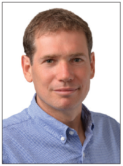
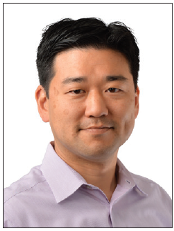
Mickey Kertesz, Ph.D., cofounded Karius in 2014 to bring the pathogen detection technology he and colleagues developed at Stanford University into the hospital. One of Kertesz’ Stanford colleagues, David Hong, M.D., is now Karius’ vice president of medical affairs and clinical development. Hong joined Karius four years ago because of the problems that he was facing on the pediatric infectious disease ward—problems common to hospitals everywhere.
Mickey Kertesz, Ph.D., Founder, Karius
In a hospital, every patient is at risk for infection, and to Hong’s frustration, the technology available for detecting infections is decades old with many limitations.
“Taking an NGS approach yields more exact information,” Hong said. “It not only tells us if an infection is present, it also tells us exactly what is causing the infection. This information is incredibly useful for a clinician who needs to know how to effectively treat infections.”
In early October, Karius released results of a clinical trial, PREDSEQ, that started roughly one year ago. Working in conjunction with St. Jude Children’s Research Hospital in Memphis, TN, the pilot study enrolled high-risk pediatric leukemia patients. Hong indicates that the study was trying to “answer the question of whether a pathogen was present before it was able to be cultured.” Indeed, the results showed that Karius could detect a pathogen two to four days prior to clinical symptoms, providing proof of concept of a tool that can identify patients who are about to get sick.
The test developed by Karius can accurately detect pathogens in diagnostically challenging or culture-negative infections or in deep-seated infections where cultures are hard to obtain. Additionally, it could allow for immunocompromised patients to be more actively monitored and for antimicrobial therapies to be more precisely determined.
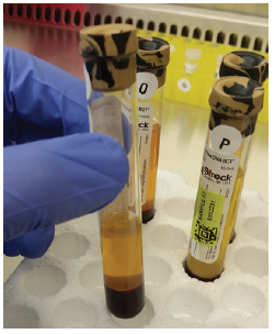
The Karius system detects traces of pathogen cfDNA in plasma samples after sifting through the enormous bulk of human cfDNA. The system also has to discount cfDNA from commensal bacteria present in the human microbiome. Essentially the system finds the proverbial needle in the haystack, and it does so by using agnostic whole-genome processing while eschewing primers and probes.
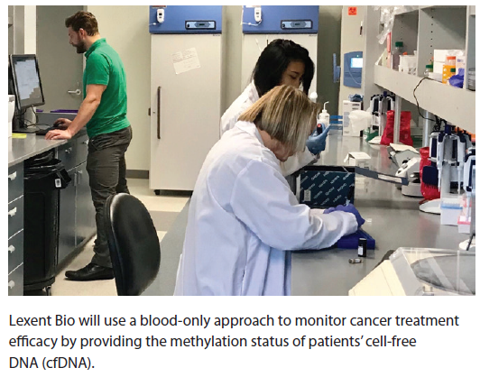
Lexent Bio will use a blood-only approach to monitor cancer treatment efficacy by providing the methylation status of patients’ cell-free DNA (cfDNA).
According to Hong, “Karius curated its database of pathogens to include common pathogens of immunocom-promised patients and others that are notoriously difficult to diagnose such as the non-Aspergillus fungal species.”
Looking ahead, Hong imagines a world where high-risk patients are treated preemptively for their infections, based on their NGS results—similar to how stem cell patients are treated now with the constant monitoring of their cytomegalovirus antibody titers.
Adding Precision to Sequencing
While the goal of some companies may be to sequence as many people as possible, Mission Bio’s goal is much smaller, but not any less significant. Charlie Silver, cofounder and CEO, said that Mission Bio is “the only company that sequences DNA mutations in every cell in the sample” and that is driven by the idea that it can “sensitively assay and determine all of the clones that are present in a tumor.” By doing that, the company can, he asserts, “better line up precision medicines and the right combinations to be able to more effectively treat patients.”
Mission Bio’s technology is derived from research that was conducted in 2014 in the laboratory of Adam Abate, Ph.D., associate professor of bioengineering at the University of California, San Francisco. The company’s platform, called Tapestri, uses microfluidics to partition thousands of cells into individual droplets. Each cell, then, occupies its own miniature reaction vessel. The cell is lysed, and its DNA is combined with barcoded beads, primers, and other reagents. The barcode indicates the cell’s identity and mutational profile.
After thermal cycling, the product that comes out of Tapestri can be processed in NGS workflows, generating clonal information that may be analyzed by Mission Bio software. The Mission Bio system results in high-throughput single-cell DNA mutation sequencing and characterizes cell-to-cell genomic heterogeneity.
In cancer patients, this information could be used to measure treatment outcomes or guide tweaks to treatment protocols. “In almost all tumor populations, there is fundamental genetic heterogeneity that exists,” said Dennis Eastburn, Ph.D., cofounder and CSO at Mission Bio. “Bulk sequencing provides an average of the genetic varients found in a sample, but you might miss the genetic diversity between cells.”
“Our method,” he asserted, “enables rapid and cost-effective targeted genome sequencing of thousands of tumor cells in parallel.” In addition, Tapestri could be used to identify clinically actionable rare cancer cells that come back after treatment in what is known as minimum residual disease.
Phase Genomics is applying a novel approach to analyzing Hi-C data.

In a recently published proof-of-concept paper in Genome Research entitled, “High-throughput single-cell DNA sequencing of acute myeloid leukemia tumors with droplet microfluidics,” Mission Bio researchers described how they sequenced longitudinally collected acute myeloid leukemia (AML) tumor populations from two patients and genotyped up to 62 disease-relevant loci across more than 16,000 individual cells.
The researchers stated that “targeted single-cell sequencing was able to sensitively identify cells harboring pathogenic mutations during complete remission and uncovered complex clonal evolution within AML tumors that was not observable with bulk sequencing.” Based on these results, the researchers proposed that the ability to characterize genetic heterogeneity in tumor cell populations “will make feasible the routine analysis of AML heterogeneity, leading to improved stratification and therapy selection for the disease.”
The potential applications for the Tapestri platform are widespread, reaching areas outside of oncology. For example, Mission Bio recently joined the National Institute of Standards and Technology (NIST) consortium for genome editing which “addresses the measurements and standards needed to increase confidence and lower the risk of utilizing genome-editing technologies in research and commercial products.”
In this role, Tapestri can perform single-cell analysis in the context of genome editing. It can identify if edits occurred in a homo- or heterozygous manner and if multiple edits have occurred in the same cell type. The information that is needed to avoid pitfalls in making gene editing safe for patients, for example, in the introduction of off-target edits or DNA rearrangements, is now possible with the Tapestri platform.
Determining the Response to Cancer Treatment
Haluk Tezcan, M.D., CMO at Lexent Bio, said that “oncologists are data driven.” If Tezcan has his way, they will get the data they need from his company.
Lexent, one of the newest companies in precision medicine, will use a blood-only approach (no tissue biopsy needed) to follow cancer over a course of time by providing the methylation status of patients’ cfDNA.
Lexent’s CEO Ken Nesmith said the company is “not focused on diagnosing disease.” Rather, the company’s “liquid biopsy technology will help oncologists and their patients understand if the patients are responding to treatment, sooner and more accurately than currently possible.”
To ensure that this information can be generated, the Lexent team makes a point of evaluating the best methods to map the cell-free methylome. In one study, company scientists compared bisulfite conversion of cfDNA followed by whole-genome bisulfite sequencing (WGBS) and bead-based fractionation of cfDNA followed by whole-genome sequencing (WGS). In applying these two approaches to cfDNA from patients with advanced-stage cancer and healthy controls, the scientists found that methylation differences in CpG islands and shores, but not differences in gene bodies or promoters, were effective at segregating cancer samples from healthy samples.
David Hong, M.D., VP of medical affairs and clinical development, Karius
The scientists also found that WGBS was a more sensitive method to detect changes in hypomethylated regions. By analyzing the WGS and methylation status of cfDNA, Lexent hopes to provide information as to whether there is progression of the cancer or not—a space where oncologists need better, faster information. Still in its infancy, Lexent has a goal of having clinical validation done over the next couple of years.
Footnotes
This story originally appeared in the November 15, 2018, issue of Genetic Engineering & Biotechnology News (GEN)