
Abstract
The hub genes and signaling pathways associated with Duchenne muscular dystrophy (DMD) were predicted by bioinformatic methods to improve the therapeutic effect and quality of life of patients. Microarray data sets GSE465, GSE1004, and GSE1007 were downloaded from the Gene Expression Omnibus (GEO) database. The differentially expressed genes (DEGs) were identified by GEO2R, and function enrichment analyses were performed by DAVID. The protein–protein interaction (PPI) network was constructed and the module analysis was performed using STRING and Cytoscape. A total of 195 DEGs were identified. The enriched functions and pathways of the DEGs include extracellular exosome, focal adhesion, extracellular matrix (ECM), focal adhesion, PI3K-Akt signaling pathway, calcium signaling pathway, and ECM–receptor interaction. Fifteen hub genes were identified. DEGs and hub genes identified in the present study help us understand the molecular mechanisms underlying the pathogenesis and progression of DMD, and provide candidate targets for treatment of DMD.
1. Introduction
Duchenne muscular dystrophy (DMD) is an inherited muscular systrophy related to the X chromosome and affects about 1 in 5000 boys (Yiu and Kornberg, 2015), which is caused by a mutation in a gene that encodes dystrophin and is the largest known human gene with a length of 2500 kb. Children with DMD, whose motor functional development is basically normal at birth or in early infancy, show progressive muscle atrophy during childhood. Most children lose the ability to walk after the age of 10, with the disease gradually progressing, died of heart failure or respiratory failure about 30 (Singh et al., 2018). At present, glucocorticoid is the most effective method in the treatment of DMD. In patients with DMD, confirmed by a long-term prospective cohort study, glucocorticoid treatment may reduce the risk of losing clinically meaningful mobility and upper limb disease progression milestones across the life span as well as risk of death (McDonald et al., 2018). However, glucocorticoids have many serious side effects, such as behavior change, osteoporosis, cataract, weight gain, and Cushing appearance. Although the FDA has finally approved the application of the drug Eteplirsen (exondy51) for the treatment of DMD, its long-term efficacy and safety still need to be further observed (Aartsma-Rus and Krieg, 2017). There is no fundamental treatment for DMD, and many methods of gene therapy, such as gene replacement therapy and antisense nucleotide method (AON), are being studied.
There are some doubts in the clinical treatment of DMD, which cannot be completely explained by the mutation of dystrophin. This suggests that there may be other genes involved in the pathogenesis of DMD. The identification and treatment of these genes may help to alleviate the suffering of patients with DMD and enhance the treatment effect. In this study, bioinformatic methods were used to identify potential hug genes related to DMD, which is hoped to improve the therapeutic effect and the quality of life of patients.
2. Methods
2.1. Microarray data
Three gene expression data sets were searched and downloaded from the Gene Expression Omnibus (GEO). Muscular Dystrophies is used as the key word of data retrieval. The study type is Expression profiling by array. The limiting species is Homo sapiens.
2.2. Identification of differentially expressed genes
The differentially expressed genes (DEGs) between DMD and normal samples were screened using GEO2R. Fold change (FC) ≥1.5 and p value <0.01 were considered statistically significant.
2.3. Kyoto Encyclopedia of Genes and Genomes and gene ontology enrichment analyses of differentially expressed genes
To analyze the function of DEGs, gene enrichment analysis was performed using the Database for Annotation, Visualization, and Integrated Discovery (DAVID, version 6.8) online database. FDR (false discovery rate) <0.05 and p < 0.05 were considered statistically significant.
2.4. Protein–protein interaction network construction and module analysis
The protein–protein interaction (PPI) network was predicted using Search Tool for the Retrieval of Interacting Genes (STRING, version 10.5) online database. The PPI networks were drawn using Cytoscape and the most significant module in the PPI networks was identified using Molecular Complex Detection (MCODE). The criteria for selection were as follows: MCODE scores >5, degree cutoff = 2, node score cutoff = 0.2, max depth = 100, and k-score = 2.
3. Results
3.1. Data retrieval results from gene expression omnibus
According to the selection criteria, three data sets were screened, namely GSE465, GSE1004, and GSE1007. There were seven different platforms in GSE465, among which the data of GPL8300 platform were excluded because there was no normal control group. There were two platforms in GPL1004, in which data based on GPL91 were removed because there were no samples of DMD. Therefore, a total of 90 cases of DMD and 75 cases of normal control samples were selected from the 3 data sets (Table 1).
Data Retrieval Results from Gene Expression Omnibus
DMD, Duchenne muscular dystrophy.
3.2. Identification of differentially expressed genes in Duchenne muscular dystrophy
Based on the standard, DEGs (2866 in GSE465GPL91, 816 in GSE465GPL92, 545 in GSE465GPL93, 415 in GSE465GPL94, 674 in GSE465GPL95, 1045 in GSE465GPL246, 2020 in GSE1004GPL8300, 1449 in GSE1007GPL92, 933 in GSE1007GPL93, 579 in GSE1007GPL94, and 986 in GSE1007GPL95) were identified. Genes appear above four results simultaneously (each GSE has at least one GPL platform) as DEGs and a total of 195 DEGs were obtained by taking the intersection of the above 11 results. Volcano plots of DEGs from GSE465 are shown in Figure 1. Volcano plots of DEGs from GSE1004 and GSE1007 are shown in Figure 2.
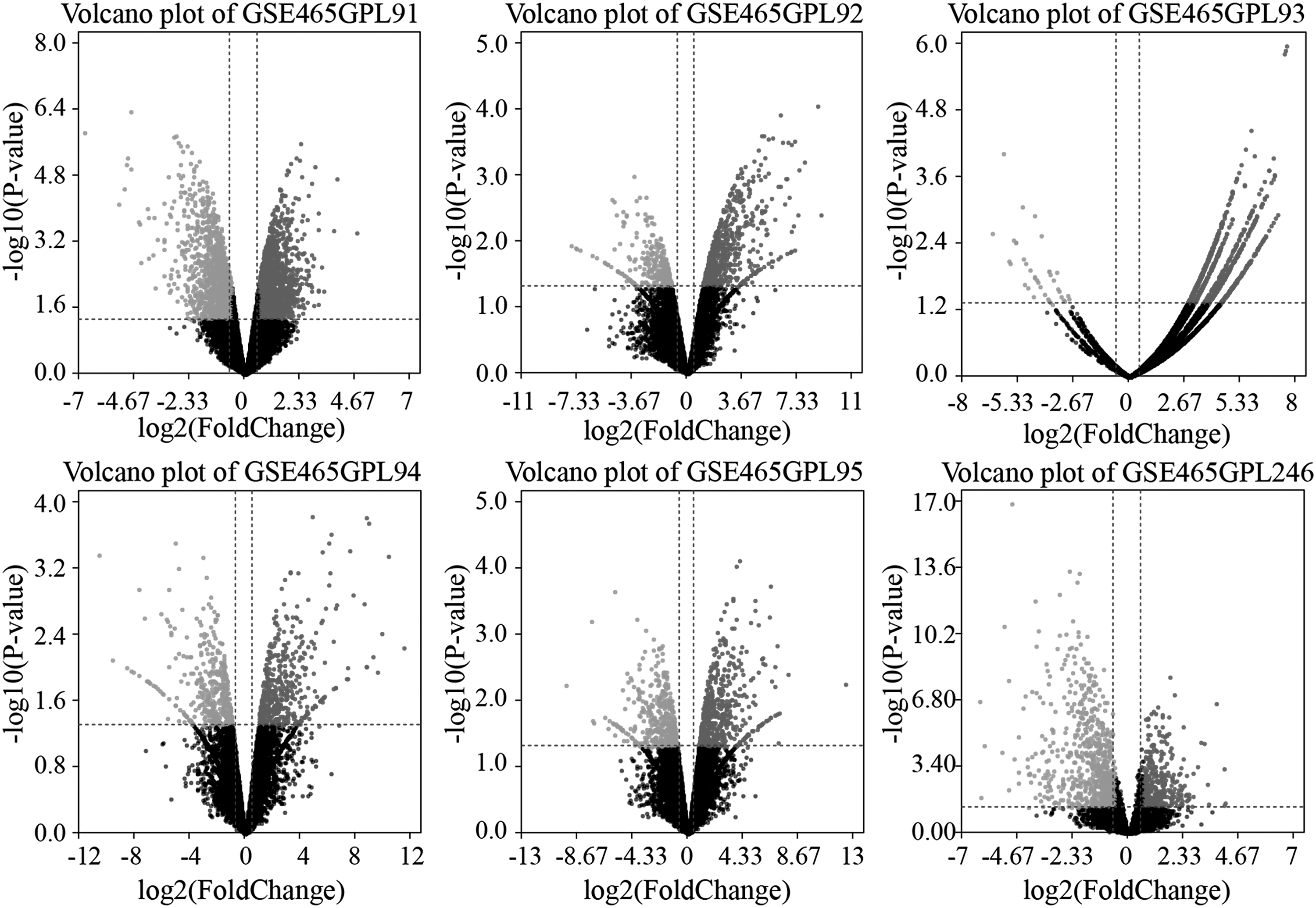
Volcano plots of DEGs from GSE465. DEG, differentially expressed gene.

Volcano plots of DEGs from GSE1004 and GSE1007.
3.3. Kyoto Encyclopedia of Genes and Genomes and gene ontology enrichment analyses of differentially expressed genes
Gene ontology (GO) analysis results (Table 2) showed that changes in the cellular component of DEGs were significantly enriched in extracellular exosome, focal adhesion, and extracellular matrix (ECM). Kyoto Encyclopedia of Genes and Genomes (KEGG) pathway analysis revealed that the downregulated DEGs were mainly enriched in focal adhesion, PI3K-Akt signaling pathway, calcium signaling pathway, and ECM-receptor interaction. Visualization of p value and quantity is shown in Figure 3.
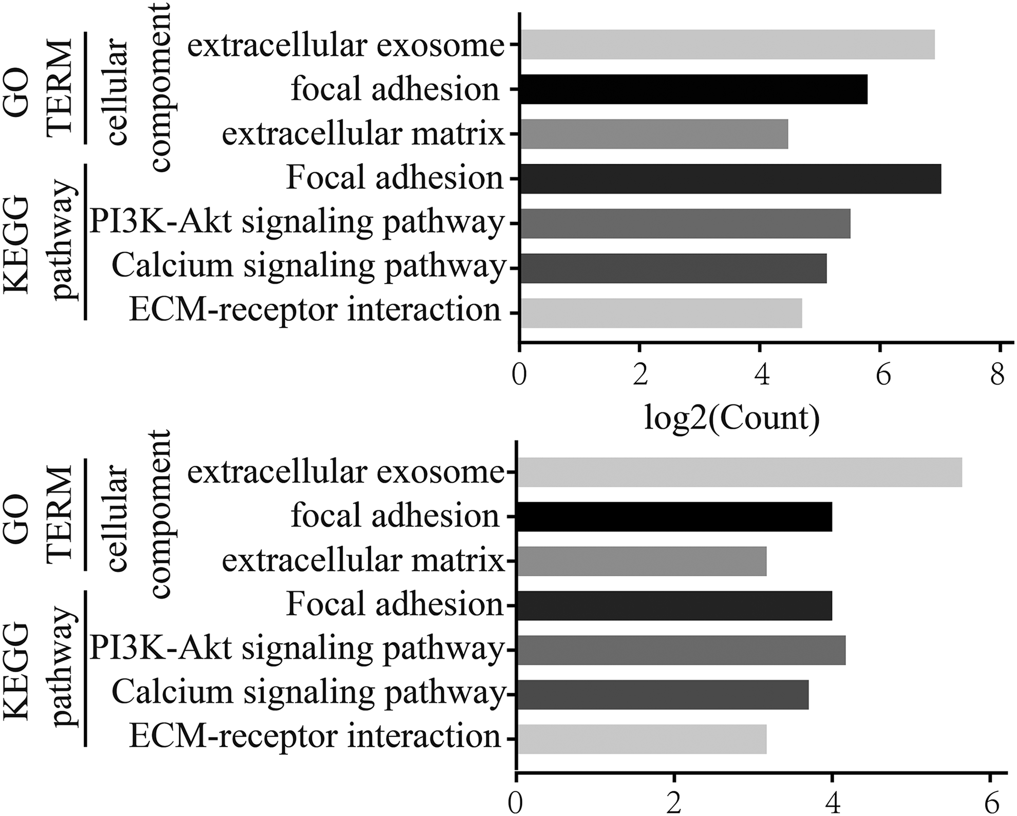
Visualization of p value and quantity.
Enriched Gene Ontology Terms and Kyoto Encyclopedia of Genes and Genomes Pathway Analysis of the Differentially Expressed Genes
GO, gene ontology; KEGG, Kyoto Encyclopedia of Genes and Genomes.
3.4. Protein–protein interaction network construction and module analysis
The PPI network of DEGs was constructed, which consists of 150 nodes and 408 interactive edges (Fig. 4A) and the most significant module, which is composed of 15 nodes and 56 interactive edges, was obtained using Cytoscape (Fig. 4B).
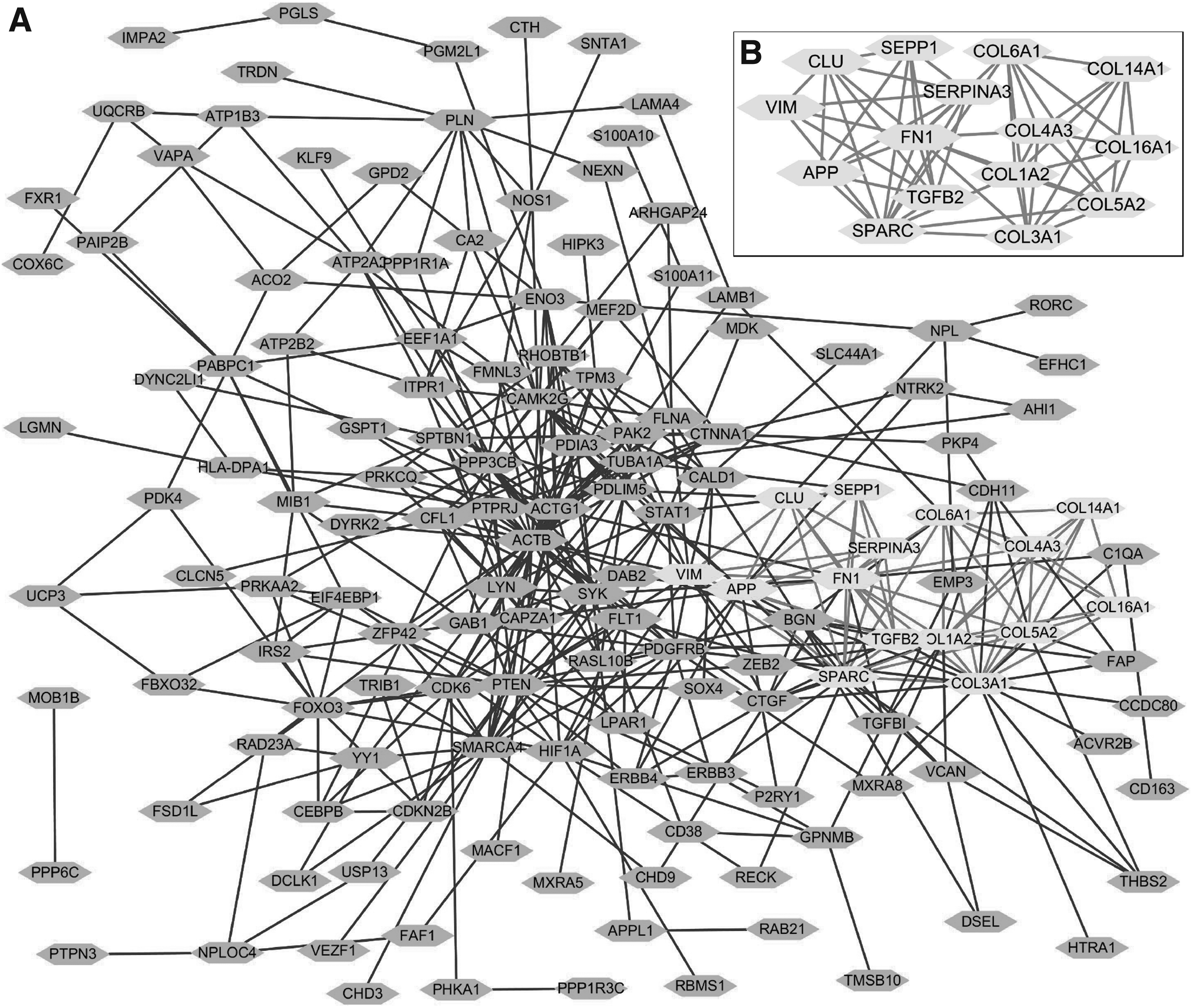
Protein–protein interaction network constructed with DEGs and results of MOCODE analysis.
4. Discussion
The cause of DMD is the mutation of the dystrophin located on chromosome 21X, which leads to the reduction of skeletal muscle cells or the lack of functional dystrophin, and eventually leads to cell death. The main purpose of DMD therapy is to improve muscle strength, exercise function, lung function, and myocardial protection. At present, the main treatment regimen is hormone therapy combined with ventilator support. There is no cure for the cause, and gene therapy is still under researching. There are gene therapy methods reported in literature, such as recombinant adeno-associated viral vector-mediated gene transfer (Odom et al., 2011), AON method with repair of specific gene mutations, Eteplirsen approved by FDA for DMD (Aartsma-Rus and Krieg, 2017), induced pluripotent stem cells that rely on the unlimited proliferation and differentiation of pluripotent stem cells (Dowling et al., 2018), and exon jump and exon excision dependent on CRISPR/CAS9 gene editing (Hotta, 2015; Ousterout et al., 2015).
There are still some questions in the clinical treatment and basic research of DMD. Some patients had complete deletion of the coding gene for the antiatrophic protein, which should be presented as the curved type of DMD, however, its clinical symptoms were consistent with the Becker muscular dystrophy. The clinical symptoms were different in two patients with the same exon deletion. The dystrophin gene has only a few base changes, resulting in slight changes in the dystrophin, whose clinical symptoms appear relatively late, however, the symptoms are very serious. These problems cannot be clearly explained by amyotrophic protein mutations. We speculate that there may be other genes involved in the pathogenesis of DMD.
GO analysis of DEGs in this study showed that changes in the cellular component of DEGs were significantly enriched in extracellular exosome, focal adhesion, and ECM. In the hub genes identified in this study, TGFB was involved in extracellular exosome, vimentin (VIM) was involved in focal adhesion, and TGFB, VIM, and COL14A1 were involved in ECM. KEGG signaling pathway analysis of DEGs showed that hub genes such as fibronectin 1 (FN1), COL1A2, and COL5A1 were involved in the focal adhesion pathway, hub genes such as COL3A1, COL6A1, and FN1 were involved in the PI3K-Akt signaling pathway, and hub genes such as COL3A1, COL1A2, and FN1 were involved in the ECM–receptor interaction.
Excessive ECM deposition gradually replaces muscle fiber as the endpoint of most serious muscle diseases. Collagens are important components of the ECM, and some studies reported that the genes COL1A1, COL3A1, COL6A1, and COL6A2 were closely related to DMD (Zanotti et al., 2015; Reddy et al., 2017). FN1 is also an important component of ECM and may be involved in the development of DMD. The elevation of TGFB2, which is related to the Wnt signaling pathway, can affect the autocrine or paracrine function of muscle satellite cells in patients with DMD (Biressi et al., 2014). SPARC is an important regulator of cytoskeletal proteins expressed during muscle development and regeneration by binding to actin. SPARC is highly expressed in muscle lesions such as DMD and has a strong correlation with DMD (Jorgensen et al., 2017). VIM is a key structure for maintaining cytoskeletal integrity, which provides the inelasticity of microtubulin and actin, and is involved in biological processes such as muscle contraction with DMD, which may be associated with DMD. It has been reported that SEPP1 is regulated by the glucocorticoid receptor (Rock and Moos, 2009), and glucocorticoids are at present the most effective drug for treating duodenal dystrophy; therefore, the SEPP1 gene may be associated with the development of DMD. Studies have shown that Serpina3n increases in cyclic release during glucocorticoid-mediated muscle atrophy (Gueugneau et al., 2018). Genes downregulated by mineralocorticoid receptor (MR) antagonism included FOS, ANKRD1, and GADD45B, with known roles in skeletal muscle; NPR3 and SERPINA3 were bona fide targets of MR, and it can be speculated that SERPINA3 may be involved in the pathologic process of DMD (Chadwick et al., 2015).
Focal adhesion pathways were significantly enhanced between DMD pathology and normal cases (Narayanan and Subramaniam, 2013). In animal studies, the sustained activation of Src kinase through PI3K/Akt phosphorylation leads to the activation of mTOR's autophagy inhibitor target, and the inhibition of Src kinase may partially rescue defective autophagy and lysosomal biogenesis (Pal et al., 2014). Inositol 1,4,5-trisphosphate (IP3)-dependent Ca2+ signaling mediates delayed myogenesis in DMD fetal muscle (Farini et al., 2016). However, the relationship between ECM receptor interaction pathways and DMD has not been reported.
In conclusion, the present study was designed to identify DEGs that may be involved in the pathogenesis or progression of DMD. A total of 195 DEGs and 15 hub genes were identified and may be regarded as biomarkers for DMD. However, further studies are needed to elucidate the biological function of these genes in DMD.
Footnotes
Acknowledgments
We sincerely appreciate the editor and reviewers of the journal for their helpful comments on this article. Funding: This work was supported by the National Natural Science Foundation of China (grant no. 81772346).
Availability of Data and Materials
The data sets used and/or analyzed during the present study are available from the corresponding authors on reasonable request.
Author Disclosure Statement
The authors declare that no competing financial interests exist.