




Abstract
The aim of this study was to investigate perceptual eye position (PEP) and to evaluate the effect of dichoptic visual perceptual training in postoperative intermittent exotropia [X(T)]. We enrolled 30 non-strabismus children (control group) and 54 postoperative X(T) children [divided into training group (33 patients) and non-training group (21 patients)]. All subjects received measurements of PEP, and the postoperative X(T) children were measured both in the third postoperative day and the third postoperative month. All patients in training group received 3-month dichoptic visual perceptual training based on a unique virtual reality platform. The results showed that the postoperative X(T) children with normal eye position still had an abnormal PEP. After a period of visual perceptual training, the PEP pixels in postoperative children dramatically decreased. The results revealed that PEP could evaluate fixation disparity and binocular visual function perceptively and precisely, and the dichoptic visual perceptive training may rebuild binocular visual function.
Introduction
Intermittent exotropia [X(T)] is the most common form of exotropia.1–3 Binocular visual function is often disturbed by X(T).4,5 Surgery is a commonly used treatment especially for patients with a large angle of deviation.6,7 But the relapse is common in postoperative X(T), even with a successful surgical alignment. 8 According to previous study, only about 30–75 percent of cases regained normal binocular visual function after a successful surgical realignment.9–13 And the abnormal postoperative binocular conditions may increase the danger of relapse. So, the assessment and reconstruction of the binocular visual function in postoperative X(T) is important for best long-term results.
Perceptual eye position (PEP), used to describe sensory binocular alignment, is a standard psychophysical approach and one of the indicators evaluating fixation disparity and binocular visual function. PEP is measured under a dichoptic vision condition. What's more, the PEP pixels are recorded by a computer so that binocular misalignment can be quantified more perceptively and precisely.
In this study, we use PEP to detect the binocular visual function of postoperative X(T) with normal eye position. Furthermore, we conduct a study on the effect of dichoptic visual perceptual training based on a virtual reality (VR) platform in postoperative X(T) patients to evaluate the potential success of this therapy.
Methods
Participants
Thirty non-strabismus children and 54 children who had undergone strabismus surgery for correction X(T) were recruited from the ophthalmology department of West China Hospital, Sichuan University, from February 1, 2018 to June 30, 2019. All 54 postoperative X(T) children had a perfect ocular alignment, which were determined through prism plus cover test by one specialist (Y.L.). Subjects with any other ocular disease were excluded from this study. Therefore, the 54 postoperative X(T) children were divided into two subgroups based on their parents' choice, one group (31 patients) were given some visual perceptual training (training group [TG]), the other not (non-training group [non-TG]). All subjects received measurements of PEPs, and the postoperative X(T) children were measured both at the third postoperative days and the third postoperative month.
Measurement of PEP
The system automatically records vertical and horizontal bias by the 360° test object to observe any ocular misalignment. The devices, used to measure PEP, include Windows XP system PC host, LG2342p polarized three-dimensional (3D) monitor with a resolution power of 1920 × 1080 and refreshing frequency of 120 Hz, and 3D polarized glass. A visual and perceptual examination evaluation system, invented by the National Engineering Research Center for Healthcare Devices, was used. PEP was measured by the cross-into-circle test, which allowed the left eye to see a cross and the right eye to see a circle (Fig. 1). The midpoint of the monitor was held 80 cm away and as high as the position of patients' eyes, with the average light source of 80 cd/m2 in white, attenuating to 50 cd/m2 when the patients wear 3D polarized glasses, and 30 cd/m2 in black, attenuating to 3 cd/m2 when the patients wear 3D polarized glasses. The stimulating template was 51 circles was 0.4° × 0.4°, whereas the size of the cross was 0.33° × 0.33° (1° fixation test object). Patients used a computer mouse to place within what they perceived to be the circle's center, and were then instructed to click the mouse. The system automatically recorded vertical and horizontal bias by the 360° test object to observe any ocular misalignment. The minimum unit of ocular misalignment observed by this computer-controlled ocular misalignment system was 1 pixel, which equals 0.04 prism. To distinguish from conventional eye position, we defined this bias pixel as PEP.
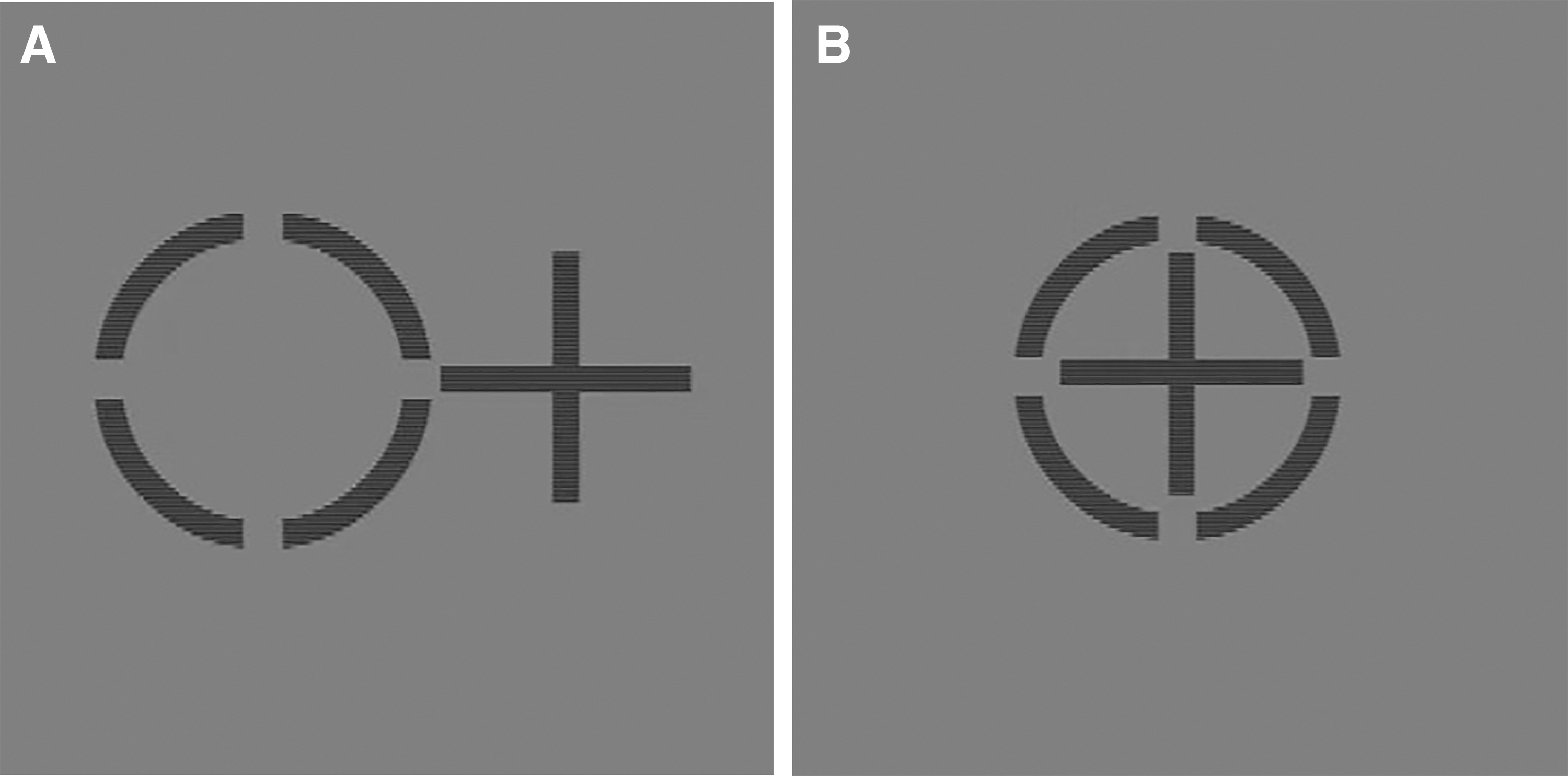
Measurement of PEP. By wearing 3D polarized glasses, patients should see a cross in their left eye and a circle in their right eye
Methods of visual perceptive training
In TG, we used an improved push–pull model training system provided by National Engineering Research Center for Healthcare Devices. In this training system, first we inspect the binocular integration relationship through a series of visual sensory examinations, then we give them a short period (about 5–10 minutes) neuroplastic training to obtain the detailed parameters. Finally, we design a personalized dichoptic visual perceptive training plan based on a VR platform for every patient of TG. All patients in TG were asked to wear VR binocular visual tools and did a 20-minute personalized dichoptic visual perceptive training two times per day for 3 months.
Statistical analysis
All data were obtained by mean ± standard deviation. Comparison among the three groups was made by using the one-way analysis of variance, and comparisons between two independent groups were made by using a two-tailed paired samples t test, with p value <0.05 being considered statistically significant. All data were analyzed with SPSS software (version 23.0; SPSS, Inc., Chicago, IL).
Results
A total of 84 subjects were recruited in the study and divided into three groups: 33 postoperative X(T) children in TG, 21 postoperative X(T) children in non-TG, and 30 non-strabismus children in control group. There were no statistically significant differences in gender (p = 0.059) or age (p = 0.156) of the patients among the three groups (Table 1). Details are shown in Tables 2 and 3 and Figures 2 and 3.
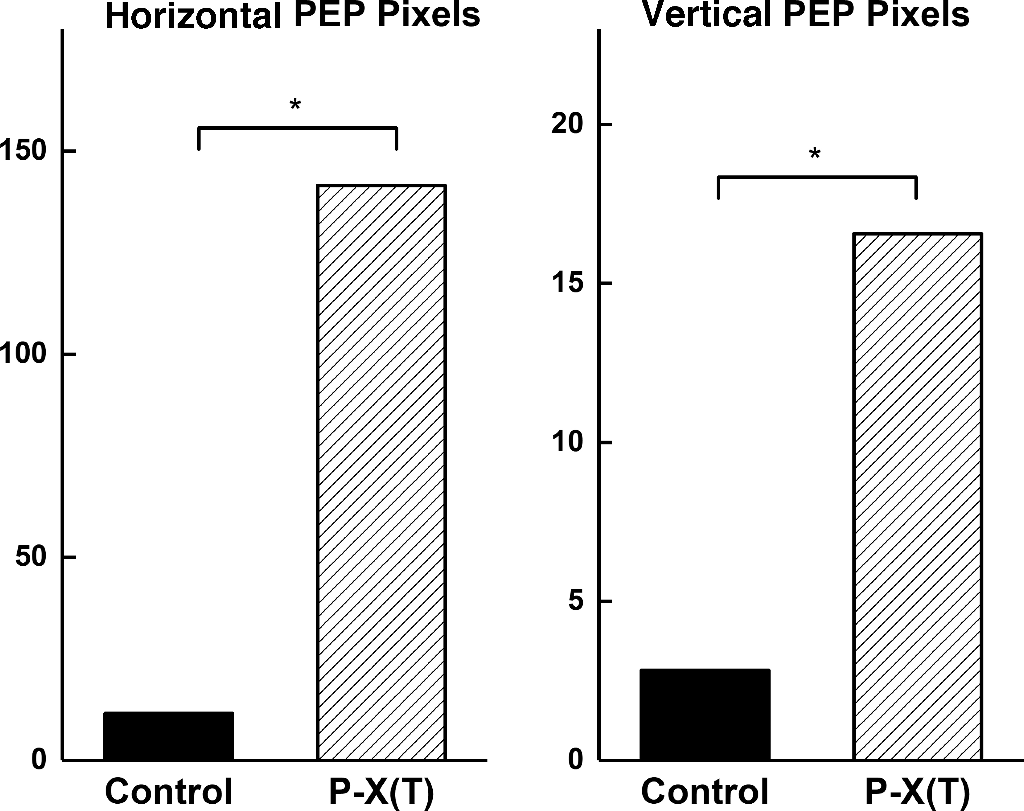
Comparisons of the PEP pixels in controls and P-X(T) patients. *P-X(T) versus control, p < 0.05. P-X(T), postoperative intermittent exotrope.
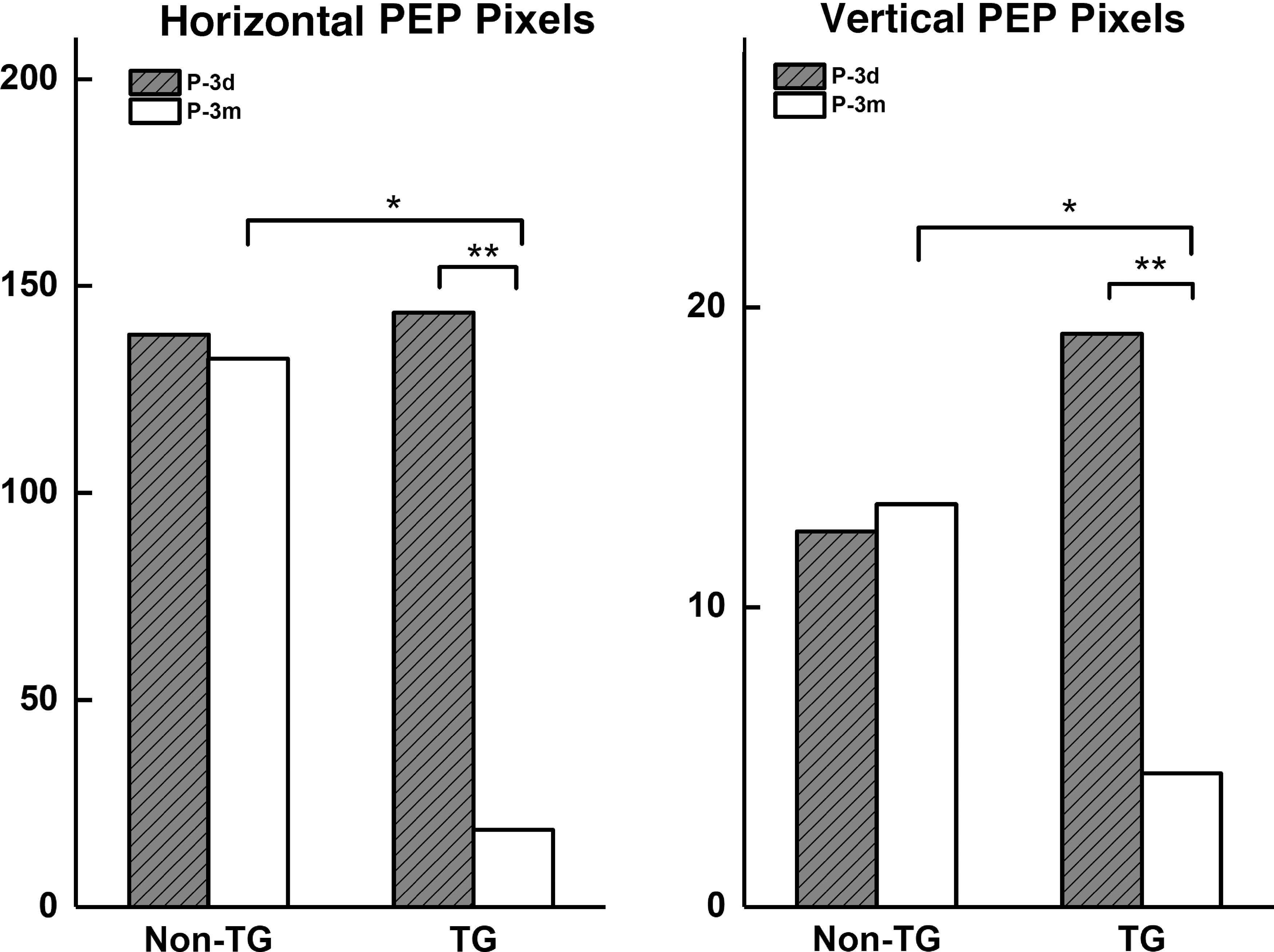
Comparisons of the PEP pixels in training group and non-training group in postoperative 3 days and postoperative 3 months. *Non-TG in postoperative 3 months versus TG in the same time, p < 0.05; **TG in postoperative 3 days versus in 3 months, p < 0.05. Non-TG, non-training group; TG, training group.
Participant Characteristics
Values are means ± standard deviations for all subjects in each group.
Control, control group; non-TG, non-training group (without visual perceptual training); Num., the number in each group; P-X(T), postoperative intermittent exotrope; TG, training group (with visual perceptual training).
The Vertical and Horizontal Perceptual Eye Position Pixels in Control Group and Postoperative Intermittent Exotrope Group
Values are means ± standard deviations for all subjects in each group. Bold p value represents <0.05.
The Vertical and Horizontal Perceptual Eye Position Pixels of Postoperative Intermittent Exotrope Patients in Postoperative 3 Days and Postoperative 3 Months
Values are means ± standard deviations for all subjects in each group. Bold p value represents <0.05.
Discussion
In clinical practice, the traditional definition of eye position is described and measured by the prism plus cover test, which is convenient for clinical assessments, but not sufficient in providing a quantitative measurement of the sensory eye balance. PEP reflects the integration of a variety of substances in the human brain after they act upon the sensory organs directly.14–16 PEP pixels can be measured by the computer-controlled perceptual examination evaluation system under a dichoptic vision condition.14–16 The previous studies have shown that the sensory eye balance is abnormal in patients with amblyopia, anisometropia, and strabismus. 14
In our study, we quantitatively assessed the sensory eye dominance of surgically corrected X(T) with normal apparent eye position. Using measurement of PEP, we show clear evidence that the two eyes are still unbalanced even though they have a normal eye position after the surgery. We found that most of X(T) after a successful strabismus surgery with normal clinical eye position also had an obvious abnormal PEP, compared with the non-strabismus children with a normal PEP. This may be attributed to anomalous retinal correspondence, which remapped the usual relationship between the two eyes.17–19 Compared with the eye position measured by traditional way, the PEP shows a more precise result.
More importantly, we divided the postoperative X(T) children into two groups, one group with some dichoptic visual perceptive training based on a unique VR platform (TG) and the other without any training (non-TG). After 3 months, we measured PEP pixels in all 54 postoperative X(T) children again. We found that, interestingly, the PEP pixels in TG dramatically decreased, whereas the PEP pixels in non-TG almost did not change any more. The training based on computer-controlled perceptual examination could rebuild sensory eye balance to obtain satisfying binocular visual function. Maybe the computer-controlled perceptual examination can reduce the relapse of X(T), by reconstructing the binocular retina corresponding points after strabismus surgery.
In summary, this preliminary study reveals that a functional eye imbalance still remains in surgically corrected cases that have clinically normal eye position with normal stereopsis. Our results demonstrate that the successful postoperative X(T) children still had an abnormal PEP when comparing with the non-strabismus children with a normal PEP, but the PEP pixels after visual perceptual training dramatically decreased. Our study provides the possibility that a further measurement of the PEP might reveal the sensory eye dominance, and a perceptive treatment, aiming at the rebalance of the ocular dominance, might be necessary for binocular stability and reconstruction of the binocular visual function in postoperative X(T).
Footnotes
Author Disclosure Statement
No competing financial interests exist.
Funding Information
This study was supported by the Major Program, International Science and Technology Cooperation Program of Science and Technology Program of Guangzhou, China (Grant No. 201704020048) and Science and Technology Planning Project of Guangdong Province of China (Grant No. 2017B010110013).