
Abstract
Background:
Advanced glycation end products (AGEs) are tissue proteins that accumulate with age and in diabetes mellitus (DM). AGEs can be measured by the AGE-Reader (DiagnOptics Technologies BV, Groningen, The Netherlands), which measures skin autofluorescence (SAF). SAF has been suggested as a measure to screen for undiagnosed DM or impaired glucose tolerance. SAF has never been investigated in gestational DM (GDM). Therefore we compared SAF at diagnosis in GDM patients with normal pregnancy. If SAF is elevated in GDM, future research could focus on the possible use of the AGE-Reader as a screening method for GDM.
Methods:
In this monocenter observational study SAF was measured in 60 GDM patients at diagnosis and 44 pregnant women without diabetes.
Results:
SAF did not differ between GDM at diagnosis (mean [SD], 1.74 [0.31] arbitrary units) and normal pregnancy (1.76 [0.32] arbitrary units). SAF was lower in white European patients than in patients with other ethnicity.
Conclusions:
This first study of tissue AGE accumulation in pregnancy shows no differences in SAF between women with GDM at diagnosis and normal pregnancy. This is most likely due to mild severity and short duration of hyperglycemia in GDM at diagnosis, but it does not exclude potential differences in SAF later in pregnancy. However, the fact that no differences are detected at diagnosis makes it unlikely that the AGE-Reader can be developed as a screening method for GDM in the future. Furthermore, we found that ethnicity should be taken into account when measuring SAF.
Introduction
Gestational DM (GDM) is considered to be a mild form of diabetes. Nevertheless, GDM is associated with an increased incidence of maternal and fetal/neonatal complications. 13 Glucose levels are associated with adverse outcome in a linear way without any obvious thresholds above which risks are elevated. 14 Recent intervention trials have shown that treatment of hyperglycemia can improve pregnancy outcome. 15,16
Prior studies found elevated serum AGEs in GDM, 17 –19 but data on tissue AGEs are lacking. The only study investigating tissue AGEs (through skin autofluorescence [SAF]) in relation to pregnancy showed elevated SAF in recently preeclamptic women; however, in that study SAF was measured 6–7 months after delivery and not during pregnancy, and patients in this study had preeclampsia and not diabetes. 20
Thus, if the AGE-Reader can be used as a screening method for undetected diabetes mellitus, this method could be applied in pregnancy as well. No data are available about tissue AGEs during pregnancy or in GDM or normal pregnancies. In this study we measured SAF in patients with GDM and in pregnant women without diabetes. If SAF would be elevated in GDM at diagnosis, future research could focus on the development of the AGE-Reader as a screening method in the detection strategy for GDM.
Patients and Methods
Study design
This single-center observational prospective study was conducted at the outpatient clinic of the Department of Obstetrics of the University Medical Center Utrecht, Utrecht, The Netherlands. In this outpatient clinic patients are seen by an obstetrician or midwife in a primary-, secondary-, or tertiary-care setting. Patients with GDM are also treated by a diabetes specialist (internal medicine) and diabetes nurse educator. Patients were included from April 2010 until December 2010. The study was approved by the local ethics committee, and all subjects gave written informed consent before measurements.
Patients
The screening and diagnostic strategy in our center is based on the recommendations of the American Diabetes Association. The diagnosis of GDM is based on an abnormal 100-g oral glucose tolerance test (OGTT), which is usually performed between week 24 and 28 of gestation. Although in the current guidelines a 75-g OGTT could be used, in this study we used the 100-g OGTT. Both the 100-g and 75-g tests have been used in The Netherlands, but during the study period only the 100-g test was used. Subjects at increased risk for GDM (based on risk factors) are first screened by a 50-g glucose test (challenge test), which is considered abnormal when the 1-h post-load value is 7.8 mmol/L (140 mg/L) or more. If this challenge test is positive, a diagnostic OGTT is performed. During this OGTT capillary blood glucose levels are measured in the fasting state and at 1, 2, and 3 h after the intake of 100 g of glucose. Patients are diagnosed with GDM if two or more of the cutoff points are met or exceeded; normal values for OGTT are as follows: 0 h <5.3 mmol/L (95 mg/L), 1 h <10.0 mmol/L (180 mg/L), 2 h <8.7 mmol/L (157 mg/L), and 3 h <7.8 mmol/L (140 mg/L). 21 In case of clinical suspicion of GDM (fetal growth acceleration, large for gestational age, or polyhydramnios), the 100-g OGTT is performed without a prior screening test. GDM patients included in our study all had a positive OGTT.
The control group of pregnant women without diabetes can be divided into two subgroups. One group consisted of pregnant women for whom an OGTT or challenge test was performed because of risk factors or clinical suspicion of GDM, but testing was negative. These women were included if an OGTT was performed in which all four glucose values were below the cutoff points or if a challenge test was negative. 21 The other subgroup consisted of pregnant women without any risk factor for GDM for whom an OGTT or challenge test was not performed.
SAF was measured in all subjects during OGTT (mean of four values), or if measurement of SAF during OGTT was not possible becasue of logistic reasons, SAF was measured after OGTT (within 3 weeks). Because it was unknown which SAF measurement we should take during OGTT, we investigated if SAF changed during OGTT in 37 patients. This group of 37 subjects consisted of 20 GDM patients, 10 patients with only one abnormal value at OGTT, and seven pregnant women without diabetes. No changes in SAF were detected during OGTT (P value for time, not significant). Furthermore, changes in SAF did not differ between the groups (P value for time×group, not significant). Therefore the mean of four SAF values was taken for further analysis. Inclusion criteria were gestational age at OGTT of 20–32 weeks and sufficient knowledge of the Dutch language. Exclusion criteria consisted of preexistent type 1 or type 2 DM, renal failure (glomerular filtration rate <30 mL/min), preeclampsia at the time of inclusion or in a previous pregnancy, serious infection or hospital admission during the last 6 months, active autoimmune disease, current use of corticosteroids, smoking, or skin reflectance <6% (if the percentage of reflected light by the skin is less than 6% [usually in patients with dark brown or black skin], then measurement by the AGE-Reader is not possible).
All patients with GDM were treated by a diabetes specialist and diabetes nurse educator following standard protocol consisting of monitoring, diet, and if necessary insulin. Pregnant women without diabetes were treated by their midwife or obstetrician, following standard protocol.
SAF (AGE-Reader)
The level of AGEs in the skin was measured noninvasively with the AGE-Reader, a desktop unit on which the patient positions the volar side of the right lower arm on a light source. The excitation light source is an ultraviolet-A black light tube, with a wavelength between 350 and 420 nm (peak wavelength of 370 nm), which illuminates around 2 cm2 of the skin. A spectrometer detects the reflected light from the skin in the 420–600 nm range. SAF is calculated as the ratio of the total emission intensity and the total excitation intensity and expressed in arbitrary units (AU). 2,3 The measurements of SAF are validated against levels of AGEs (pentosidine, carboxymethyllysine, and carboxyethyllysine) in skin biopsy specimens in healthy controls, in patients with diabetes, and in patients on hemodialysis. 2,4 Prior reproducibility studies of repeated measurements in 25 healthy volunteers have shown a mean relative error of 5%. 22 Calculations of within-subject reproducibility from our own results using four consecutive measurements of 37 patients (during OGTT) showed comparable results (coefficient of variation: 4.9%). The AGE-Reader has been validated in patients with skin reflectance ≥6%. A correction is made to the SAF value if the reflectance is between 6% and 12%. If the reflectance is below 6%, mostly in patients with a dark brown and black skin, measurement with the AGE-Reader is not possible.
Clinical data
At the first visit a questionnaire was completed for baseline characteristics such as age, ethnicity, body mass index (BMI), obstetric history, medical history, medication, and family history. Using standard laboratory techniques, blood was analyzed for HbA1c (reference value, 20–42 mmol/mol [4.0–6.0%]) and fructosamine (reference value, <270 μmol/L) at diagnosis.
Statistics
Results are presented as mean and SD for normally distributed continuous parameters. Differences in baseline characteristics were tested using Student's t test for continuous variables and using a χ 2 test for categorical variables. Differences in mean SAF between GDM and normal pregnancy (all pregnant women without diabetes) were tested using Student's t test. Differences between the GDM group and both control subgroups were investigated using a one-way analysis of variance. Differences in SAF between GDM and pregnant women without diabetes were adjusted for prespecified factors (age, BMI, time since last meal) and significant differences in baseline characteristics, using a linear regression model. Factors possibly associated with SAF (age, BMI, ethnicity [white European or other], time since last meal, category [GDM or control], HbA1c, fructosamine at diagnosis, and gestational age at SAF measurement) were analyzed using a univariate and multivariate linear regression model. A P value of <0.05 was considered significant. Changes in SAF during OGTT were investigated using analysis of variance for repeated measurements.
The sample size was calculated before start of the study using a study in recently preeclamptic women, which showed a difference in SAF of 0.4 AU (SD 0.5). 20 If the expected AGE accumulation in GDM patients would be comparable with preeclampsia, then at least 26 patients per group (α of 0.05 and a power of 80%) to show a 28% difference of SAF level in GDM patients compared with non-GDM were required. Because this study was part of a larger ongoing study investigating association of SAF levels during pregnancy and adverse pregnancy outcome, we calculated a minimum of patients required for this substudy. The actual recruited number of patients in this study is higher (60 GDM vs. 44 control patients), and this sample size is sufficient to detect an even smaller difference in SAF (0.28 AU instead of 0.4 AU).
Results
A total of 153 patients signed informed consent. We included 60 GDM patients and 44 control patients. The control group consisted of 44 pregnant women without diabetes, including 21 women who were confirmed by a negative OGTT, five women who were confirmed by a negative challenge test, and 18 pregnant women without risk factors for GDM. Twenty patients had only one abnormal value and could therefore not be diagnosed with GDM but could also not be considered a control; data from these subjects were only used for analysis of SAF during OGTT. Furthermore, we excluded 29 women who had one or more exclusion criteria: smoking (n=14), weeks of gestation <20 or >32 (n=5), skin reflectance <6% (n=2), withdrew consent (n=2), active autoimmune disease (n=1), current use of corticosteroids (n=1), serious infection or hospital admission during the last 6 months (n=1), and control patients with risk factors, but without confirmatory OGTT (n=3).
Baseline characteristics of GDM and control patients are presented in Table 1. BMI was significantly lower in pregnant women without diabetes, compared with GDM. Previous GDM was more common in the GDM group as expected, and glucose levels at OGTT of GDM patients were significantly higher than in controls. Gestational age at SAF measurement was 1 week longer in GDM patients.
All data are expressed as mean (SD).
Significant difference compared with control: * P<0.05, ** P<0.001.
BMI, body mass index; GDM, gestational diabetes mellitus; HbA1c, hemoglobin A1c; OGTT, oral glucose tolerance test; SAF, skin autofluorescence.
SAF at diagnosis did not differ between GDM and pregnant women without diabetes: GDM, SAF=1.74 AU (0.31); controls, SAF=1.76 AU (0.32) (Fig. 1). These results remained unchanged after adjustment of SAF for age, BMI, time since last meal, and gestational age at SAF measurement in a linear model. Exclusion of the outlier in the GDM group (Fig. 1) did not change these results. No differences were detected if SAF of GDM patients was compared with that of either control groups (confirmed by OGGT and/or challenge test, 1.73 [0.33]; or pregnant women without diabetes without risk factors, 1.81 [0.30]) with one-way analysis of variance (P=0.67).
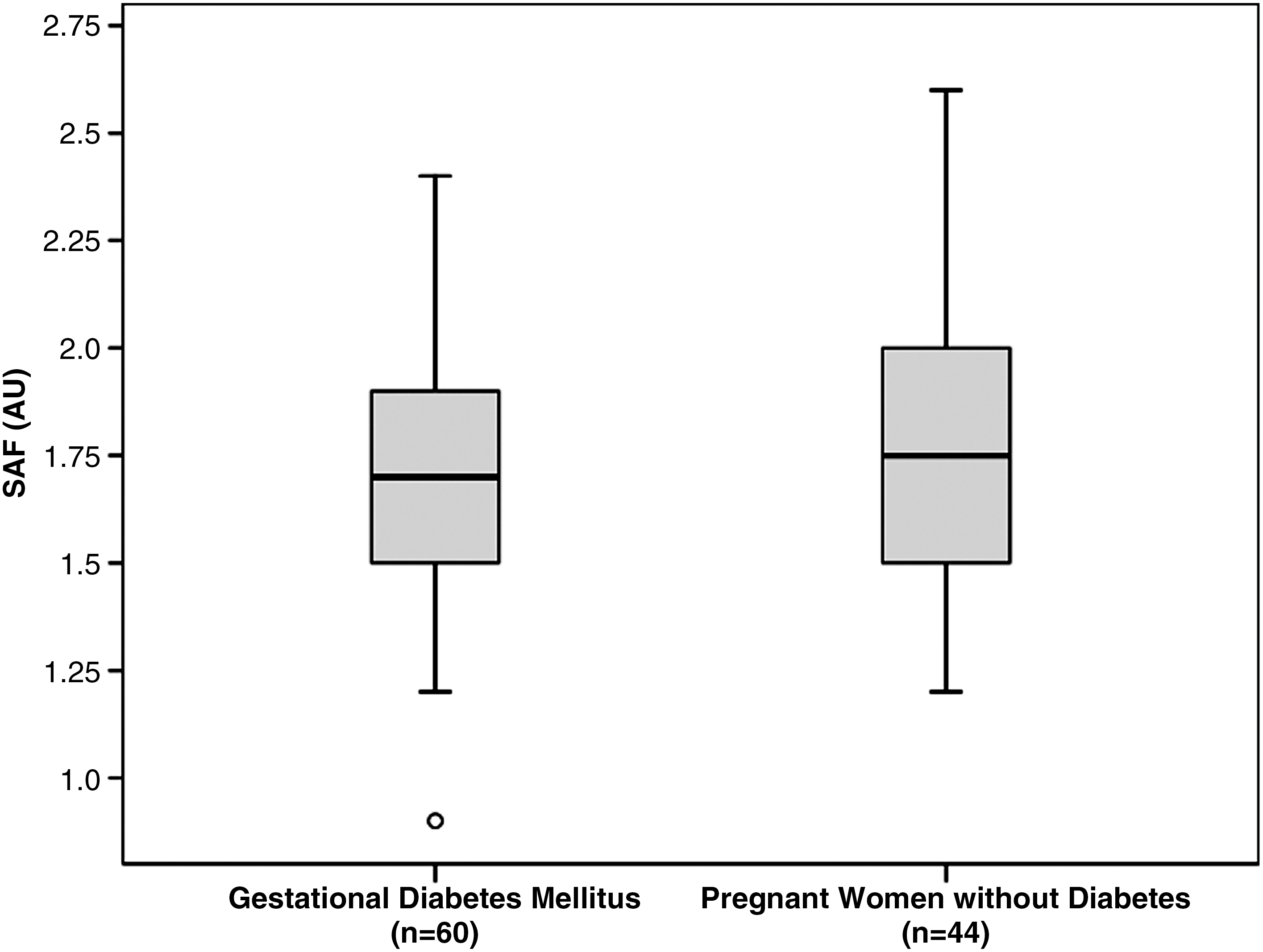
Skin autofluorescence (SAF) values in GDM patients versus pregnant women without diabetes. AU, arbitrary units.
In a linear model containing age, BMI, ethnicity (white European or other), time since last meal, category (GDM or control), HbA1c, fructosamine at diagnosis, and gestational age at SAF measurement, only age and ethnicity were significantly associated with SAF (Table 2). With every year a patient gets older, SAF increases 0.02 AU (P<0.001). Patients with ethnicity other than white European had a mean SAF level that was 0.26 higher (P<0.001). Reflectance in other ethnicities than white Europeans was lower. Therefore differences in SAF with ethnicity were corrected for reflection in a multivariate linear model, but this did not change the result. The differences in SAF level between different ethnicities are also shown in Figure 2. SAF levels in white Europeans were lower compared with other ethnicity in both patients with GDM and pregnant women without diabetes: GDM group, white Europeans (1.6 AU) versus patients with another ethnicity (1.9 AU), P<0.001; pregnant women without diabetes, white Europeans (1.7 AU) versus patients with other ethnicity (1.9 AU), P=0.07. Factors such as time since last meal did not have any significant effect on SAF (β=0.03, P=0.43).
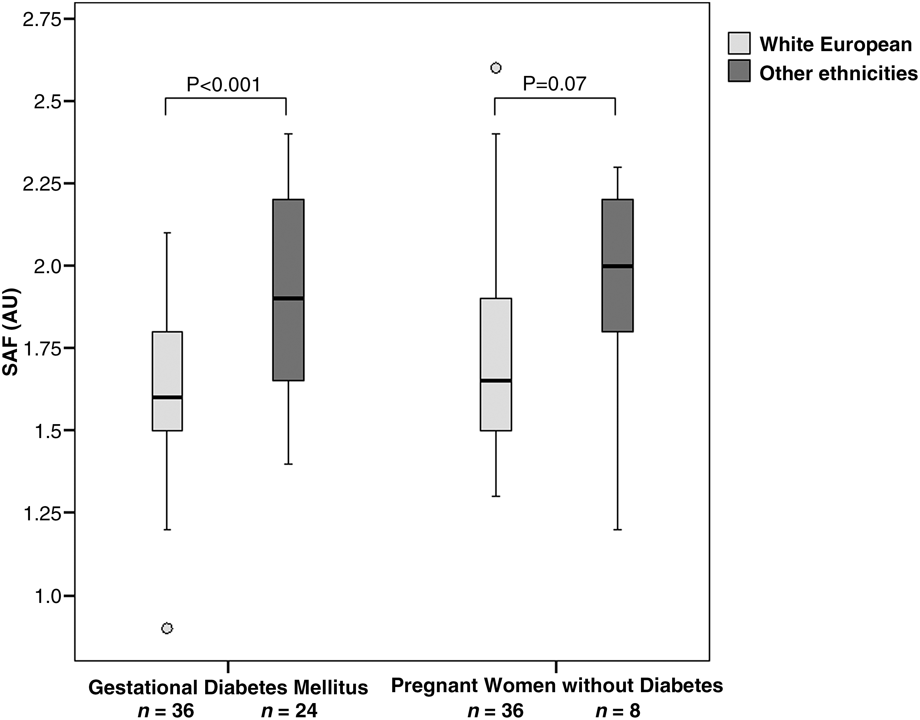
Differences in skin autofluorescence (SAF) between patients of white European origin and patients with other ethnicities. AU, arbitrary units.
BMI, body mass index; GDM, gestational diabetes mellitus; HbA1c, hemoglobin A1c.
Discussion
This is the first study investigating tissue accumulation of AGEs, measured as SAF, in GDM patients compared with pregnant women without diabetes. No accelerated accumulation of AGEs was found in GDM at the time of diagnosis. SAF was lower in white European patients compared with other ethnicities.
Unlike serum AGEs, tissue AGEs have never been measured in pregnant patients before. In contrast to our findings, elevated serum levels of AGEs during pregnancy have been described in GDM patients compared with pregnant women without diabetes. 17 –19 However, studies did not always show consistent results: for example, in one study total serum AGE levels in GDM patients were found to be increased, but levels of a single specific AGE (carboxymethyllysine) were actually decreased. 17 In another study serum AGE levels were elevated in GDM patients but not in pregnant patients with preexistent DM, 19 which seems contradictory because hyperglycemia is generally more severe and longer present in preexistent DM than in GDM. In addition to this, the use of different assays can be a problem in the measurements of serum AGEs, 23 and serum AGEs are not necessarily a representation of tissue AGEs, whereas SAF has been validated by actual tissue AGEs in skin biopsy specimens. 2,4 Finally, most of the studies investigating serum AGEs measured serum AGEs in the third trimester and one also in the second trimester. 17 –19 The mean gestational age at measurement in our study was around 27 weeks (second trimester). Therefore we can only conclude from our study that no differences in SAF exist between control subjects without diabetes and GDM patients at diagnosis (second trimester), but we cannot exclude that differences may appear later in pregnancy.
But why did we not find elevated SAF in GDM in our study? Considering that AGE accumulation, measured as SAF, has been found in patients with diabetes (type 1 and type 2) in many studies, 8 –10,24 the absence of a difference between GDM patients and the control population was unexpected. Furthermore, the absolute SAF level we found for GDM patients as well as for pregnant women without diabetes was similar to reference levels for SAF in control subjects as has been published before (SAF levels for control subjects at an age of 30–40 years, 1.73 AU). 25 There are two possible explanations for the fact that SAF levels were not elevated in GDM patients at the time of diagnosis. First, the hyperglycemia may not have been severe enough to cause accelerated AGE accumulation in these GDM patients at diagnosis. GDM mostly results in mild hyperglycemia (many patients can be treated with diet only). However, accumulation of AGEs has been described before in studies with mild hyperglycemia such as in adequately regulated patients with type 2 diabetes and in patients with only impaired glucose tolerance. 8,12 Second, the duration of glycemic exposure may have been too short. The exact duration of glycemic exposure in GDM patients at diagnosis is unknown. It is probably no longer than several weeks because GDM patients usually only become insulin resistant and hyperglycemic in the second trimester. Most studies in patients with type 1 and type 2 diabetes have shown that duration of diabetes is directly related to SAF. 8 –11 Accumulation of AGEs has always been thought to be a slow process, but short-term accumulation of AGEs has also been found to occur. SAF was markedly elevated in patients admitted to the intensive care unit (patients without diabetes) compared with healthy controls. 26 However, the clinical setting of the intensive care unit suggests that AGE accumulation was due to severe non–hyperglycemia-related oxidative stress rather than to hyperglycemia. The absence of evidence of accelerated AGE accumulation at diagnosis in GDM patients does not exclude accelerated accumulation later in pregnancy. Therefore it would be interesting to measure SAF levels during pregnancy in GDM patients. But the fact that SAF is not elevated at the time of diagnosis makes it very unlikely that the AGE-Reader can be developed as a screening method for GDM in the future.
Two factors were significantly associated with SAF: age and ethnicity. The association between age and SAF is well known, and although women in our study did not represent a very wide age range, this association was still found. No prior data exist on SAF in patients with different ethnicity. We found that women with ethnicity other than white European had higher SAF levels. Whether genetic factors or environmental factors (for instance, nutritional habits) contribute to this difference is unknown. In this study the differences in SAF between ethnicity was an unexpected finding for which this study was not designed or powered. However, because differences were very consistent, ethnicity seems to be a factor to take into account when measuring SAF. We did not find an association between HbA1c and SAF. This is in contrast to many studies in patients with type 1 and type 2 diabetes, which have reported a positive association between these two parameters, 8 –10,27 although an absence of this association has been described too. 11 The fact that we did not find this association could be due to the fact that SAF and HbA1c were measured at diagnosis, and HbA1c levels were usually normal (mean HbA1c in our study was 35 mmol/mol [5.3%]).
We found that AGE levels did not change during OGTT, which confirms prior literature on this topic. 28 There was also no relation between SAF and time since last meal (this was measured in patients within 3 weeks after OGTT), which is in contrast to prior literature in which a 10% elevation of SAF was described following an AGE-rich meal. 28,29 However, in that study the meal contained an abnormally high AGE content, whereas in our study levels were measured in patients measured after a normal breakfast. We showed that SAF can be measured at any time during OGTT and at any time after breakfast.
A few limitations of this study have to be addressed. SAF was not measured at the day of OGTT in all patients; some measurements were done days after diagnosis (maximum of 3 weeks) because of logistic reasons. However, separate analyses showed no differences in SAF between patients included on the same day of the diagnosis and patients included within 3 weeks, so it seems that SAF did not change in this short period of time. Furthermore, the exact start of hyperglycemia in GDM is hard to establish, and at a maximum of 3 weeks post-OGTT none of the patients included within 3 weeks had already started insulin treatment.
Serum AGE levels were not investigated in this study. So we cannot compare our results with serum AGE levels. This study was specifically addressing the question if tissue AGEs (measured by SAF) are elevated in GDM at diagnosis.
In conclusion, we showed that SAF (a measure of tissue AGE accumulation) is not elevated in GDM pregnancies at diagnosis compared with pregnancies without diabetes. This could be due to the mild and short duration of hyperglycemia present in this condition at diagnosis and does not exclude potential differences in SAF later in pregnancy. Because we did not find differences in SAF at diagnosis, it is unlikely that the AGE-Reader can be developed as a screening method for GDM in the future, although this study was not specifically designed or powered to investigate this. Furthermore, we found that not only age, but also ethnicity, should be taken into account when measuring SAF.
Footnotes
Author Disclosure Statement
No competing financial interests exist.