
Abstract
Background:
Advanced glycation end products (AGEs) and oxidation products (OPs) play an important role in diabetes complications, aging, and damage from sun exposure. Measurement of skin autofluorescence (SAF) has been promoted as a noninvasive technique to measure skin AGEs, but the actual products quantified are uncertain. We have compared specific SAF measurements with analytically determined AGEs and oxidative biomarkers in skin collagen and determined if these measurements can be correlated with chronological aging and actinic exposure.
Methods:
SAF at four excitation (ex)/emission (em) intensities was measured on the upper inner arm (“sun protected”) and dorsal forearm (“sun exposed”) in 40 subjects without diabetes 20–60 years old. Skin collagen from the same sites was analyzed by liquid chromatography–tandem mass spectrometry for three AGEs—pentosidine, carboxymethyllysine (CML), and carboxyethyllysine (CEL)—and the OP methionine sulfoxide (MetSO).
Results:
There was poor correlation of AGE-associated fluorescence spectra with AGEs and OP in collagen, with only pentosidine correlating with fluorescence at 370ex/440em nm. A little-studied SAF (440ex/520em nm), possibly reflecting elastin cross-links, correlated with all AGEs and OPs. Levels of CML, pentosidine, and MetSO, but not SAF, were significantly higher in sun-exposed skin. These AGEs and OPs, as well as SAF at 370ex/440em nm and 440ex/520em nm, increased with chronological aging.
Conclusions:
SAF measurements at 370ex/440em nm and 335ex/385em nm, except for pentosidine, which correlated with fluorescence at 370ex/440em, correlate poorly with glycated and oxidatively modified protein in human skin and do not reflect actinic modification. A new fluorescence measurement (440ex/520em nm) appears to reflect AGEs and OPs in skin.
Introduction
In addition to the direct structure–function changes that they induce, AGEs can act as photosensitizers when exposed to ultraviolet radiation, which can induce further protein cross-linking, oxidation, and peroxide formation. 9,10 In addition, damage may be further amplified by signal transduction receptors for AGEs, which have been identified in several cell types and can lead to inflammation and oxidative stress. 1 This cascade of oxidative stress from these sources can produce structural and functional damage in skin and accumulation of related end products with aging.
Some AGEs, such as pentosidine, exhibit autofluorescence, which can be detected at excitation (ex)/emission (em) wavelengths of 335ex/385em nm. 11 Other autofluorescent products have been found in collagen (370ex/440em nm) 12 and elastin (440ex/520em nm), 13 although their chemical structures have yet to be fully elucidated (Table 1). The chemical structures of many AGEs have now been identified, however, and these quantitatively account for most of the known products, but the majority of these are not autofluorescent, 14 –18 potentially limiting the value of autofluorescence as indicators for their levels in tissues.
In vivo skin autofluorescence (SAF), particularly that reflecting a broad spectrum of ex and em wavelengths, has been proposed as a simple noninvasive technique to measure skin levels of AGEs and oxidation products (OPs) and has been shown to be a marker of aging and photoaging. 2,19 Accumulation of AGEs in skin reflects damage in the vascular system in diabetes, and SAF has therefore been used in assessing the risk of diabetes complications. 20
The aims of the present study were twofold. First, we sought to establish whether specific wavelengths of SAF can be correlated with levels of individual chemically quantified AGEs in skin collagen. Second, we wished to determine whether SAF measured with a specifically designed fluorescence spectrophotometer and analytically measured AGEs correlate with chronic sun exposure and aging in subjects without diabetes. To achieve this we studied these parameters from the same sun-exposed and sun-protected skin sites from 40 subjects without diabetes between the ages of 20 and 60 years.
Subjects and Methods
Subjects
All aspects of this study conformed to the Code of Federal Regulations, Title 21 Section 50. Forty white female volunteers with skin type II–III 21 and approximately the same color tone of light–medium skin with L* (reflectance) values between 65 and 72 were recruited for the study. The subjects were nonsmokers of general good health. They were overweight with a body mass index of 25–35 kg/m2 as measured by a body fat analyzer electronic scale. The subjects did not have diabetes and were not pregnant or breast feeding.
The 40 subjects were distributed into four age range groups: 20–30 years old (n=15), 30–40 years old (n=5), 40–50 years old (n=5), and 50–60 years old (n=15). Informed consent was obtained from each subject, the details and risks of the procedure were discussed with them, and the subjects completed a short medical history form. The upper inner arms and the dorsal forearm test areas were examined to exclude sunburn, rashes, scratches, burn marks, etc., as well as warts, nevi, scars, and active skin lesions in the test site. Subjects were also instructed to not use any skin creams.
The most uniformly pigmented areas from the test areas were chosen for marking by 1.5-inch-diameter circles on the skin sites. These sites were used to measure skin color and fluorescence by spectroscopy, followed by a punch biopsy procedure.
Color measurements
A Minolta (Ramsey, NJ) Chromameter was used to measure skin color from all sites. For each site, five measurements were obtained from a 1.5-inch-diameter area and averaged. Skin color was quantified by performing L*a*b* colorimetric measurements (the L* value is a light–dark saturation value [luminance], whereas a* and b* values are measurements of red and blue, respectively) and calculating the individual typology angle (ITA°) using the formula ITA°=(arc tangent [(L*−50)/b*])×180/3.1415, where arc tangent is expressed in radians, according to the COLIPA guidelines (see guideline for the colorimetric determination of skin colour typing and prediction of the minimal erythemal dose, 2007 edition 22 ).
Fluorescence excitation spectroscopy
Fluorescence ex spectroscopy was performed with a Spex Skin Skan® (Jobin Yvon, Edison, NJ) fluorescence spectrophotometer.
Skin biopsies
After the sites were anesthetized with xylocaine/epinephrine (AstraZeneca, London, UK), a 4-mm-diameter punch biopsy was obtained from each of the four test sites, and a scalpel was used to remove tissue above the subdermal fat. The wound was closed with a suture and covered with an adhesive bandage. Each subject returned after 5–7 days to have the sutures removed. The punch biopsy specimens were placed in plastic vials, which were immediately flash-frozen in a mix of ethanol and dry ice and stored at −80°C until processed for analysis.
Sample preparation
Epidermis and subdermal fat were removed from the skin biopsy specimens, and after mincing the tissue was homogenized (Polytron, Brinkmann Instruments, Inc., Westbury, NY) for 30 s in ice-cold phosphate-buffered saline. Following centrifugation at 2,000 g at 4°C for 10 min, the pellet was washed twice with 5 mL of deionized water and then delipidated.
All liquid reagents used in the experiment from this point were degassed and purged with nitrogen immediately before use. After preparation, the samples were digested with proteolytic enzymes.
Following digestion, hydrolysates were centrifuged at 750 g to remove unhydrolyzed material, and the supernatant was filtered through 3,000-molecular-weight cutoff filters. Filters were washed with 100 μL of water, and the combined filtrates were dried with a rotary evaporator. The sample was reconstituted in 50 μL of water and frozen at −80°C until analysis by liquid chromatography–tandem mass spectrometry. Several controls were assayed with each set of samples.
Analytical methods
Pentosidine was determined by high-performance liquid chromatography as previously described, 4 using an Agilent (Santa Clara, CA) model 1100 series instrument fitted with a postcolumn 23 flow fluorimeter.
CML, CEL, and methionine sulfoxide (MetSO) were quantified on an Agilent model 6410 triple quadrupole liquid chromatograph/mass spectrometer using a modification of the method of Thornalley et al. 24 Heavy or deuterated internal standards of CEL and CML (d4CEL and d4CML, respectively) and MetSO (d3MetSO) were used as noted above, and similar ion transitions were used for quantification. A 2.1-×250-mm 5-μm (particle size) Hypercarb (Thermo Fisher, Waltham, MA) column was used with a mobile phase of 26 mM ammonium formate (pH 3.8) and acetonitrile. A linear gradient from 0% to 15% acetonitrile in 6 min, followed by a 6-min hold at 15% acetonitrile, was used in analysis. Total analysis time was 40 min.
Results were expressed relative to collagen protein content determined by the measurement of hydroxyproline, assuming collagen contains 14% of this amino acid by weight as previously described. 25
Two internal standard blanks and blank samples containing all ingredients except collagen were assayed thorough the entire digestion process.
To confirm that no artifactual conversion of methionine to MetSO occurred, deuterated (d3) methionine was added to a mix of subject tissues and to a blank during digestion, and measurements were performed for d3 MetSO.
Statistical methods
AGEs and SAF were compared between sun-exposed and sun-unexposed skin as described in Tables 2 and 3. The association of SAF and AGEs/OPs was examined using Spearman correlations. In addition, we used multiple linear regression analyses to regress SAF on AGEs and OPs while adjusting for the variation in SAF explained by ITA°. We report partial R values from this regression. To account for repeated measures from subjects (lower and upper arms), the method of generalized estimating equations was used to estimate these regression models. Log transformations were used as appropriate to normalize distributions.
Products were measured in skin collagen, obtained from skin biopsy specimens from 40 subjects without diabetes by liquid chromatography–tandem mass spectrometry as described in Subjects and Methods. Following delipidation with chloroform/methanol (2:1 vol/vol) overnight on an orbital shaker at room temperature, the collagen pellet was then digested for 72 h at 37°C at pH 7.5 with collagenase (type VII, 140 U) in 20 mM HEPES buffer containing 5 mM CaCl2. Catalase (Sigma Chemical Co., St. Louis, MO) (1,000 U) was also added to each sample incubation mixture, as was thymol. Following collagenase digestion, sequential digestions at 37°C with Pronase (24 h) and aminopeptidase/prolidase (48 h) were performed as previously described 26 with all digestions performed under a nitrogen atmosphere. The skin sites measured were right and left upper inner arms (sun unexposed) and lower outer arms (chronically sun exposed).
Advanced glycation end products and oxidation products were compared between sun-exposed and -unexposed sun-protected skin using paired t tests. P values were corrected for individual typology angle.
NS, not significant.
A Spex Skin Skan fluorescence spectrophotometer was used on the same test sites used for the skin biopsy to measure fluorescence. Three consecutive readings were obtained from each site and averaged. Readings were taken at excitation wavelength of 370 nm and emission wavelength of 440 nm and at excitation of 335 nm and emission at 385 nm to quantify advanced glycation end products and pentosidine, respectively, 27 and putative collagen/elastin cross-linking was measured using excitation at 440 nm and emission at 520 nm. 13,28 The skin sites measured were right and left upper inner arms (sun unexposed) and lower outer arms (chronically sun exposed).
P values were corrected for individual typology angle.
The following statistical relationships were studied: (1) AGEs/OPs and age; (2) SAF and age; (3) AGEs/OPs in sun-exposed and sun-protected skin; (4) SAF in sun-exposed and sun-protected skin; and (5) relationship between SAF at the four ex/em wavelengths putatively associated with specific AGEs and the ratio of AGEs/OPs.
Results
Relationship between skin collagen AGEs/OPs and SAF
The major objective of these studies was to investigate the relationship between SAF at three ex/em intensities—370ex/440em nm (putative collagen cross-links), 335ex/385em nm (pentosidine), and 440ex/520em nm (putative elastin–collagen cross-links)—with AGEs and OPs measured in skin collagen. In all of these analyses SAF was corrected for skin pigment by multivariate regression analysis using the generalized estimating equations method.
As shown in Table 4, only pentosidine (P=0.01) showed significant correlation with SAF at 370ex/440em nm, whereas CEL, CML, and MetSO showed no statistical relationship with 370ex/440em (P values of 0.073, 0.27, and 0.63, respectively).
Advanced glycation end products and oxidation products were measured in skin collagen by liquid chromatography–tandem mass spectrometry as described in Subjects and Methods. Skin autoflluorescence was corrected for skin pigment (individual typology angle) by multivariate regression analysis using the generalized estimating equations method, while the association of SAF and advanced glycation end products/oxidation products was examined using Spearman correlations.
Statistically significant relationships are indicated in bold.
Significant but inverse relationship.
No AGE (pentosidine [P=0.62], CML (P=0.17], or CEL [P=0.33]) correlated with SAF at 335ex/385em nm, but the oxidation product MetSO showed a significant inverse relationship (P=0.012) with SAF intensity at 335ex/385em nm.
SAF measured at 440ex/520em nm showed positive correlations with the AGEs pentosidine, CML, and CEL (P=0.02, 0.02, and 0.014, respectively) and also showed a statistically significant relationship with the OP MetSO (P=0.043).
Sun-exposed and -protected skin and collagen AGEs/OPs
Relative to sun-protected skin, skin from sites chronically exposed to sun showed significantly higher levels of both pentosidine and CML and the OP MetSO, whereas CEL showed no difference. The increase in MetSO in sun-exposed skin was most significant (P=0.000004), although increases in pentosidine (P=0.009) and CML (P=0.022) were also statistically significant but of somewhat lesser magnitude (Table 2).
Sun-exposed and -protected skin and SAF
For each SAF ex/em pair putatively associated with specific AGE products, a t test was used to compare the fluorescence in sun-exposed and -protected skin. As seen in Table 3, the increases in chemically quantified AGEs and OPs seen in sun-exposed skin were not observed for any fluorescent spectrum. Conversely, the average SAF at 335ex/385em nm and 440ex/520ex nm was actually two- to fourfold higher in sun-protected relative to sun-exposed skin.
Change in chemically determined skin collagen AGEs/OPs with age
Pentosidine, MetSO, and CML levels all increased with age and showed significant correlations with increasing age on regression analysis (Fig. 1A–C), whereas that of CEL did not (Fig. 1D). More specifically, the correlation between both pentosidine and MetSO with age was highly significant (P values of 0.0001 and 0.0002, respectively), as were CML levels (P value of 0.011), whereas CEL was not correlated with age (P=0.20).
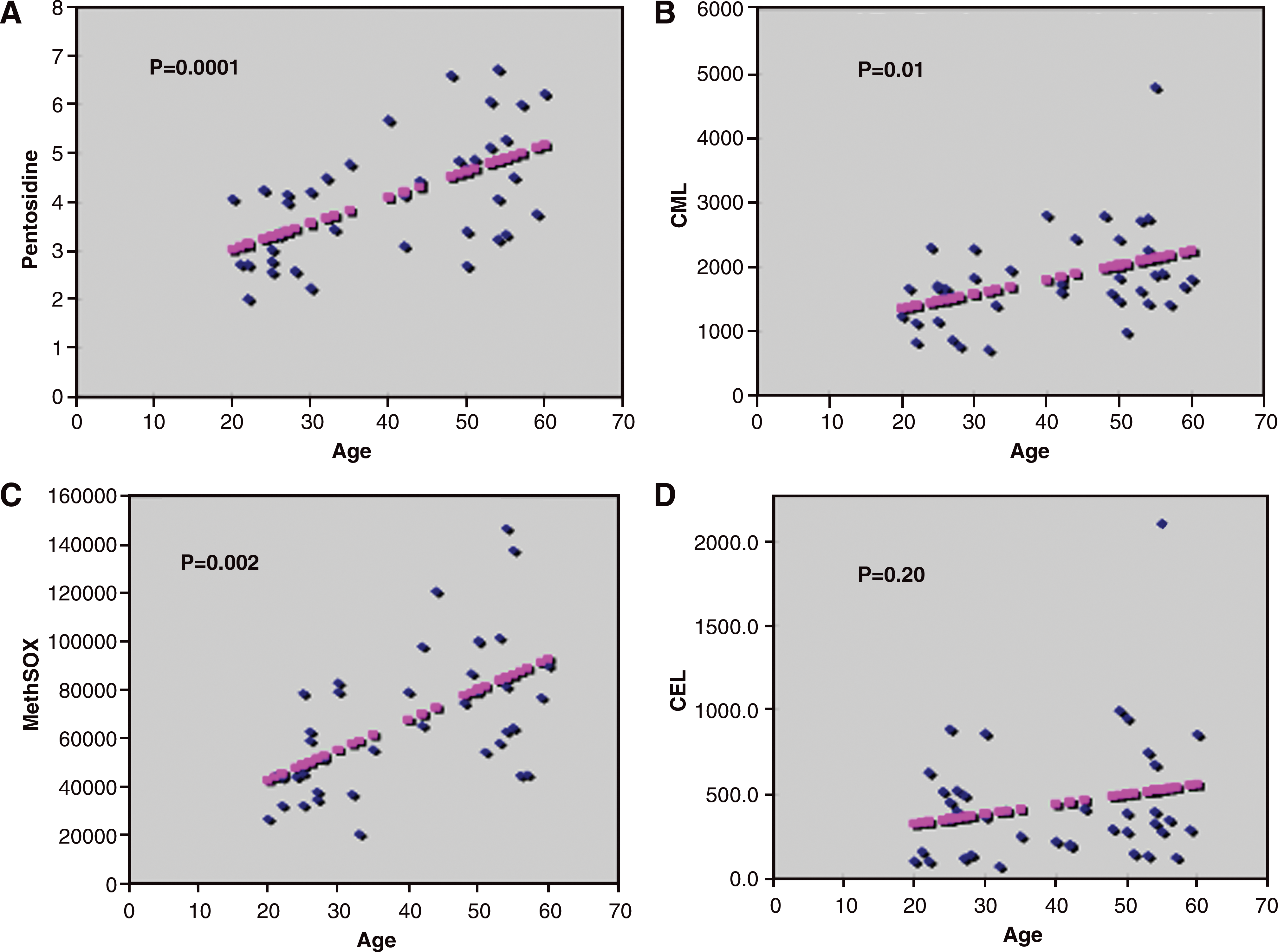
Statistical relationships by regression analysis of the advanced glycation end products (
Change in SAF with age
Of the three SAF ex/em intensities, two (370ex/440em nm and 440ex/520em nm) showed significant correlations with age with similar P values of<0.0001 (Fig. 2), whereas SAF at 335ex/385em nm did not (P value of 0.48).

Statistical relationship by regression analysis of skin fluorescence and age for the excitation/emission pairs reflecting advanced glycation end products in skin collagen and elastin. Panels (
Discussion
SAF has been proposed to noninvasively measure the tissue accumulation of AGEs. Because SAF readings correlated with micro- and macrovascular disease in diabetes and with overall vascular mortality in several clinical trials, 19,20,29,30 these studies have documented an apparently causal relationship between AGEs and vascular disease.
The major aim of these studies was to determine if SAF correlates with specific AGEs and OPs in skin from individuals without diabetes. This is important because many cosmetic companies are using SAF to support claims on the effect of their products on skin AGEs. In these studies we observed that in skin collagen only levels of pentosidine correlated with a fluorescence known to measure AGEs (370ex/440em nm), whereas no AGEs or OPs correlated with another fluorescence spectrum known to measure pentosidine (335ex/385em nm). We did find for the first time, however, that a less-studied fluorescence intensity of 440ex/520em nm, possibly reflecting elastin–collagen cross-links, 13 correlated with all AGEs and OPs measured in chronologically aged skin.
Our findings show a relatively weak association of the primary SAF wavelengths known to reflect AGEs and chemically quantified AGEs/OPs in skin collagen, with only one ex/em wavelength pair correlating with one AGE (pentosidine). These results did not confirm a prior study by Meerwaldt et al., 31 who found uniformly statistically significant relationships between SAF and levels of the AGEs pentosidine, CML, and CEL in skin. Because chemically measured levels of these AGEs were quantitatively similar in the two studies, the different outcomes are more likely due to factors relating to measurement of SAF. For example, we chose to measure specific fluorescent ex/em wavelengths that have been shown to reflect AGEs in prior studies, 11 –13 whereas Meerwaldt et al. 31 used an excitation light source of 300–420 nm (peak ex∼350 nm) and measured the average light intensity emitted per nm over the 420–600 nm range to calculate autofluorescence. The spectrophotometers used in the two studies also differed in design. We used a Skin Skan instrument with a fiber optic probe to deliver the ex wavelengths and read the em values, whereas Meerwaldt et al. 31 used an AGE Reader (DiagnOptics, Groningen, The Netherlands) with a concave cradle for the forearm that illuminates a skin area of 1 cm2. The Skin Skan fiber optic probe measures a more limited area of SAF, and at the ex wavelengths used in this study the depth of penetration is relatively shallow, 27 whereas the AGE Reader reads from a greater area and penetrates deeper into the dermis.
The two studies also took different approaches to correcting for skin pigmentation. To eliminate the known quenching effect of skin pigmentation on SAF we selected 40 white subjects without diabetes with light–medium skin reflection color tone, used a narrow range of L* (reflectance) values for our study, and corrected the fluorescence for slight differences in skin pigment (ITA°) by multivariate analysis. Conversely, Meerwaldt et al. 31 used 46 multiethnic subjects and corrected for skin pigment by comparing reflectance with that of a white Teflon® (Dupont, Wilmington, DE) block (assuming 100% reflectance). 28 Because head-to-head studies were not performed, it is difficult to estimate the effects of these differences in instrumentation and protocol on the respective outcomes.
Both studies measured levels of three specific AGEs in skin collagen, but we also included collagen levels of the important oxidation product, MetSO. This gave us the additional advantage of determining the effect of age and sun exposure on oxidative stress in skin, as well as the correlation of oxidative stress with traditional SAF spectra.
Because various skin fluorophores, including NADH, 14 FAD, 15,16 porphyrins, 17 and the fluorescent oxidation adduct N-formylkynurenine 18 have a major impact on SAF, we chose to use specific ex/em wavelengths that have been previously shown ex vivo to measure isolated AGEs, rather than the broad nonspecific spectrum of ex/em wavelengths used in the study by Meerwaldt et al. 31
Our failure to find correlations among AGEs, OPs, and SAF is not surprising because most AGEs do not exhibit fluorescent properties. 32 Even the fluorescent AGE pentosidine did not correlate with its known fluorescent wavelengths (335ex/385em nm), but was the only AGE to correlate with the wavelength pair 370ex/440em nm, which reflects a lysine-pyridinium product that has not yet been fully chemically defined. 26 Levels of most other nonfluorescent AGEs and OPs in skin, on the other hand, did not show correlations with these commonly used SAF wavelengths. Skin levels of specific AGEs, such as pentosidine, CML, and CEL have previously been measured by high-performance liquid chromatography and mass spectroscopy and have been shown to increase with age in diabetes 4,6,11,23 and also to be predictive of diabetic microvascular disease in the Diabetes Control and Complications Trial/Epidemiology of Diabetes Interventions and Complications population. 33 Skin levels of the OP MetSO, in subjects with diabetes, has also been shown to correlate with aging and progression of diabetic nephropathy. 23
The present study also provides analytical evidence that skin from individuals without diabetes in sun-exposed areas contains significantly higher levels of two AGEs, CML and pentosidine, and the OP MetSO, confirming the prior studies of Jeanmarie et al. 34 These increases were not detected by measuring SAF at any wavelengths with the Skin Skan device, however, with these measurements actually showing a paradoxical decrease with sun exposure. Our finding that SAF values measured on the sun-protected site were higher than the values obtained on the sun-exposed area agrees with earlier findings by Sandby-Møller et al. 35 The differences observed between directly quantified advanced chemical products and SAF in detecting actinic skin changes could also be partially due to the fact that the chemical analysis of AGEs in this study measured AGEs per unit insoluble collagen, whereas the fluorescent values have no such denominator. In the current studies we did confirm that dermal collagen levels of CML, pentosidine, and MetSO, but not CEL, correlate with aging in a population without diabetes, supporting previous observations that some AGEs and OPs accumulate with chronological aging in human skin in diabetes. We also observed that SAF measured at two wavelengths (370ex/440em nm and 440ex/520em nm) correlated with chronological aging.
One unique aspect of the current study was our measurement of a little-studied fluorescence (SAF) intensity of 440ex/520em nm 13 and showing that it was highly correlated with all chemically quantified AGEs and OPs measured. It is unlikely that this SAF spectrum accounts for the correlations between AGEs and SAF in observed in the study of Meerwaldt et al., 31 however, because the maximum excitation wavelength in their studies did not exceed 420 nm. Although further studies are needed to identify the putative elastin/collagen products reflected by measurements at these wavelengths, the current study suggests that measuring SAF at 440ex/520em nm may provide a noninvasive way of measuring skin AGEs and OPs and should be examined in future studies.
Footnotes
Acknowledgments
This work was supported by grants from The Estee Lauder Companies, Melville, NY, and from the Juvenile Diabetes Research Foundation, New York, NY. Work was done in Hanover, NH, and Philadelphia, PA.
Author Disclosure Statement
None of the authors has commercial or financial interests in the use of skin autofluorescence as a diagnostic tool. H.C., N.M., and M.S.M. are employees of The Estee Lauder Companies.