
Abstract
Microneedle array devices provide the opportunity to overcome the barrier characteristics of the outermost skin layer, the stratum corneum. This novel technology can be used as a therapeutic tool for transdermal drug delivery, including insulin, or as a diagnostic tool providing access to dermal biofluids, with subsequent analysis of its contents. Over the last decade, the use of microneedle array technology has been the focus of extensive research in the field of transdermal drug delivery. More recently, the diagnostic applications of microneedle technology have been developed. This review summarizes the existing evidence for the use of microneedle array technology as biosensors for continuous monitoring of the glucose content of interstitial fluid, focusing also on mechanics of insertion, microchannel characteristics, and safety profile.
Introduction
Impact of diabetes mellitus
Intensive treatment of diabetes reduces the risk of complications, 2,3 and glucose monitoring is a core component of successful management, especially for those who are insulin-treated. Monitoring facilitates intensification of insulin therapy, with a subsequent reduction in diabetes-related complications, while minimizing the risk of hypoglycemia. There is evidence for improvement in glycemic control with increased frequency of self-monitoring of blood glucose (SMBG) and with the use of continuous glucose monitoring (CGM) for 6 or more days per week. 4 –6 A recent study showed that the use of CGM is also associated with reduced time spent in hypoglycemia in children and adults with type 1 diabetes. 7
Evolution of glucose monitoring devices and the development of microneedle technology
Since 1971, when the first glucose monitor was used, the most common method of glucose monitoring has been the use of intermittent capillary blood glucose monitoring using standard finger-prick methods. This method does not provide the user with information about duration, direction, and magnitude of glucose change over time. Furthermore, the procedure is invasive and painful, resulting in reduced concordance with the recommended frequency of monitoring and a subsequent negative impact on diabetes control. Alternative monitoring methods have been developed for CGM. Since 1999, when the first CGM system was approved, the technology has been largely used as a diagnostic tool, implanted for a limited period to identify periods of unrecognized hypo- or hyperglycemia and guide modification of insulin therapy.
There is evidence to demonstrate the therapeutic benefits of CGM devices, with real-time (RT) glucose information (RT-CGM) leading to a glycated hemoglobin (HbA1c) reduction without increase in the incidence of hypoglycemia. 4,8,9 A meta-analysis showed an overall mean HbA1c reduction of 0.3% in type 1 diabetes subjects using RT-CGM in comparison with SMBG. This meta-analysis also highlighted the correlation between compliance with the frequency of CGM device use and its effectiveness; for every 1 day increase in sensor usage per week, there was an additional HbA1c reduction of 0.15%. 5 In the landmark Juvenile Diabetes Research Foundation study, only subjects who used the device 6 or more days per week managed to achieve the desired therapeutic benefit. 4
The use of CGM technology in subjects with type 2 diabetes is limited by a paucity of data. A randomized controlled trial in subjects with type 2 diabetes demonstrated CGM reduces HbA1c without increasing incidence of hypoglycemia compared with SMBG. As in type 1 diabetes, the study also demonstrates the positive correlation between compliance with the frequency of CGM device use and effectiveness. 10,11
Commercially available CGM devices depend on amperometric electrochemical technology using glucose oxidase (EC 1.1.3.4) for detection of glucose in interstitial fluid (ISF). Currently the most commonly used CGM devices in clinical practice contain a needle-type electrochemical glucose sensor with the sensor tip in the subcutaneous tissue. As these sensors use ISF as the diagnostic biofluid, they require calibration to capillary blood glucose. Glucose concentration in the ISF is dependent on blood flow, metabolic rate, and the rate of change of glucose concentration in the blood. 12 Comparison of glucose levels in dermal ISF and finger capillary blood showed high correlation with estimated lag time between 0 and 45 min. 13 Studies have suggested that the mean lag time is 6–7 min. 14
Despite their clinical benefits, these devices remain invasive and are associated with discomfort. This can negatively affect concordance and therefore the effectiveness of CGM and is a potential limitation to continuous use. This is corroborated by the high dropout rate in the clinical studies. 15 Moreover, data for CGM sensor accuracy have demonstrated clinically relevant reduced accuracy in the critical hypoglycemic range. 16 –19 Ongoing calibration against blood glucose values is required as subcutaneous sensors are susceptible to signal drift from surface deposition of proteins and cells followed by connective tissue encapsulation (biofouling) affecting sensor accuracy and life span. 20 To overcome the challenges of CGM, emerging technologies, including microneedles, aim to minimize the level of invasiveness without compromising on sensor accuracy or lifespan.
Microneedle use was first suggested in 1976 by Gestel and Place, 21 who proposed the use of multiple needle-shaped projections with a needle length of 5–100 μm to penetrate the stratum corneum layer for drug delivery. Thanks to advances in microfabrication in the 1990s, microneedle manufacture was feasible. In 1998, Henry et al. 22 showed that using microneedles 150 μm long to penetrate stratum corneum can dramatically increase skin permeability to calcein. Since then, research in microneedle array technology has developed rapidly, particularly in the field of transdermal drug delivery. Microneedles can be manufactured as in-plane or out-of-plane, where the needles are either parallel or perpendicular to the fabrication surface, respectively. Out-of-plane microneedles can be either solid (Fig. 1) or hollow. Microneedles of different geometries, designs, and materials (including polymers, metals, glass, carbohydrates, or silicon) have been manufactured, and microneedle technology has been extensively investigated as a therapeutic tool for transdermal drug delivery. Other applications for microneedle technology have been investigated including for biopsy, 23 for light delivery to deeper skin layers for diagnosis and treatment of epithelial cancers, 24 and for measurement of electrical potentials (for example, electrocardiography). 25 Several research groups have investigated the use of microneedle technology as a diagnostic tool allowing access to biofluids (ISF or blood) for analysis of variable analytes, including glucose. For CGM using ISF, an array containing several microneedles can be used to penetrate the stratum corneum and access the ISF without impinging on the dermal nerve fibers or blood vessels. ISF can then be either extracted to the surface (using hollow microneedles) or analyzed in situ using microneedle sensors. In comparison with the smallest needle-type glucose sensor used in clinical practice for CGM, which has a length of 9 mm and a width of 420 μm (27 gauge), microneedles used for the same purpose have a length of less than 1,000 μm and a tip diameter of less than 50 μm. In vivo analysis of ISF using multiple microsensors compared with one macrosensor provides a larger surface area for the enzymatic electrochemical reaction with potential to improve accuracy of glucose sensing even in the hypoglycemic range by improving the signal:noise ratio.
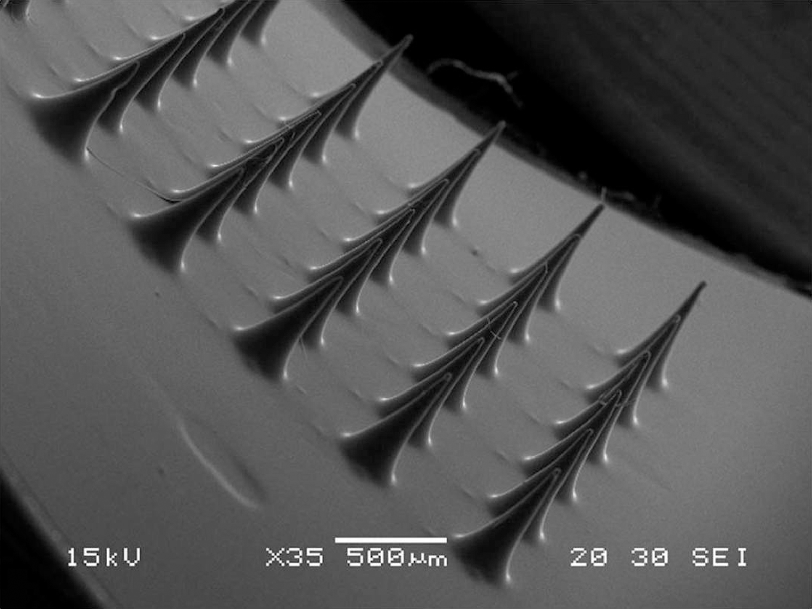
Scanning electron microscopy image showing a 500-μm-high microneedle array.
This review aims to summarize the existing evidence for the use of microneedle array technology in CGM, focusing on mechanics of insertion, penetration characteristics, potential safety, and use in glucose sensing.
Skin microanatomy
Human skin comprises the epidermis, dermis, and subcutaneous tissue. The outermost layer of epidermis, called the stratum corneum, is primarily made of dead tissue and is responsible for skin barrier characteristics. The viable epidermis, below the stratum corneum, contains living cells but is devoid of blood vessels and contains few nerves. Below the viable epidermis lies the dermis, 800–1,500 μm in thickness, which contains nerves and blood vessels at approximately 400 μm depth from skin surface 26 (Fig. 2).

Diagram showing a cross section of skin layers.
One of the main aims of microneedles accessing the ISF compartment is to penetrate the layer of stratum corneum without impinging on dermal microcirculation or dermal nerve fibers that lie at approximately the same level.
The stratum corneum represents the main mechanical barrier to microneedle insertion. 27 Knowledge of stratum corneum thickness is therefore essential to determine microneedle length and ensure adequate penetration. There is a marked site variation in mean stratum corneum thickness, ranging from 22.6±4.33 μm for volar forearm, 29.3±6.84 μm for back of the hand, and 173.0±36.96 μm for palm. The stratum corneum thickness tends to become age-dependently greater at the forearm. 28
Mechanics of Microneedle Insertion
The mechanics of microneedle insertion into skin is of paramount importance to device performance and safety. Successful insertion depends on geometry (microneedle length, wall thickness, tip radius, base diameter, density, and wall angle) and type of material. It also depends on skin resistance to penetration. All these factors determine the force needed for insertion or fracture of microneedles. Microneedles of the correct geometry and physical properties allow for a small insertion force. If the force required for insertion exceeds fracture force, needles will break or bend before insertion occurs.
The mechanics of microneedle insertion were first investigated by Davis et al., 29 who measured the force required for fracture, the force required for insertion, and their ratio (termed the margin of safety). To determine the effect of microneedle geometry on the force of insertion, individual hollow metal microneedles with tip radii of 30–80 μm, wall thicknesses of 5–58 μm, and a constant length of 720 μm were pressed against human subjects' skin using a displacement–force test station until a preset maximum load of 500 g was reached. Measured insertion forces ranged from 0.1 to 3 N, which is sufficiently low to permit insertion by hand. Forces of insertion showed an approximately linear dependence on the interfacial area of the needle tip but are independent of wall thickness. To determine the effect of microneedle geometry on the force of fracture, individual hollow microneedles were pressed against a hard surface using an axial load test station until a preset maximum displacement of 500 μm was reached. Fracture force increased significantly with increasing wall thickness and increased modestly with increasing wall angle and tip radius. The ratio of the fracture force to the insertion force can be considered the margin of safety; values greater than 1 identify needles that will insert into the skin without breaking. The largest margin of safety was achieved using needles with small tip radius to reduce insertion force and large wall thickness to provide strength and increase fracture force. 29 A similar method used to determine the margin of safety of microneedle arrays rather than individual microneedles has shown a safety margin of 6–9 when arrays of silicon solid microneedles, pyramidal in shape, with a tip radius of 5.5 μm, a base diameter of 250 μm, and 308 μm high were assessed. 30
In another study 27 to evaluate the relationship between microneedle geometry and fracture force, the force required to cause microneedle failure by axial loading increased with decreasing microneedle length, increasing base diameter or using microneedles of larger Young's modulus (a measure of stiffness of materials). The force required to cause microneedle failure by a transverse load was also measured. This showed that the transverse load failure force is smaller than the axial load failure force for microneedles of the same geometry and material. This indicates that if microneedles experience significant transverse load due to incorrect axial insertion, microneedles could fail by bending. 27 Microneedles with geometry below a 12:1 aspect ratio of length-to-equivalent diameter and a polymer with more than 3 GPa of Young's modulus were recommended to avoid microneedle mechanical failure by axial force. 31
Therefore, to ensure successful skin insertion of microneedles without breaking, appropriate microneedle geometry and materials are required to minimize insertion force and to maximize fracture force achieving the largest possible safety margin. Insertion is best achieved by small tip radius and sufficient length to overcome skin resistance, while mechanical strength is increased by increasing wall thickness, needle base diameter, and Young's modulus.
Skin Penetration
Several methods have been used to assess the ability of microneedles to successfully penetrate stratum corneum and to evaluate the characteristics of the microchannels created. Application of a staining dye to the insertion site allows en face visualization of the created micropores. Alternatively, as intact stratum corneum is a barrier against water diffusion, interruption of the layer results in increased water loss, and measurement of transepidermal water loss (TEWL) can be used as a measure of successful skin penetration by microneedles. Infrared spectroscopy and electrical impedance spectroscopy have also been used. 32 Although those techniques confirm whether the stratum corneum has been penetrated, they do not provide information on other skin penetration characteristics. Assessment of depth of penetration and its relation to microneedle array geometry require transverse visualization of microchannels, which can be obtained by biopsy of the microneedle-penetrated skin for histological examination. However, this may alter the biomechanical characteristics of the punctured skin and alter the macro- or microscopic appearance. The use of confocal laser scanning microcopy for in vivo imaging is limited to a penetration depth of only 200 μm. 33,34 Optical coherence tomography (OCT) has emerged as a powerful tool to obtain in vivo transverse images of microneedles in situ and of the created microchannels after device removal. OCT is a noninvasive optical imaging technique analogous to ultrasound, mapping the variations of reflected light rather than sound from biological samples. The major advantage of OCT is that it is capable of penetrating to a depth of 2 mm, providing cross-section imaging of epidermis and upper dermis for accurate in vivo assessment of microneedle penetration and of the created microchannels without the need for skin biopsy. Furthermore, no prior sample preparation is needed as with histological examination. When compared with histology, OCT images showed that the histological techniques overestimate the dimensions of the created microchannels. 34
Another factor influencing the mechanics of microneedle insertion is skin resistance. Because of the elasticity of skin, application of microneedles results in skin indentation at the insertion site prior to penetration of the stratum corneum layer, which may result in incomplete or even failure of penetration. 35 –37 A study using Trypan blue staining and TEWL to evaluate the relationship between microneedle length and skin penetration showed that manual application of short (300-μm-long) microneedles was not successful in penetrating the stratum corneum. Microneedles of 550 μm or more were needed to overcome skin elasticity and resistance. 36 Subsequently, successful insertion was achieved by using an electrical applicator at a velocity of 1 or 3 m/s. 37
Skin resistance to microneedle insertion was also demonstrated by OCT images showing stratified skin tissue is compressed during microneedle application, leading to partial penetration (700-μm-long microneedles created 300-μm-long microchannels when inserted in the palmar skin of human subjects). This skin resistance to microneedle insertion seems to increase as microneedle length increases, as shown in data demonstrating that the distance between the lower microneedle array base plate and stratum corneum was significantly lower with microneedle arrays of relatively short length (250 and 350 μm) compared with those of greater length (600 and 900 μm). 38
Skin resistance also increases with increased stratum corneum thickness; a positive correlation has been demonstrated between the force needed for microneedle insertion and stratum corneum thickness. 39 Various approaches have been used to reduce skin deformation at the microneedle insertion site and to overcome skin resistance, including insertion using rotation, 35 vibration, 40 or at high velocity. 37
Microchannel Characteristics
Assessment of the characteristics of the resulting microchannels following successful insertion and establishing the relationship to microneedle geometry and insertion mechanics is equally important. For example, with the use of microneedles as biosensors for in situ monitoring of ISF glucose, the larger the surface area in contact with ISF, the better signal:noise ratio and the more accurate the results obtained.
OCT has been used to evaluate the effect of microneedle geometry and force of application on skin penetration characteristics. A spring-activated applicator has been used to insert silicon microneedle arrays into neonatal porcine skin in vitro using defined insertion forces, showing that increasing the microneedle length or the force of application results in a significant increase in the depth of penetration. Moreover, alteration of the microneedle interspacing (density) had no effect on penetration depth achieved at a constant microneedle length and force of application. 38 OCT was also used to evaluate the diameter of the created microchannels. It showed that following removal of microneedles, the created microchannels collapsed secondary to the elastic properties of human skin, suggesting that microneedles are possibly even less invasive than previously suggested. 34
Previous studies using TEWL to evaluate the relationship between microneedle geometry and skin penetration characteristics show a positive correlation between microneedle length and TEWL. 36,41 Measuring the TEWL of rat skin before and after treatment with silicon microneedles of variable length and density showed that TEWL increased when arrays with longer microneedles and lower microneedle density were used. 42 This supports the hypothesis that increasing microneedle density reduces their skin piercing capacity because of proportional pressure reduction at the tips of individual microneedles, referred to as the “bed of nails” effect. 43 However, other studies showed no relation between microneedle array density and skin penetration characteristics. 37,38
Safety profile
Skin reaction
Skin irritation is a reversible local inflammatory reaction that leads to erythema and oedema. It can also be associated with local heat and pain and may be immediate or delayed. The International Organization for Standardization, responsible for setting the requirement for medical device manufacture and design, defines irritation as a “localized inflammatory response to single, repeated, or continuous application of the test substance, without involvement of an immunological mechanism.” If irritation occurs, the resulting erythema can be assessed clinically or measured by different methods including chromametry or laser Doppler imaging methods. Chromametry measures superficial color change, whereas laser Doppler imaging measures blood flow deeper in the skin. 44
The minimally invasive nature of microneedle arrays suggests that they are likely to be associated with less skin irritation compared with invasive devices provided that a biocompatible material is used. The safety of microneedles with regards to skin irritation has been demonstrated in a double-blinded, sham-controlled, randomized study in 54 human subjects. 45 An 8-point skin reaction score was used to assess skin irritation following application of 35 solid microneedles (pyramidal, 700 μm long) to facial skin or a sham device. Microneedle application was associated with minimal self-limiting facial skin irritation that rapidly resolved in less than 1 h. 45
The relationship between different variables of microneedle array manufacture and skin irritation has been evaluated in several studies. The relationship between microneedle geometry and skin redness was investigated using metal microneedles of variable length (200–550 μm) and shapes (assembled hollow and solid) in 18 human subjects. 46 Skin redness was measured by assessment of skin color and laser Doppler imaging. This showed that assembled hollow microneedle arrays resulted in less skin irritation compared with solid ones and that skin blood flow increased with longer microneedles. For all microneedles studied, irritation was minimal and lasted less than 2 h. 46
To study the effect of microneedle application time on skin irritation, polymer microneedles 500 μm long were inserted for 2, 10, 60, 120, and 240 min in five human subjects. 44 Skin redness was measured by reflectance spectrophotometry as a determinant of the degree of skin irritation. Redness score increased for all microneedles, with the highest initial value noted for microneedles applied only for 2 min, and was maintained for 30 min, indicating that redness was greater when application time was shorter. Regardless of the duration of microneedle application, redness was maintained for 30 min and then rapidly resolved between 30 min and 2 h after removal. 44
Pain
Needle insertion, whether for drug administration or diagnostic fluid sampling, can cause undesirable pain and trauma. This is particularly relevant in diabetes, where frequent needle use is part of the daily routine for people with diabetes either for insulin administration or for monitoring. This can result in declining treatment adherence and is particularly challenging in people with needle phobia. Microneedle array technology represents a minimally invasive alternative to traditional needles and lancets, resulting in reduced pain and tissue trauma.
The first study to evaluate pain related to microneedle application was carried out in 12 healthy human subjects challenged by silicon microneedle arrays containing 400 solid microneedles (length, 150 μm; base diameter, 80 μm; tip radius, 1 μm). A 26-gauge hypodermic needle was used as a positive control (inserted to 2 mm), and a smooth silicon surface was used as a negative control. Pain was assessed using a 100-mm visual analog scale. The mean score was 0.67 mm for microneedles compared with 0.42 mm for the negative control (P=0.09) and 23.9 mm for the positive control (P=0.001). 47
The relationship between microneedle geometry and the degree of pain resulting from microneedle insertion has also been evaluated. An expected positive correlation between pain and microneedle length was demonstrated in several studies. 32,47,48 As microneedle length increases, the chances of stimulating dermal nerve fibers lying at an approximate penetration depth of 400 μm increases. However, even with long microneedles, the resulting pain seems to be minimal, presumably because of the small diameter of the microneedles. In a study using 2-mm-long microneedle to extract blood for glucose monitoring, the insertion was described as barely noticeable by the majority of subjects. 49 In another study, 48 single microneedles with variable geometry (variable tip angles, thickness, width, and lengths that ranged from 480 to 1,450 μm) and arrays containing five or 50 microneedles were compared with a negative control teflon rod (McMaster-Carr, Atlanta, GA, USA) and a 5-mm insertion of a 26-gauge hypodermic needle as a positive control in 10 human subjects. Pain intensity was assessed using a 100-mm visual analog scale. The study showed that all microneedle arrays were significantly less painful than the hypodermic needle with a pain score varying from 5% to 40% of the pain from the hypodermic needle use. Microneedle length has the strongest effect on pain, with a threefold increase in length increasing the pain score sevenfold. The number of microneedles also affects pain score, but to a lesser extent, with a 10-fold increase in number of microneedles increasing the pain score 2.5-fold. 48
Pain and sensation were assessed in 12 human subjects in response to microneedle platinum-coated silicon arrays of two different lengths (180-μm-long and 280-μm-long microneedle) and a 25-gauge hypodermic needle. 50 Pain intensity was assessed using a visual analog scale, and sensory perception was determined by using the McGill Pain Questionnaire short form. Microneedles were perceived to be significantly less painful compared with hypodermic needles. Subjects described a pressing and heavy sensation rather than a sharp or stabbing one in response to microneedle application. 50
Infection
The skin acts as the main barrier against environmental organisms, and any breach in the skin barrier carries an infection risk. Because of the advantage of being minimally invasive, microneedles may be expected to be associated with less damage to skin barrier characteristics and therefore lower risk of local infection.
The ability of three different microorganisms to traverse microneedle-induced holes was investigated in vitro using porcine skin and using a silicon membrane (Silescol®; Barloworld Scientific Ltd., Stone, Staffordshire, United Kingdom). 51 A 21-gauge hypodermic needle was used as a positive control. The study showed that microorganisms can traverse microchannels created following microneedle application. However, microbial penetration was significantly less in the microneedle-created holes, and no microorganism crossed the viable epidermis. The study also demonstrated that microorganisms could adhere to microneedle arrays, which reflects the importance of microneedle sterilization during manufacturing and safe disposal after use. 51
Fabrication of antimicrobial microneedles has been demonstrated by the use of composite materials that contain biocompatible polymer and gentamicin 52 or by coating microneedles with a thin film of silver. 53
Despite the lack of studies that were designed to investigate the risk of infection in subjects with diabetes or those who are immunocompromised, none of the animal or human studies investigating microneedles has reported microneedle-related infection.
Bleeding
The most superficial skin microcapillaries are located in the dermis near the dermal–epidermal junction at a depth of approximately 400 μm from the skin surface. Therefore, microneedle arrays resulting in penetration below 400 μm can impinge on cutaneous microcirculation and cause bleeding. However, despite the use of longer microneedles, bleeding is rarely observed in human studies. 54
In one study, tiny droplets of blood (e.g., 1 μL) were observed at the insertion site of 1,450-μm-long microneedle; shorter needles (400 and 700 μm) did not result in bleeding. 48
Skin recovery
Markers used to assess disruption of skin barrier characteristics can be used to assess recovery of those characteristics. In general, recovery of the barrier characteristics of the stratum corneum is rapid following its disruption. 55
Several studies have assessed skin recovery following microneedle application. In 12 human subjects, TEWL increased significantly following insertion of microneedles 180 μm and 280 μm long and a 25-gauge hypodermic needle; in each case, TEWL recovered to baseline within 24 h with no significant difference between devices. 50 Similar results were obtained in animal studies. 42
The relationship between skin recovery and microneedle length was also demonstrated using TEWL measurement in human subjects. TEWL declined rapidly to reach baseline in 30 and 60 min following application of arrays of solid microneedles 300 μm and 400 μm long, respectively. 46
Electrical impedance spectroscopy has been used to analyze the resealing of skin barrier characteristics following microneedle insertion in human subjects. 32 Stainless steel microneedles of five different geometries were used in the absence and presence of occlusive covering to the microneedle-treated areas. In the absence of occlusion, skin barrier characteristics recovered within 2 h for all insertion sites regardless of microneedle geometry. This extended up to 40 h in the presence of occlusion. 32 OCT has also been used to assess microchannel closure rate. 33 Arrays of silicon microneedles 280 μm long were manually inserted in human subjects in the forearm and fingernail folds; after 85 min of microneedle removal the microchannel depth had been reduced from 158±20 μm down to a depth of 76±13 μm. 33
Microneedles for Glucose Sensing
Glucose sensors vary with regard to degree of invasiveness, sensing technique, and type of analyzed biofluid (blood or ISF). 12
Microneedle technology has been investigated for detection of physiologically relevant analytes such as glucose, lactic acid, hydrogen peroxide, or glutamate in biological fluids. 56 –58 Microneedle arrays can be used for either extraction of biological fluid followed by offline analysis or as sensors for in situ analysis. This will impact microneedle array geometry, material, and therefore mechanics of insertion. For example, microneedle array-based sensors that target ISF should be able to penetrate the stratum corneum layer and achieve a penetration depth of less than 400 μm to access ISF without impinging on cutaneous microcirculation or nerve fibers. Those targeting blood for analysis of its glucose content should be able to achieve penetration depth of 400–900 μm. 25,59 Microneedle arrays used for biofluid extraction will also need to have a specific design to allow for this.
Microneedles for blood extraction
The concept of the “electronic mosquito” blood sampling system consisting of silicon hollow microneedles, microactuators, and microsensors has been described. 60 A mechanism for biofluid extraction depending on volume change of a copolymerized gel caused by temperature and pH changes has been described. This results in a pressure change in a flow channel connected to a hollow microneedle to withdraw fluid for analysis by a glucose microsensor. The system was not upgraded to a practical level because of challenges in miniaturization and system integration. 61,62 Another blood extraction system incorporates titanium hollow microneedles (1 mm long, 25 μm internal diameter, and 60 μm external diameter), a micropumping system, and a glucose sensor; the system has not been tested in vivo. 63 A 2-mm microneedle blood extraction system operated using a microprocessor to advance the microneedle into the skin has been assessed 49 with blood drawn from the dermal microcirculation by capillary force to fill a microcuvette containing glucose assay reagent, allowing one-step testing without the need to transfer blood from skin surface to a testing machine. The microneedle insertion force was 0.038 N, whereas the fracture force was 0.134 N. Out of 268 tests, only one needle fracture was reported. Challenges to the system include the reagent formulation, sterilization procedure, shelf life, and integration of an electrochemical glucose sensor into their system. 49
Microneedle for ISF extraction
The use of microneedle arrays has also been demonstrated for ISF extraction. Single microneedles manufactured from glass (700–1,500 μm in length, 15–40 μm in tip radii) were inserted and removed 10 times in a 1-cm2 area of the skin of 15 rats and six human subjects before applying a vacuum to extract ISF and measure glucose concentration compared with that of capillary blood. 64 Penetration was confirmed by the mean of confocal microscopy and histology. Analysis of extracted ISF glucose showed good correlation to capillary blood glucose with a lag time of less than 20 min following an insulin injection. 64 In another study, 65 a device containing an array of hollow silicon microneedles with internal diameter of 10–15 μm and length of 200–350 μm and microfluidic channels was used for ISF extraction and subsequent glucose measurement from human skin. The complexity in obtaining the ISF sample, extraction time, erythema at the treated skin site, biocompatibility of glass or silicon, and the possibility of tip clogging when using hollow microneedles represent challenges to clinical use of these extraction systems.
A novel microneedle based system for ISF extraction has been demonstrated in vivo for measurement of glucose area under the curve 2 h following a glucose load. 66 Two hydrogel patches were applied for 2 h to a microneedle-treated skin area to extract ISF. The hydrogel was then placed in water overnight for glucose extraction. The study showed a good correlation between ISF and plasma glucose area under the curve. The complex procedure of ISF collection and glucose measurement represents a challenge for this system to be used clinically. Moreover, glucose area under the curve has not been widely used as an index for hyperglycemia and does not replace the need for glucose monitoring. 66
Microneedles for CGM
To use microneedle array technology for continuous monitoring of biofluid analytes, including CGM, microneedles need to be incorporated in a system that extracts ISF continuously for subsequent in vitro analysis or to be modified for use as sensors for in situ analysis.
Microdialysis techniques have been used for CGM consisting of a semipermeable microdialysis membrane inserted into the subcutaneous tissue, allowing diffusion of glucose from ISF into a dialysate that is collected for glucose analysis. The use of microdialysis-based microneedles for CGM has been reported. 67,68 In one study, 68 the microdialysis system consisted of an array of eight 200-μm-long hollow silicon microneedles, an integrated porous polysilicon dialysis membrane, and an integrated glucose oxidase-based flowthrough glucose sensor. Following penetration of stratum corneum, the microneedles fill with ISF by capillary force. Glucose diffuses through the integrated dialysis membrane into a dialysate that is pumped past the integrated glucose sensor for glucose measurement. The system recalibrates automatically through periodic pumping of reference glucose solution past the sensor avoiding the need for finger-prick testing. The estimated sensor operation time was 72 h with an instrumental lag of only 2 min. However, device assessment showed capillary forces are not sufficient to maintain a constant flow of ISF past the sensor. 68
In vitro studies assessing the use of two novel microneedle-based sensors for sensing of ISF analytes in situ have been simultaneously published. In one study, 69 the use of microneedle array-based carbon paste amperometric sensors was demonstrated for simultaneous detection of pH, glucose, and lactate. An acrylate-based polymer hollow microneedle array was aligned with wells placed on a flexible flat cable. Each well had been filled with carbon paste material that was tailored to detect pH, glucose, or lactate. The microneedle sensors were able to selectively detect changes in any of those analytes when tested in complex solutions and showed suitable performance when tested over physiologically relevant concentration range for each analyte. This study also addressed the problem of biofouling, demonstrating that coating the acrylate-based polymer microneedle arrays with a copolymer of butyl methacrylate and 2-methacryloyloxyethyl phosphorylcholine (Lipidure®; NOF Corporation Life Science Products Division, Tokyo, Japan) limited macrophage adhesion over a 48-h period. 69 Studies of the other device 57,70 demonstrated the performance of microneedle array-based glucose and lactate biosensors in vitro using a novel microfluidic-based platform. Using covalent coupling chemistry, solid microneedle arrays were modified with glucose oxidase or lactate oxidase. An epoxy-polyurethane-based membrane was used to extend the linear working range of the biosensors. Glucose biosensors performance was optimum over glucose concentrations ranging from 0 to 25 mmol/L and was consistent for over 48 h. The study has highlighted the potential to improve sensor accuracy by demonstrating that a glucose-dependent signal over 100 times that of commercially available CGM, even in the hypoglycemic range. This in turn improves the signal:noise ratio and therefore sensor accuracy. 57,70 Safety and performance (including accuracy, precision and life span) of these two systems are yet to be demonstrated in vivo in human studies.
Discussion
The use of microneedles (Table 1) as biosensors for CGM shows great potential, overcoming several disadvantages related to the currently available sensors. The minimally invasive nature of the technology allows for pain-free continuous monitoring with minimal tissue trauma and inflammation and allows for rapid skin recovery. By providing a large surface area for the enzymatic electrochemical reaction, functionalized microneedles may improve the accuracy of glucose sensing, even in the hypoglycemic range, and it is also possible to partition microneedle arrays into multiple subarrays to sense glucose simultaneously or providing a multiple analyte platform. The use of multiple simultaneous glucose sensors improves accuracy and precision of glucose monitoring. 71 Advances in microfabrication allow mass production of microneedles at low cost with economies of scale. This is particularly important as the high cost of clinically available CGM devices represents one of the important limitations in approved clinical uses. 72
AUC, area under the curve; CBG, capillary blood glucose; ISF, interstitial fluid; LDI, laser Doppler imaging; MN, microneedle; OCT, optical coherence tomography; SMBG, self-monitoring of blood glucose; TWEL, transepidermal water loss; VAS, visual analog scale.
Several in vitro and in vivo studies have been conducted to investigate the safety of microneedle arrays in transdermal drug delivery. These studies have demonstrated the advantages of minimally invasive microneedles. These advantages were highlighted by public and healthcare professionals in an exploratory research study to capture their perception of microneedle technology. 73
A microneedle biosensor of appropriate geometry and material is required to ensure a mechanically robust structure, allowing for successful stratum corneum penetration. Several studies have highlighted the importance of application method in insertion. The need for an applicator to deliver a defined insertion force or velocity to ensure successful skin penetration without mechanical failure depends on the force needed for insertion or fracture, which is determined by microneedle geometry and material. Microneedle arrays with appropriate microneedle length that require a small insertion force and have an acceptable safety margin can be successfully inserted manually. 74 However, the use of an applicator allows for reproducible skin penetration by limiting inter- and intra-individual variability in skin resistance to penetration and force applied for insertion. 75 Another potential advantage to the use of a microneedle applicator device is that it provides the user (and the healthcare professional) with the assurance that a successful painless penetration has occurred by producing a marker such as an audible click.
OCT allows for in vivo assessment of stratum corneum penetration, microchannels' characteristics, and skin recovery without the need for skin biopsy and histological examination. It has become an important tool for researchers in this field and, it is hoped, will assist the progression of this novel technology into mainstream clinical medicine.
In comparison with needle-type continuous glucose sensors, which are well established in clinical practice, microneedle array technology provides the potential for sensation-less monitoring with sensor precision and accuracy extending into the hypoglycemic range manufactured at low cost. However, many questions remain unanswered regarding life span, calibration, risk of fracture, and sensor accuracy, and comparative studies demonstrating the performance of microneedle systems against commercially available devices have not been performed.
The growing interest in microneedle-array technology for continuous analyte sensing is promising. However, further work is required before microneedle-based sensors are embedded in clinical practice. Despite the description of several microneedle-based glucose monitoring systems in the literature, only four systems have managed to reach human studies. 49,64 –66 This highlights the need for more work to address the challenges of miniaturization, system integration, biocompatibility, biofouling, system accuracy, and lifespan extension before demonstration of the safety and efficacy of microneedle-based sensors in vivo through comprehensive clinical studies.
Finally, combining the therapeutic and diagnostic uses of microneedle array technology opens up the possibility to close the loop between insulin delivery and glucose monitoring in people with type 1 diabetes.
Conclusions
The growing evidence from CGM studies correlating compliance with frequency of device use and effectiveness has highlighted the need for a painless, noninvasive (or minimally invasive), accurate continuous glucose monitor. This is particularly important in view of recent advances in diabetes technology, including the use of sensor-augmented pump therapy and rapid progress in closed-loop research. Research in the use of microneedle technology in CGM is promising and has demonstrated the favorable safety profile of the microneedle. It has also demonstrated the importance of proper microneedle geometry and material to ensure successful insertion of microneedles without mechanical failure. However, more research efforts are needed to develop and clinically assess a minimally invasive microneedle-based CGM that is accurate, safe, user-friendly, and cost-effective.