Dimethyl-5-acetyl-4-methyl-6-(4-methylphenylimino)-6H-thiopyran-2,3-dicarboxylate (4) has been synthesized and its antiproliferative activity is reported here. Compound 4 inhibited the growth of human colon cancer cell line HCT-15 with an IC50 value of 3.5 μM and of breast cancer cell line MCF-7 with an IC50 value of 1.5 μM in a dose/time-dependent manner using a sulforhodamine B assay. Moreover, suppression of clonogenic activity occurred after exposure to 4 at a concentration of 4 μM for HCT-15 and 1.7 μM for MCF-7. The results also showed tumor cell invasion through matrigel and cell adhesion. The effect of ligand complexation on DNA structure led to overall affinity constant of K4-DNA=9.8×10⁴ M−1.
Introduction
Thiopyrans are used as versatile building blocks in bioorganic synthesis (Vedejs and Krafft, 1982; Nedolya et al., 1998; Yoshimura et al., 2009) and natural products (Casy et al., 1986; Casy and Taylor, 1988; McDonald et al., 1988; McAllister and Taylor, 2001; Ward et al., 2006) with various pharmaceutical activities such as anti-inflammatory (Hamadi et al., 2011), antibacterial (Brown et al., 2002), antihyperplasia (Quaglia et al., 2002), antipsychiatric (Van Vliet et al., 2000), and antiviral activities (Zhang et al., 2009). Appropriately substituted thiopyrans are potent and selective inhibitors of DNA-protein kinase (DNA-PK) (Hollick et al., 2003, 2007), which play an essential role in the physiological process of V(D)J recombination and is also involved in the repair of DNA double-strand breaks (DSBs) induced by ionizing radiation and certain cancer chemotherapeutic agents (Smith and Jackson, 1999; Boulton et al., 2000; Kim et al., 2002). Studies of anticancer activities of thiopyran derivatives proved that they are one of most active antiproliferative agents against tumor cell lines (Sugita et al., 2001; Wang et al., 2006); however, they possess excessive lipophilicity and cause insufficient biomembrane permeability. Docking studies of thiopyran derivatives with selected anticancer targets (peroxisome proliferator-activated receptor [PPAR-γ] receptor, Bcl-XL-BH3 peptides complex and tubulin) resulted in a set of quantitative structure-activity relationship (QSAR) models with statistical significance and satisfactory predictive ability (Lesyk et al., 2006). In view of their wide range of biological activities, we decided to synthesize dimethyl-5-acetyl-4-methyl-6-(4-methylphenylimino)-6H-thiopyran-2,3-dicarboxylate (4) and study in detail the dose/time-dependent antiproliferative activity and the anticlonogenic properties of 4 on two cancer cell lines, MCF-7 (breast cancer) and HCT-15 (colon cancer). The present study also investigated the effect of 4 on cell invasion and adhesion. The effect of ligand complexation on DNA structure was investigated, as DNA-binding molecules represent a valuable portion of the clinically useful antitumor drugs (Weinstein et al., 1997; Hurley, 2007). Most of the drugs that noncovalently bind to DNA (Minotti et al., 2004; Hurley, 2002) selectively interact with the nucleic acid along the minor groove or by intercalation.
Materials and Methods
Chemicals and materials
Trypsin, trypan blue, antibiotic and antimycotic agent, fetal bovine serum (FBS), sulforhodamine B (SRB), and dimethyl sulfoxide (DMSO) were purchased from Sigma Chemical Co. Chemicals and solvents for synthesis of 4 were purchased from Sigma-Aldrich Italia. NMR spectra were obtained with a Varian Gemini 300 MHz spectrometer. Chemical shifts (d) are reported in parts per million downfield from tetramethylsilane and were referenced from solvent references. Electron impact (70 eV) mass spectra were obtained on a ThermoQuest Finnigan GCQ Plus mass spectrometer. Chromatographic separations were performed on silica gel columns by flash (Kieselgel 40, 0.04–0.06 mm; Merck) or gravity column (Kieselgel 60, 0.063–0.200 mm; Merck) chromatography. Reactions were followed by thin-layer chromatography on Merck aluminum silica gel (60 F254) sheets that were visualized under an ultraviolet (UV) lamp. Evaporation was performed in vacuo (rotating evaporator).
Chemical synthesis of 4
To a stirred solution of dimethyl acetylenedicarboxylate (DMAD [2], 2 mmol) and arylisothiocyanate (1, 2 mmol) in 10 mL of acetonitril, a mixture of 1,3-dicarbonyl (2 mmol) and sodium hydride (2 mmol) in acetonitril was added at room temprature. The reaction mixture was stirred for 8 h. The solvent was removed under reduced pressure and the residue was separated by silica gel column chromatography (Merck 230–400 mesh) using petroleum ether-EtOAc (7:1) as eluent to give 4. Oil; yield: 0.68 g (92%).
Cell culture and cell morphology
Human breast cancer MCF-7 and colon cancer HCT-15 cell lines were supplied from ATCC and grown as a monolayer in Dulbecco's modified Eagle's medium (DMEM) containing 10% FBS, 2 mM glutamine, 100 units/mL penicillin, and 100 mg/mL streptomycin. Cultures were maintained at 37°C with 5% CO2 in a humidified atmosphere. Cell shape and morphology of treated and untreated cells were viewed using an inverted phase-contrast microscope model Zeiss and photographed using a Nikon camera attached to the microscope.
In vitro evaluation of cytotoxic activity
Growth activity of 4in vitro was evaluated by the SRB assay (Mehrzad et al., 2010; Rajabi et al., 2010). The 4 stock solution (10 mM in DMSO) was stored at 4°C and diluted with DMEM to 0.1–1 μM at room temperature before treatment. The final percentage of DMSO in the reaction mixture was <1% (v/v). Cells (2×10³ cells/well) were plated in 96-well plates and incubated in medium for 24 h. Serial dilutions of 4 were added and incubated at 37°C for 96 h. The assay was terminated by the addition of 50 μL of ice-cold trichloroacetic acid (final concentration, 10% TCA) and incubated for 60 min at 4°C. The plates were washed five times with distilled water and air-dried. SRB solution (50 μL) at 0.4% (w/v) in 1% acetic acid was added to each of the wells, and plates were incubated for 30 min at room temperature. The residual dye was removed by washing five times with 1% acetic acid. The plates were air dried under a hood. Bound stain was subsequently eluted with 10 mM Trizma base, and the absorbance was read on an enzyme-linked immunosorbent assay plate reader at a wavelength of 540 nm and used as a relative measure of viable cell number. The percentage of growth inhibition was calculated using the following equation: percentage growth inhibition (1−At/Ac)×100, where At and Ac represent the absorbance in treated and control cultures, respectively. IC50 was determined by interpolation from dose–response curves.
Clonogenic assay
MCF-7 and HCT-15 cells were plated at a density of 700 cells/well in 12-well plates and treated with 0, 5, 10, 25, 50, 80, and 100 μM 4 for 14 days. The plates were rinsed in PBS and colonies were fixed in methanol, stained with 10% Giemsa, and counted under a light microscope.
Cell invasion
Briefly, 6.5-mm-diameter polycarbonate filters of 8 μm pore size (Transwell; Costar) were coated with 20 μg of basement membrane Matrigel, dried, and reconstituted at 37°C with DMEM before use. The lower chambers were filled with a conditioned medium of fibroblasts as chemoattractant, supplemented with 2 mg/mL bovine serum albumin (BSA) and 10 ng/mL basic fibroblast growth factor. After a brief exposure to ethylenediaminetetraacetic acid, the cells were collected and then added on the upper chamber (105 cells/chamber in DMEM containing 0.2 mg/mL BSA and 10% FBS). 4 was also added to the upper chamber. After 24 h of incubation at 37°C, the filters were removed and stained with a Diff-Quick kit (DADE). The number of cells that had spread on the lower surface of the filter was counted with a microscope.
Cell adhesion
Adhesion assay of tumor cells to basement membrane was performed in 96-well plates (Costar) using substrate for cell endogenous phosphatases. The wells were coated overnight at 4°C with Matrigel (20 μg/100 μL) or 1% BSA as control. Nonspecific binding sites were blocked with 1% BSA for 1 h at 37°C and washed three times with adhesion buffer (10 mM HEPES, 140 mM NaCl, 5.6 mM glucose, 5.4 mM KCl, 2 mM CaCl2, 1 mM MgCl2, and 1 mM MnCl2 [pH 7.4]). The cells (5×10⁴/well in adhesion buffer), pretreated or not with 4 were allowed to adhere for 20 min at 37°C. Then, nonadhered cells were removed by washing three times with adhesion buffer. The substrate for phosphatases (p-nitrophenyl phosphate; Sigma) in acetate buffer containing 0.1% Triton X-100 (pH 5.5) was added to each well and incubated for 1 h at 37°C. The reaction was stopped with 1 N NaOH, and absorbance was measured at 405 nm. Each point was determined in triplicate.
DNA titration experiments
The absorbance at 260 and 280 nm was recorded to check the protein content of DNA solution (Nafisi et al., 2008). DNA (5 mg/mL) was dissolved in distilled water (pH 7) at 4°C for 24 h with occasional stirring to ensure the formation of a homogeneous solution. The final concentration of the DNA solution was spectrophotometrically determined at 260 nm using molar extinction coefficient ɛ260=6600 cm−1 M−1 (expressed as molarity of phosphate groups). The UV absorbance at 260 nm of a diluted solution (1/187.5) of DNA used in our experiments was 0.666 and the final concentration of the DNA solution was 12.5 mM. Solutions of 4 (0.05–12.5 mM) were prepared in distilled water and added dropwise to DNA solution in order to attain the desired ligand/DNA molar ratios (r) of 1/40, 1/20, 1/10, 1/5, 1/2, and 1 with a final DNA concentration of 6.25 mM. The pH of the solutions was adjusted at 7.0±0.2 using NaOH solution.
Results and Discussion
Chemical synthesis of 4
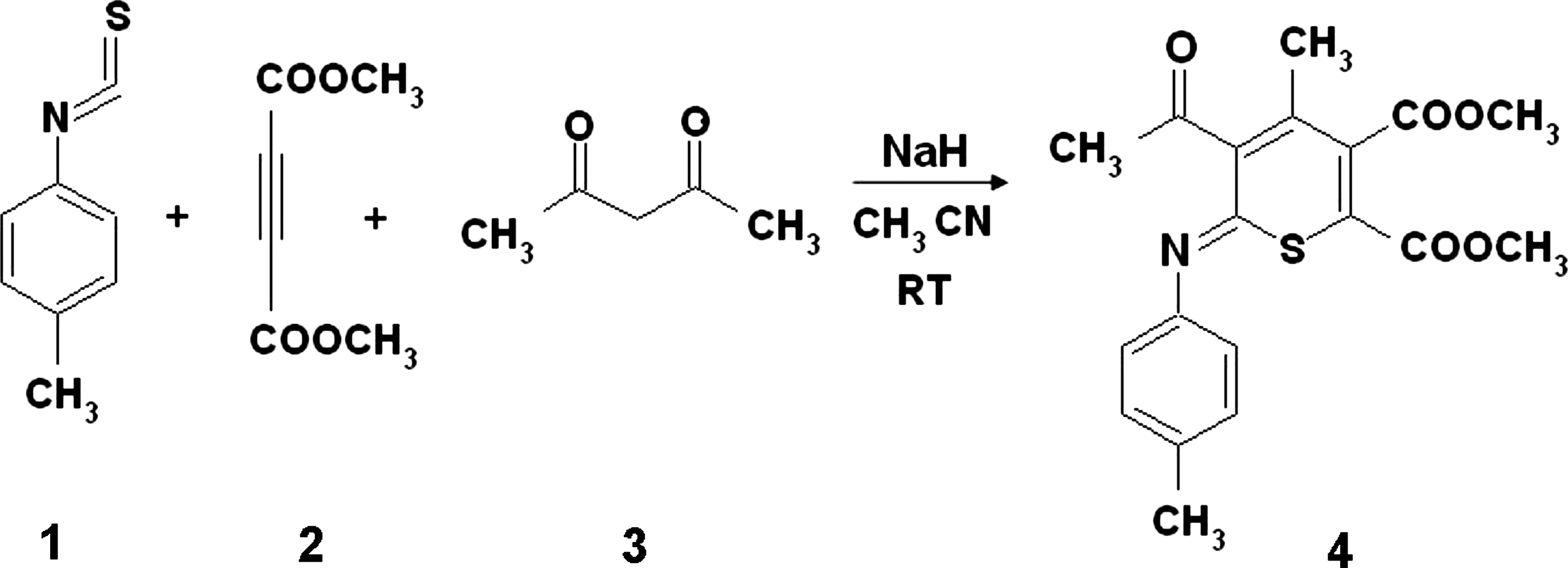
A facile synthesis of 4 is described via reaction between 1,3-dicarbonyls, electron-deficient acetylenic compounds such as DMAD, and aryleisothiosyanate in the presence of sodium hydride as a base in acetonitril at room temperature (Fig. 1). The advantages of this protocol are that no catalyst is required for this reaction and the simplicity of the present procedure makes it an attractive alternative to the complex multistep approaches. The reaction mixture was stirred for 8 h. The solvent was removed under reduced pressure and the residue was separated by silica gel column chromatography to give 4.
Synthesis of dimethyl-5-acetyl-4-methyl-6-(4-methylphenylimino)-6H-thiopyran-2,3-dicarboxylate (4).
Cytotoxic evaluation in vitro and clonogenic assay
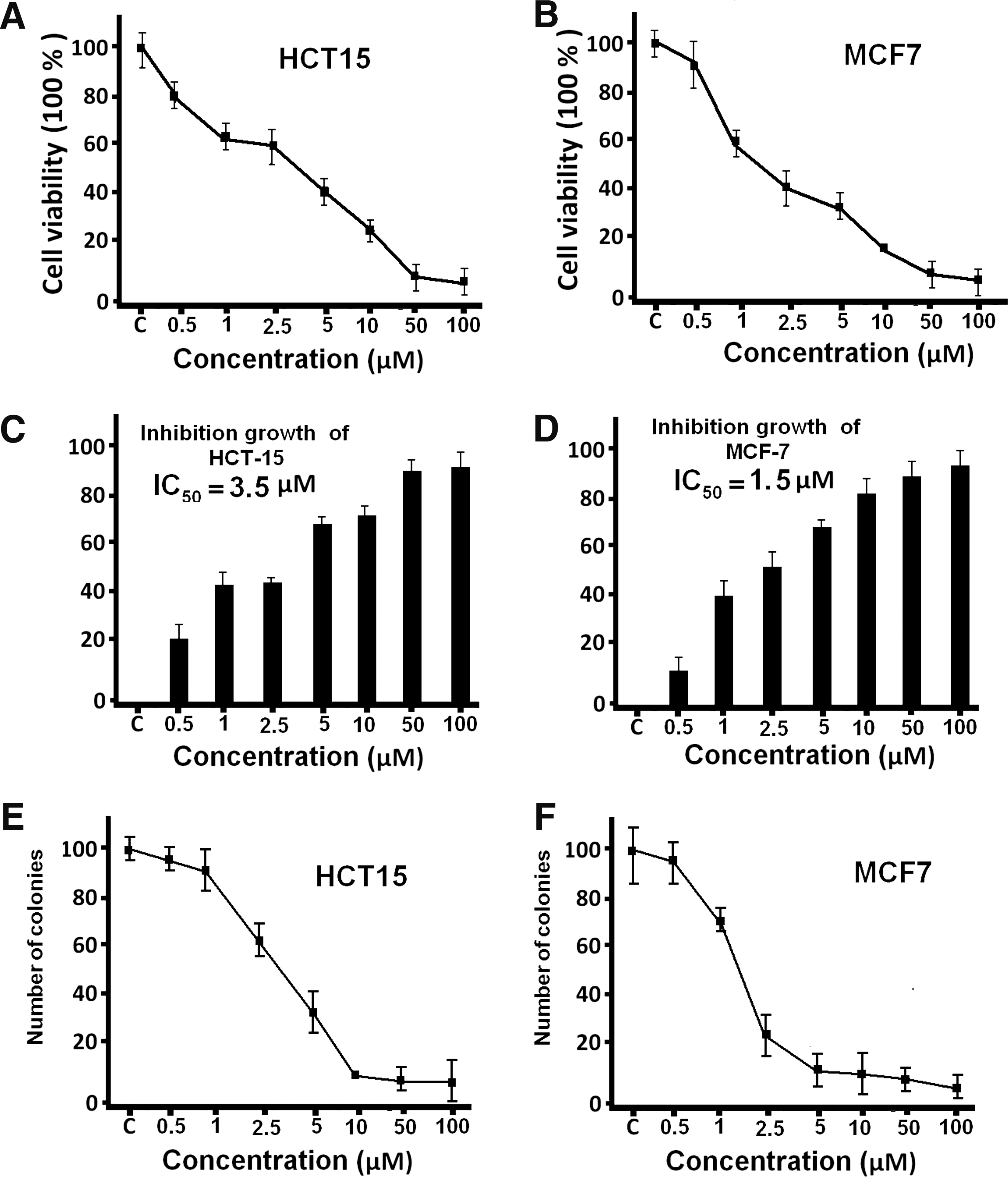
One of the factors of importance in determining the killing of mammalian cells following exposure to a variety of anticancer agents is the proliferative state of cell population (Valeriote and van Putten, 1975). The compound 4 was evaluated for its growth inhibitory effect on HCT-15 colon and MCF-7 breast cancer cell lines. The cells were treated with 4 at different concentrations, ranging from 1 to 100 μM, for 4 days using the SRB assay (Fig. 2A, B). In the presence of different doses of 4, the cells were inhibited ranging from 10% to 90% with a loss of viable cells (Fig. 2C, D). The compound 4 inhibited the proliferation of MCF-7 and HCT-15 cells in a concentration- and time-dependent manner. The IC50 was determined by interpolation from dose–response curves. The percentage of growth inhibition was calculated by comparison of the absorbance of treated cells versus control cells. The viability of the HCT-15 cells was unaffected by less than 0.2 μM of 4, but changed to 80% in response to 0.5 μM 4. However, the viability of HCT-15 decreased to 50% (IC50) in response to 3.5 μM of 4 and continued to decrease as the concentration of 4 was increased; viability was 20% and 5% with exposure to 10 μM and 50 μM of 4, respectively. Subsequently, the viability of the MCF-7 cells was also unaffected by less than 0.5 μM of 4 treatment but changed to 60% in response to 1 μM 4, decreased to 50% (IC50) in response to 1.5 μM 4, and continued to decrease as the concentration of 4 was increased; viability was 20% with 10 μM. Therefore, the IC50 calculated for HCT-15 was 3.5 and 1.5 μM for MCF-7 (Fig. 2C, D). Control groups showed regular polygonal shape, and cell antennas were short. Cell morphology was affected by 4 treatment and included loss of adhesion, rounding, cell shrinkage, and detachment from the substratum. Analysis of clonogenic activity in HCT-15 and MCF-7 cells treated with 4 at concentrations ranging from 0.5 to 100 μM of 4 for 14 days revealed a complete inhibition of colony formation at 4 μM for HCT-15 and 2 μM for MCF-7 cells, whereas concentrations lower than 5 μM were ineffective (Fig. 2E, F).
Effects of 4 on the proliferation of HCT-15 and MCF-7 cells. (A) Effect of 4 on the proliferation of HCT-15; (B) effects of 4 on the proliferation of MCF-7; (C) inhibition of 4 on HCT-15 growth; (D) inhibition of 4 on MCF-7 growth. The percentage of growth inhibition was calculated using the following equation: (1−At/Ac)×100, where At and Ac represent the absorbance in treated and control cultures, respectively. IC50 was determined by interpolation from dose–response curves. Dose–response curve of 4-mediated inhibition of HCT-15 (E) and MCF-7 (F) cell colony formation.
Effects of 4 on cell invasion and cell adhesion
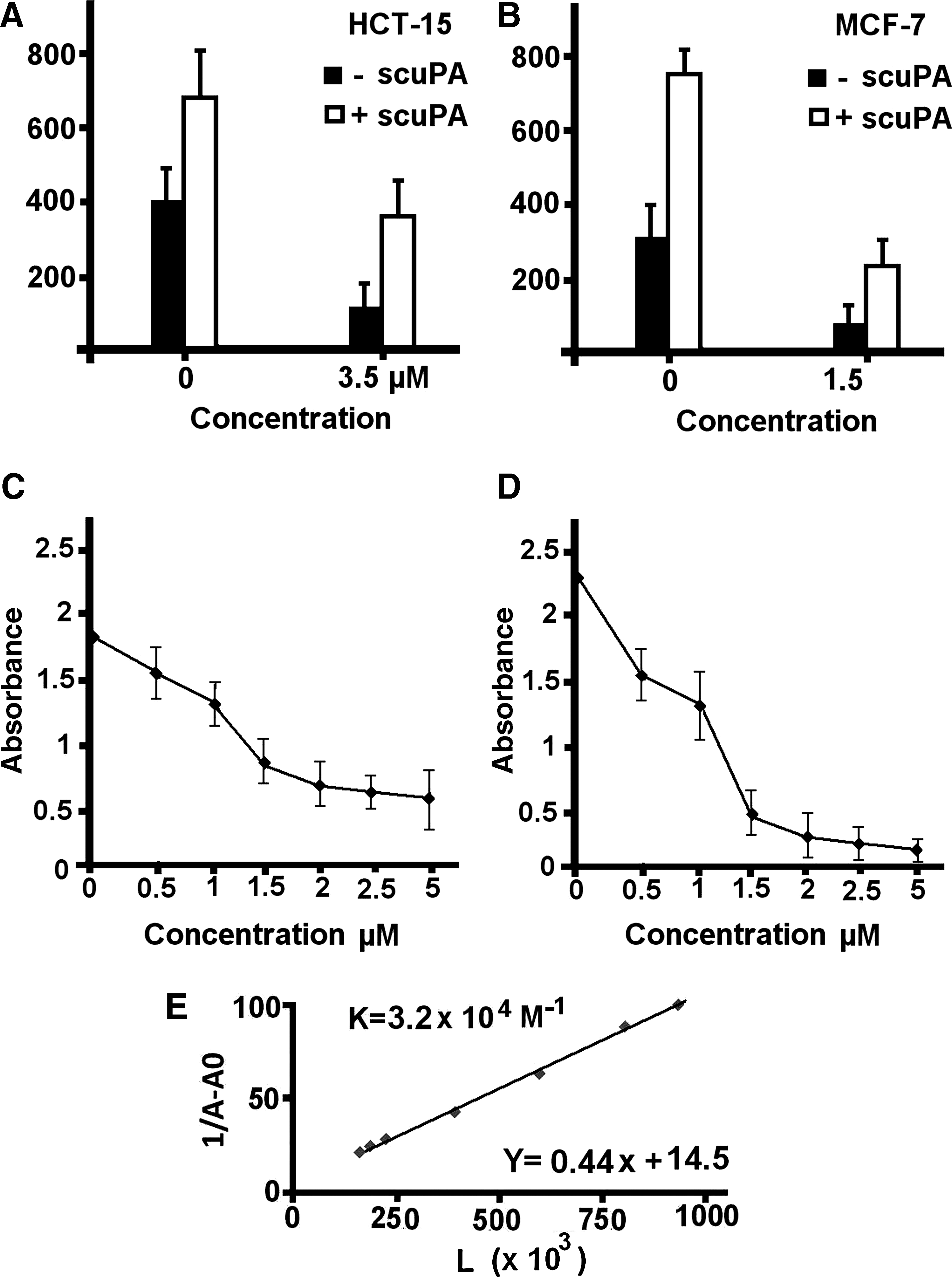
We next examined the effect of 4 on HCT-15 and MCF-7 cell invasion through Matrigel, a reconstituted extracellular matrix (ECM). Cell invasion involves a series of proteolytic events involving proteolytic enzymes such as urokinase-plasminogen activator (u-PA) and matrix metalloproteinases (MMPs). The concept that these proteases are involved in invasion and metastasis, as well as tumor angiogenesis, has been well accepted (Mignatti and Rifkin, 1996). As shown in Figure 3A, 3.5 μM and 1.5 μM of 4 greatly reduced the invasiveness of HCT-15 cells and MCF-7, respectively (Fig. 3B): 60% inhibition for HCT-15 and 75% inhibition for MCF-7 compared with control. Therefore, compound 4 reduced tumor cell invasiveness through Matrigel, and we propose that this may relate to a decrease in protease production (Shao et al., 1998; Lindenmeyer et al., 2001). However, the reduced invasiveness and migration of cells induced by 4 was not simply due to insufficient protease-dependent matrix degradation. Although cell surface saturation by exogenous scu-PA did increase the invasion capacity of the 4-treated cells, it was still reduced compared with that of scu-PA–saturated control cells.
(A, B) Invasion through Matrigel. Cells were added in upper chamber with 4 and assay time was 24 h. Inhibitory effect of 4 on HCT-15 cell (C) and MCF-7 cell (D) adhesion to Matrigel. Pretreated cells were allowed to adhere for 20 min, and absorbance was measured at 405 nm. (E) The plot of 1/(A−A0) versus 1/[L] for DNA and iPA complexes at different drug concentrations.
Indeed, 4 also altered both cells' adhesive behavior in ECM proteins and our results showed that the ability of 4-treated cells to adhere to Matrigel was reduced in a dose-dependent manner, demonstrating 50% inhibition at 1.25 μM 4 for both cell lines (Fig. 3C, D). Pretreated cells were allowed to adhere for 20 min, and absorbance was measured at 405 nm. Values are means±standard deviation of three separate experiments each carried out in triplicate. As 4 altered the cells' adhesive behavior in ECM proteins, this inhibitory effect on cell adhesion seems not to be related to the u-PA system, inasmuch as it was not influenced by saturating the cells with scu-PA. The reason for this reduced adhesion is unclear, and further study is needed to identify the cell surface adhesive molecules and the matrix proteins involved. Our results reveal that 4, which inhibits the expression of proteases involved in degradation of the ECM, may also regulate other cellular activities associated with the invasive phenotype, including cell migration and cell-matrix adhesion. The decrease in invasiveness may be also due to modification of the ultrastructure of these cells, which was not restored by u-PA.
Calculation of binding constant with DNA
Small molecules that bind reversibly to DNA are among the antitumor drugs currently used in chemotherapy. In the pursuit of a more rational approach to cancer chemotherapy based upon these molecules, it is necessary to exploit the interdependency between DNA-binding affinity, sequence selectivity, and cytotoxicity. Although there are previous studies considering that large and more complex molecules are more potent antitumor agents (Huang et al., 2006), there is no clear association between biological potency and the strength of binding to DNA, but there are some examples illustrating that changes in DNA binding among structurally related molecules can be accompanied by abrupt changes in biological activity (Denny et al., 1983). The strategy followed here is the calculation of the overall binding constant, which was carried out on the basis of UV absorption, and the equilibrium for 4 and DNA complex can be described as follows:\documentclass{aastex}\usepackage{amsbsy}\usepackage{amsfonts}\usepackage{amssymb}\usepackage{bm}\usepackage{mathrsfs}\usepackage{pifont}\usepackage{stmaryrd}\usepackage{textcomp}\usepackage{portland,xspace}\usepackage{amsmath,amsxtra}\pagestyle{empty}\DeclareMathSizes{10}{9}{7}{6}\begin{document}\begin{align*} [ { \rm DNA} + {\bf 4} ] \leftrightarrow [ { \rm DNA} - {\bf 4} ] K_{\bf 4} \\ K_{\bf 4} = [ { \rm DNA} - {\bf 4} ] / [ { \rm DNA} ] [ {\bf 4} ] \end{align*}\end{document}
The double reciprocal plot of 1/[A−A0] versus 1/[ligand] is linear and the association binding constant (K) is calculated from the ratio of the intercept on the vertical coordinate axis to the slope (Tajmir-Riahi et al., 1995; Mandeville et al., 2010; Froehlich et al., 2011). Concentrations of the complexed ligand were determined by subtracting absorbance of free DNA at 260 nm from those of the complexed DNA. Concentration of the free ligand was determined by subtraction of complexed ligand from total ligand used for the experiment. Our data show that 1/[complexed ligand] almost proportionally increases as a function of 1/[free ligand] (Fig. 3E). Therefore, an overall binding constant K4-DNA=9.8×10⁴ M−1 was estimated for 4-DNA.
Conclusion
The preventive effect of heterocyclic compounds on cancer incidence makes them attractive candidates for the development of anticancer agents. We demonstrated here the synthesis and characterization of compound 4. The antiproliferative effects mediated by 4 on the growth of HCT-15 and MCF-7 cell lines were monitored at different concentrations, ranging from 1 to 100 μM for a total of 4 days, and IC50, calculated by SRB assay, were 3.5 μM for HCT-15 and 1.5 μM for MCF-7. Cell shape and morphology of treated cells showed regular polygonal shape, cell antennas were short, and the treated cells affected by 4 showed loss of adhesion, rounding, cell shrinkage, and detachment from the substratum. The present analysis of these two human cancer cell lines revealed suppression of clonogenic activity after exposure to 4 at a concentration of 4 μM for HCT-15 and of 2 μM for MCF-7. We next examined the effect of 4 on HCT-15 and MCF-7 cell invasion through Matrigel, a reconstituted ECM; 3.5 μM and 1.5 μM of 4 greatly reduced the invasiveness of HCT-15 cells and MCF-7, respectively. Compound 4 also inhibits expression of adhesion proteins in both cells.
The connection between DNA damage and antiproliferation activity is supported by a large body of literature. At present, no clear evidence of 4-induced DNA damage has been demonstrated, and our result from structural analysis showed interaction of 4 with DNA and the binding constant value K4-DNA=9.8×10⁴ M−1 suggests that 4 interacts at DNA in good mode. Although there are some examples illustrating that changes in DNA binding among structurally related molecules can be accompanied by abrupt changes in biological activity (Denny et al., 1983), a deeper understanding of the mechanism of action of drugs is needed, which includes the dissection of forces driving their noncovalent binding to DNA. This information can be used to develop new anticancer agents with higher activity (Portugal et al., 2005; Portugal, 2009). Therefore, further research is required to demonstrate that binding effect may be related to DNA damage or apoptosis in cancer cells.
Footnotes
Acknowledgment
Dr. M. Rajabi greatly appreciates the financial support from the Molecular Medicine Ph.D. Program at the University of Milan.
Disclosure Statement
No competing financial interests exist.
References
1.
BoultonS., KyleS., DurkaczW.2000. Mechanisms of enhancement of cytotoxicity in etoposide and ionising radiation-treated cells by the protein kinase inhibitor wortmannin. Eur J Cancer, 36:535–541.
2.
BrownM.J., CarterP.S., FenwickA.E., FosberryA.P., HamprechtD.W., HibbsM.J., ArvestR.L., MensahL., MilnerP.H., O'HanlonP.J., PopeA.J., RichardsonC.M., WestA., WittyD.R.2002. The antimicrobial natural product chuangxinmycin and some synthetic analogues are potent and selective inhibitors of bacterial tryptophanyl tRNA synthetase. Bioorg Med Chem Lett, 12:3171–3174.
3.
CasyG., LaneS., TalorR.J.K.1986. The preparation of thiathromboxane analogues and a formal total synthesis of dithiathromboxane A2 based on conjugate addition reactions of thiin-4-ones. J Chem Soc Perkin Trans, 1:1397–1404.
4.
CasyG., TaylorR.J.K.1988. Facile Ramberg-Bäcklund reactions for the synthesis of 2,3-disubstituted cyclopentenones; A short synthetic route to tetrahydrodicranenone B. J Chem Soc Chem Commun, 454–455. 10.1039/C39880000454. http://pubs.rsc.org/en/content/articlelanding/1988/C39880000454
5.
CasyG., TaylorR.J.K.1989. The synthesis of 2,3-disubstituted cyclopentenones using Ramberg-Bäcklund reaction in conjunction with organocopper chemistry. Tetrahedron, 45:455–466.
6.
DennyW.A., AtwellG.J., BaguleyB.C.1983. Potential antitumor agents. 39. Anilino ring geometry of amsacrine and derivatives: relationship to DNA binding and antitumor activity. J Med Chem, 26:1625–1630.
7.
FroehlichE., MandevilleJ.S., WeinertC.M., KreplakL., Tajmir-RiahiH.A.2011. Bundling and aggregation of DNA by cationic endrimers. Biomacromolecules, 12:511–517.
8.
HamadiH., KhoobiM., HossainiZ., ShafieeA.2011. Facile and efficient synthesis of functionalized iminothiopyran and isothiochromen via one-pot multicomponent reactions. Mol Divers, 15:35–40.
9.
HollickJ.J., GoldingB.T., HardcastleI.R., MartinN., RichardsonC., RigoreauL.J., SmithG.C., GriffinR.J.2003. 2,6-disubstituted pyran-4-one and thiopyran-4-one inhibitors of DNA-dependent protein kinase (DNA-PK)Bioorg Med Chem Lett, 13:3083–3086.
10.
HollickJ.J., RigoreauL.J., Cano-SoumillacC., CockcroftX., CurtinN.J., FrigerioM., GoldingB.T., GuiardS., HardcastleI.R., HicksonI., HummersoneM.G., MenearK.A., MartinN.M., MatthewsI., NewellD.R., OrdR., RichardsonC.J., SmithG.C., GriffinR.J.2007. Pyranone, thiopyranone, and pyridone inhibitors of phosphatidylinositol 3-kinase related kinases. Structure-activity relationships for DNA-dependent protein kinase inhibition, and identification of the first potent and selective inhibitor of the ataxia telangiectasia mutated kinase. J Med Chem, 19:1958–1972.
11.
HuangR., WallqvistA., CovellD.G.2006. Assessment of in vitro and in vivo activities in the National Cancer Institute's anticancer screen with respect to chemical structure, target specificity, and mechanism of action. J Med Chem, 49:1964–1979.
12.
HurleyL.H.2002. DNA and its associated processes as targets for cancer therapy. Nat Rev Cancer, 2:188–200.
13.
KimC.H., ParkS.J., LeeS.H.2002. A targeted inhibition of DNA-dependent protein kinase sensitizes breast cancer cells following ionizing radiation. J Pharmacol Exp Ther, 303:753–759.
14.
LesykR., ZimenkovskyB., AtamanyukD., JensenF., Kieć-KononowiczK., GzellaA.2006. Anticancer thiopyrano[2,3-d][1,3]thiazol-2-ones with norbornane moiety. Synthesis, cytotoxicity, physico-chemical properties, and computational studies. Bioorg Med Chem, 14:5230–5240.
15.
LindenmeyerF., LiH., MenashiS., SoriaC., LuH.2001. Apigenin acts on the tumor cell invasion process and regulates protease production. Nutr Cancer, 39:139–147.
16.
MandevilleJ.S., N'soukpoé-KossiC.N., NeaultJ.F., Tajmir-RiahiH.A.2010. Structural analysis of DNA interaction with retinol and retinoic acid. Biochem Cell Biol, 88:469–77.
17.
McAllisterG.D., TaylorR.J.K.2001. The synthesis of polyoxygenated, enantiopure cyclopentene derivatives using Ramberg-Bäcklund rearrangement. Tetrahedron Lett, 42:1197–1200.
18.
McDonaldB.P., SteeleR.W., SutherlandJ.K.1988. Synthetic approaches to thiathromboxanes. Part 2. Synthesis of structural isomers of thiathromoxane A2. J Chem Soc Perkin Trans, 1:675–679.
19.
MehrzadJ., RajabiM., KhalilzadehM.A.2010. Design and antiproliferative activity of 2-(3,5-Dihydroxyphenyl)-6-hydroxybenzothiazole (DHB) on PC-3 prostate cancer cell line. Iran J Org Chem, 2:498–500.
20.
MignattiP., RifkinD.B.1996. Plasminogen activators and angiogenesis. Curr Top Microbiol Immunol, 213:33–50.
21.
MinottiG., MennaP., SalvatorelliE., CairoG., GianniL.2004. Anthracyclines: molecular advances and pharmacologic developments in antitumor activity and cardiotoxicity. Pharmacol Rev, 56:185–229.
22.
NafisiS., HashemiM., RajabiM., Tajmir-RiahiH.A.2008. DNA adducts with antioxidant flavonoids apigenin, morin and naringin. DNA Cell Biol, 27:433–442.
23.
NedolyaN.A., BrandsmaL., VerkruijsseH., TrofimovB.A.1998. 2-Iminothiopyrans from methylthiobutadiene and isothiocyanates. Chem Heterocycl Comp, 34:550–553.
24.
PortugalJ.2009. Evaluation of molecular descriptors for antitumor drugs with respect to noncovalent binding to DNA and antiproliferative activity. BMC Pharmacol, 9:11.
25.
PortugalJ., CashmanD.J., TrentJ.O., Ferrer-MirallesN., PrzewlokaT., FoktI., PriebeW., ChairesJ.B.2005. A new bisintercalating anthracycline with picomolar DNA binding affinity. J Med Chem, 48:8209–8219.
26.
QuagliaW., PiginiM., PiergentiliA., GiannellaM., GentiliF., MarucciG., CarrieriA., CarottiA., PoggesiE., LeonardiA., MelchiorreC.2002. Structure-activity relationships in 1,4-benzodioxan-related compounds. 7. Selectivity of 4-phenylchroman analogues for alpha(1)-adrenoreceptor subtypes. J Med Chem, 45:1633–1643.
27.
RajabiM., SignorelliP., GorincioiE., GhidoniR., SantanielloE.2010. Antiproliferative activity of N6-isopentenyladenosine on MCF-7 breast cancer cells: cell cycle analysis and DNA-binding study. DNA Cell Biol, 29:687–691.
28.
ShaoZ.M., WuJ., ShenZ.Z., BarskySH.1998. Genistein exerts multiple suppressive effects on human breast carcinoma cells. Cancer Res, 58:4851–4857.
29.
SmithG.C.M., JacksonS.P.1999. The DNA-dependent protein kinase. Genes Dev, 13:916–934.
30.
SugitaY., HosoyaH., TerasawaK., YokoeI., FujisawaS., SakagamiH.2001. Cytotoxic activity of benzothiepins against human oral tumor cell lines. Anticancer Res, 21:2629–2632.
31.
Tajmir-RiahiH.A., NeaultJ.F., NaouiM., DiamantoglouS.1995. The effects of HCl on the solution structure of calf thymus DNA: a comparative study of DNA denaturation by proton and metal cations using Fourier transform IR ifference spectroscopy. Biopolymers, 35:493–501.
32.
ValerioteF., Van PuttenL.1975. Proliferation-dependent cytotoxicity of anticancer agents: a review. Cancer Res, 35:2619–2630.
33.
Van VlietL.A., RodenhuisN., DijkstraD., WikstromH., PugsleyT.A., SerpaK.A., MeltzerL.T., HeffnerT.G., WiseL.D., LajinessM.E., HuffR.M., SvenssonK., SundellS., LundmarkM.2000. Synthesis and pharmacological evaluation of thiopyran analogues of the dopamine D3 receptor-selective agonist (4aR,10bR)-(+)-trans-3,4,4a,10b-tetrahydro-4-n-propyl-2H,5H [1]b enzopyrano[4,3-b]-1,4-oxazin-9-ol (PD 128907)J Med Chem, 43:2871–2882.
34.
VedejsE., KrafftG.A. 1982. Cyclic sulfides in organic synthesis. Tetrahedron, 38:2857–2881.
35.
WangW., LiH., WangJ., ZuL.S.2006. Enantioselective organocatalytic tandem Michael-Aldol reactions: one-pot synthesis of chiral thiochromenes. J Am Chem Soc, 128:10354–10355.
36.
WardD.E., JheengutV., BeyeG.E.2006. Thiopyran route to polypropionates: an efficient synthesis of serricornin. J Org Chem, 71:8989–8992.
37.
WeinsteinJ.N., MyersT.G., O’ ConnorP.M., FriendS.H., FornaceA.J., KohnK.W., FojoT., BatesS.E., RubinsteinL.V., AndersonN.L.1997. An information-intensive approach to the molecular pharmacology of cancer. Science, 275:343–349.
38.
YoshimuraY., YamazakiY., SaitoY., TakahataH.2009. Synthesis of 1-(5,6-dihydro-2H-thiopyran-2-yl)uracil by a Pummerer-type thioglycosylation reaction: the regioselectivity of allylic substitution. Tetrahedron, 65:9091–9102.
39.
ZhangX., LiX., FanX., WangX., LiD., QuG., WangJ.2009. Ionic liquid promoted preparation of 4H-thiopyran and pyrimidine nucleoside-thiopyran hybrids through one-pot multi-component reaction of thioamide. Mol Divers, 13:57–61.