
Abstract
The aim of this study was to clone the heart-type fatty acid binding protein (H-FABP) gene of Xuhuai goat, to explore it bioinformatically, and analyze the subcellular localization using enhanced green fluorescent protein (EGFP). The results showed that the coding sequence (CDS) length of Xuhuai goat H-FABP gene was 402 bp, encoding 133 amino acids (GenBank accession number AY466498.1). The H-FABP cDNA coding sequence was compared with the corresponding region of human, chicken, brown rat, cow, wild boar, donkey, and zebrafish. The similarity were 89%, 76%, 85%, 84%, 93%, 91%, 70%, respectively. For the corresponding amino acid sequences, the similarity were 90%, 79%, 88%, 97%, 95%, 94%, 72%, respectively. This study did not find the signal peptide region in the H-FABP protein; it revealed that H-FABP protein might be a nonsecreted protein. H-FABP expression was detected in vitro by reverse transcription-polymerase chain reaction (RT-PCR), and the EGFP-H-FABP fusion protein was localized to the cytoplasm. The gene could also be transiently and permanently expressed in mice.
Introduction
Heart-type fatty acid binding protein (H-FABP) is a member of the family with wide distribution and strong biological functions (Li et al., 2010). The main function of H-FABP is to take part in cellular fatty acid transportation. H-FABP specifically binds hydrophobic ligand molecules, then transfers fatty acids from the cell membrane to the triglycerides and phospholipid synthesis sites. Therefore, H-FABP is a key fatty acid carrier protein (Wei et al., 2010). H-FABP may be a candidate gene affecting intramuscular fat content and meat quality of livestock. The intramuscular fat is the main precursor of muscle's flavor, and is positively correlated with meat quality and taste, as it affects the meat's tenderness, flavor, especially the meat juice. Modification of H-FABP at the molecular level could improve the taste and flavor of meat, providing a new application for animal breeding.
At present, the H-FABP gene cDNA of cattle, pigs, donkeys, humans, rats, mice, and chicken have been cloned (Chmurzyńska, 2006; Zhong et al., 2006; Li et al., 2010), but that of Xuhuai goat has not been reported yet. In this study, Xuhuai goat H-FABP gene cDNA sequences were cloned for the first time and its subcellular location determined.
Materials and Methods
Experimental material
Xuhuai goat heart tissue was collected from Erling goat slaughterhouse Zhenjiang City, Jiangsu Province. Escherichia coli DH5α strain, plasmid pEGFP-C1, and goat embryo fibroblast (GEF) culture was preserved by the laboratory.
Reagents
First Strand cDNA Synthesis Kit, HindIII, EcoRI restriction enzymes, T4 DNA Ligase were purchased from MBI Company; LA Taq DNA polymerase, Taq DNA polymerase, pMD19-T kit, Takara Agrose Gel DNA purification Kit were purchased from Takara Company; mini plasmid kit and polyethylene imide were from Beijing Dingguo company; 1 kb DNA Ladder and 3000 bp DNA Ladder were from Genescript; Lipofectamine LTX and PLUS Reagents were from Shanghai Hesheng.
Cloning and expression of H-FABP and subcellular localization of the products
Primer design and synthesis
Primers for the Xuhuai goat H-FABP gene were designed according to the sequences in GenBank (accession number: AY466498.1), with the restriction sites of HindIII and EcoRI at the 5′ end. The primer was synthesized by Sangon Biotech Company, the sequences was shown as follows:
F:5′ CG
R:5′ CCC
Amplification of H-FABP gene CDS
Total RNA was extracted from Xuhuai goat heart tissue using Trizol reagent (Invitrogen) according to the manufacturer's protocol. The quality of total RNA was assessed with a Nanodrop 1000 (Thermo Scientific) and 1% agarose gel electrophoresis. cDNA was synthesized from l μg of total RNA, using First Strand cDNA Synthesis Kit (MBI) according to the manufacturer's recommended protocol. The cDNA was used as a template to amplify the full-length H-FABP gene by polymerase chain reaction (PCR), The PCR products were detected by 0.8% agarose gel electrophoresis, purified by gel extraction, and ligated into the pMD19-T vector.
The ligation products were transformed into E. coli DH5α competent cells, cultured on the LB (Amp+) solid medium for 12–16 h, and plasmid was extracted using Plasmid Minispin Kit (Dingguo Biotech Company). Plasmids were characterized by double enzyme digestion and sequenced by Sangon Biotech Company. Plasmid containing the correct insertion was named pMD19-T-H-FABP.
Sequence analysis of H-FABP gene
The homology of the H-FABP CDS were checked by the BLAST software between Xuhuai goat and other species; human (NM_004102.3), chicken (NM_001030889), rat (NM_024162.1), cow (NM_174313), boar (NM_001099931), donkey (GQ244482.1), and zebrafish (NM_152961), respectively. DNAStar was used to analyze the phylogenetic relationships among these species. SignalP 3.0 Server (
Construction of the pEGFP-H-FABP expression vector
pMD19-T-H-FABP and the vector were digested with HindIII and EcoRI, separated by electrophoresis on a 0.8% agarose gel. The bands were excised and purified, then ligated. The ligation products were transformed into E. coli DH5α and cultured on LB (Amp+) solid medium for 12–16 h. Positive clones were cultured in LB (Amp+) liquid medium for 12–16 h, then plasmids extracted by Plasmid Minispin Kit (DingGuo Company). Constructs were sequenced by Sangon Biotech Company and the plasmid containing the right insertion was named pEGFP-H-FABP. This plasmid will result in the coexpression of H-FABP and enhanced green fluorescent protein (EGFP).
H-FABP primary structure and subcellular localization analysis
ExPASy ProtParam tool (
Transfection of the recombinant plasmid pEGFP-H-FABP into GEF cells
Cell culture and LipofectamineTM LTX&PLUS (LTX) was used for transfection. Twenty-four hours post-transfection, the transfected cells were observed under fluorescence inverted microscope.
Detection of the expression of H-FABP at the mRNA level by reverse transcription-PCR (RT-PCR)
Forty-eight hours post-transfection, total RNA was extracted from transfected GEF cells with Trizol reagent (Invitrogen) as per manufacturer's protocol. cDNA was synthesized from l μg of total RNA using First Strand cDNA Synthesis Kit (MBI) according to the manufacturer's recommended protocol. Then cDNA was used as a template to amplify H-FABP sequences.
Detection of fusion protein products by western blot
Forty-eight hours post-transfection, cells were harvested, and washed with phosphate-buffered saline (PBS) three times. Total protein was extracted by cell lysate according to the manufacturer's instructions. About 10 μL protein samples were processed by 10% sodium dodecyl sulfate–polyacrylamide gel electrophoresis and then transferred to polyvinylidene fluoride (PVDF) membrane (80 mA, 2 h). The protein on PVDF membrane was hybridized with EGFP antibody (1:1000), and goat anti-rabbit IgG (1:2000) was used as the secondary antibody and proteins detected with diaminobenzidine (DAB).
Preparation of transgenic mice mediated by testicular injection
Tail vein injection of recombinant plasmid
A total of six healthy, 6-week-old mice were selected for tail injection. The pEGFP-H-FABP plasmid was wrapped by PEI in a ratio of 3:1. Twelve micrograms of pEGFP-H-FABP plasmid was injected into the tail vein of three mice.
Detection of the target gene expression from frozen mouse tissue sections
Forty-eight hours after injection, the mice were sacrificed. The heart, liver, kidney, testis, and muscles were collected for frozen sections, and observed under fluorescence inverted microscope.
Testicular injection of recombinant plasmid
Four healthy, 6-week-old male mice were chosen for testis injection. The pEGFP-H-FABP plasmid was wrapped by PEI in a ratio of 3:1. Twelve micrograms of pEGFP-H-FABP plasmid was injected into the testis. After five days, one treated mouse and three healthy adult, nonpregnant female mice were put in one cage and the F1 generation obtained.
Detection of transfection efficiency
Twenty days after testicular injection, the epididymis of the male mouse was cut into pieces, and incubated in PBS for 10 min at room temperature so that the sperm could be freed. Ten μL was observed by fluorescence microscope for sperm expression of green fluorescent protein. The remaining tissue was filtered, and the semen collected and centrifuged to extract sperm DNA, and the construct detected by PCR.
Detection of gene expression in the F1 generation
Fifteen mice of F1 generation survived. Both the experiment and control group mice were killed and parts of heart tissues were collected for DNAand protein extraction. The heart DNA was used as template for PCR; the PCR products were detected by agarose gel electrophoresis. The protein was extracted and detected by western blot. Untreated mouse DNA and protein were used as negative controls.
Results
Cloning of H-FABP gene
To amplify the H-FABP gene, RT-PCR was performed. A band of the expected size (402 bp) was amplified (Fig. 1). This fragment was ligated with pMD19-T to produce a new plasmid named pMD19-T-H-FABP. The pMD19-T-H-FABP plasmid was characterized by PCR (Fig. 2) and enzyme digestion (Fig. 3). The amplified fragment size was 402 bp. Single enzyme digestion with EcoRI produced a single band of 3.1 kb; results of double enzyme digestion with HindIII and EcoRI were 2.7 kb and 402 bp, consistent with our expectations. The sequencing results showed there was a mutation at 383 (C-T), but it was a synonymous mutation. The sequence was submitted to GenBank (accession number HQ895788).
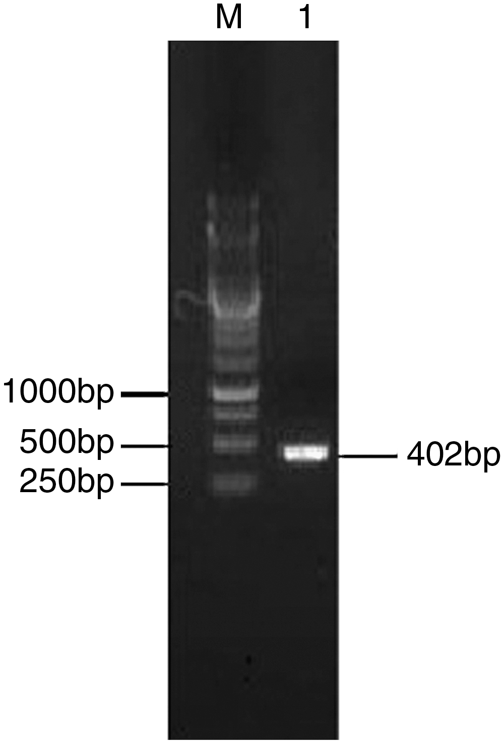
PCR amplification of H-FABP cDNA. M, 1 kb DNA marker; 1, PCR amplification of target gene cDNA. H-FABP, heart-type fatty acid binding protein; PCR, polymerase chain reaction.
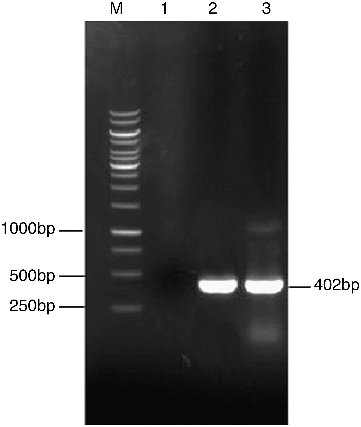
PCR amplification of pMD19-T-H-FABP bacterial suspension. M, 1 kb marker; 1, negative control; 2, positive control; 3, experimental group.
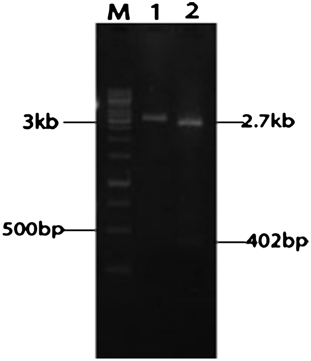
Restriction enzyme digestion of pMD19-T-H-FABP plasmid. M, 1 kb marker; 1, single-enzyme results; 2, double digestion results.
Bioinformatics analysis of H-FABP gene
DNAStar software was used to analyze the Xuhuai goat H-FABP gene. The results showed that the length of the CDS was 402 bp, including the initiation codon ATG and the termination codon TAG, and encoded 133 amino acids.The H-FABP cDNA coding sequence of Xuhuai goat was compared with the corresponding regions of human, chicken, brown rat, cow, wild boar, donkey, and zebrafish with Blast software. The similarity of the H-FABP gene with the above were 89%, 76%, 85%, 84%, 93%, 91%, 70%, respectively. This result showed that H-FABP cDNA sequence of Xuhuai goat had the highest homology with wild boar, followed by donkey, and lowest with the zebrafish. The amino acid similarity were 90%, 79%, 88%, 97%, 95%, 94%, 72%, respectively (Table 1).
Identification of fusion expression vector pEGFP-H-FABP
To examine the construction of fusion expression vector pEGFP-H-FABP, PCR was used. The PCR product showed a 402 bp band. Single or double enzyme digestion was also performed. EcoRI digestion resulted in 5.1 kb band, while HindIII and EcoRI digestion resulted in 4.7 and 402 kb band respectively (Fig. 4). All were consistent with the predicted construction, and sequence confirmed the right construction of fusion expression vector pEGFP-H-FABP.
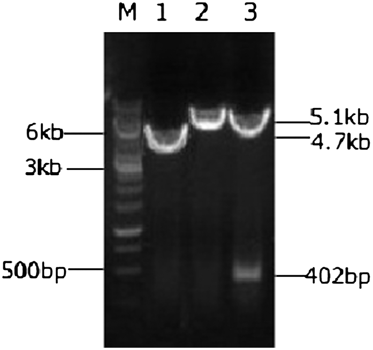
Restriction enzyme digestion of pEGFP-H-FABP plasmid. M, 1 kb marker; 1, pEGFP-H-FABP plasmid; 2, single-enzyme results; 3, double digestion results. EGFP, enhanced green fluorescent protein.
H-FABP protein primary structure and subcellular localization
ExPASy ProtParam tool and PSORT Protein Sorting Prediction tool was used to analyze the primary structure and subcellular localization, and the results showed that there was no signal peptide sequence for H-FABP. Primary structure analysis showed that the predicted molecular weight was 14.76 kDa with theoretical isoelectric point of 6.369. Subcellular localization analysis showed that H-FABP mostly expressed in the cytoplasm (Table 2).
Detection results of GEF cells transfected with pEGFP-H-FABP
The pEGFP-H-FABP plasmid was transfected into GEF cells. Green fluorescence was seen 24 h post-transfection and was distributed in the cytoplasm, not the nucleus (Fig. 5A, a). GEF cells were transfected with pEGFP-C1 as a positive control, and the green fluorescence was distributed throughout the cell (Fig. 5B, b). Untransfected GEF cells were the negative control and did not show any fluorescence (Fig. 5C, c). There were no significant differences for the cells selected by G418 for 14 days and 24 h post-transfection when both were transfected with pEGFP-H-FABP (Fig. 5D, d).
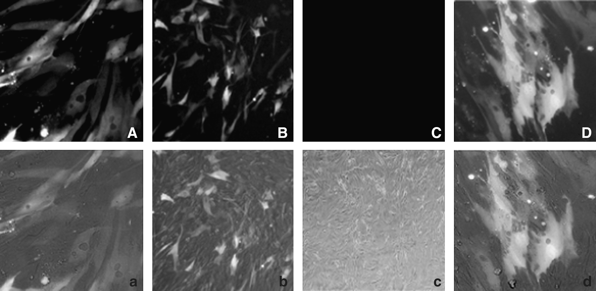
Image of goat fibroblasts under light and dark field 24 h after transfection of pEGFP-H-FABP, pEGFP-C1, and nontransfected (200 times;).
Detection of H-FABP by RT-PCR
H-FABP cDNA from GEF cells at 48 h post-transfection was used as a template to amplify the H-FABP gene, together with GAPDH as an internal control. Water and pEGFP-H-FABP were used as the negative and positive templates respectively. There was a specific band at 402 bp for the experimental group and positive control group, while there was no specific band for the negative control (Fig. 6). The GAPDH control group had a band at the predicted size (593 bp). These data showed there was H-FABP gene expression after GEF cells were transfected with pEGFP-H-FABP.
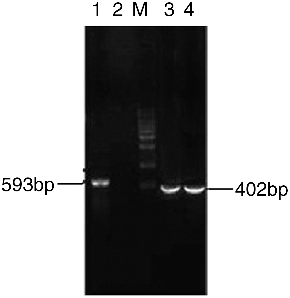
Detection of H-FABP by RT-PCR. 1, GAPDH; 2, negative control; 3, positive control; 4, The experimental group; M, marker.
Detection of H-FABP protein expression by western blot
Western blot results showed that the experimental group transfected with pEGFP-H-FABP expressed a single band of 42 kD which was the sum of EGFP (27 kD) and H-FABP (14.76 kD); the negative control group transfected with pEGFP- C1 expressed a single band of 27 kD, whereas there was no band for the blank control (Fig. 7)
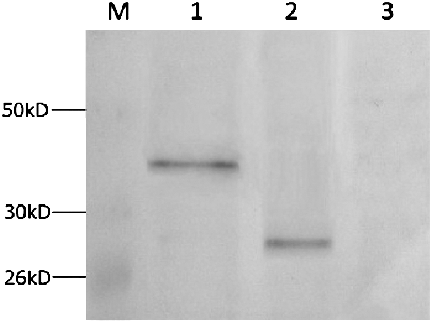
Detection of transfected protein by western blot. M, prestained protein marker; 1, transfection of pEGFP-H-FABP; 2, transfection with pEGFP-H-C1; 3, transfection without vector.
Fluorescence detection of frozen tissue sections after tail vein injection in mice
Forty-eight hours after tail vein injection, the mice were killed and heart, liver, kidney, testis, and muscle samples were collected for frozen sections and observed under fluorescence microscope, The H-FABP gene expressed in these tissues at different levels (Fig. 8).

Fluorescence detection of frozen section of mice tissue 48 h after the intravenous injection.
Fluorescence detection of sperm smear
Twenty days after testicular injection, male mice sperm smears were made and observed under the fluorescence microscope; it showed that in part of sperm there was fluorescence expression in the experimental group but no fluorescence in the negative control mice sperm (Fig. 9).
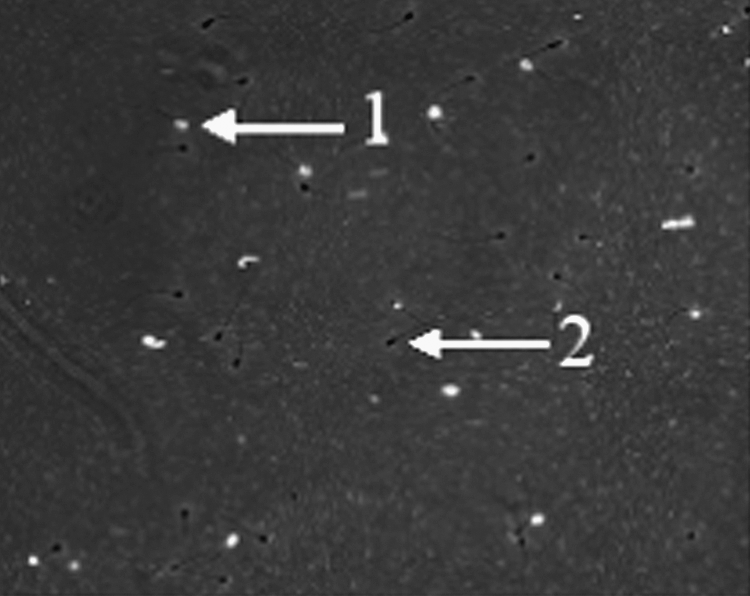
Fluorescence detection of experimental sperm smear. 1, positive sperm with fluorescence; 2, negative sperm without fluorescence.
Detection of sperm DNA
Mouse sperm DNA was extracted from experimental and control groups, and PCR was conducted to amplify H-FABP. Agarose gel electrophoresis results showed a band of 402 bp for the experimental group and the positive control group, while there was no band in the negative control group and blank control (Fig. 10).

PCR amplification result of sperm DNA. M, 1 kb marker; 1, experimental group; 2, positive control; 3, negative control; 4, blank control.
Heart tissue DNA testing of F1 generation
DNA was extracted from F1 mice hearts and used as templates for H-FABP specific PCR. Untreated mice were used as negative controls. Nine of 15 mice in the experimental group were positive for a 402 bp band; there were no bands for the negative control group (Fig. 11).
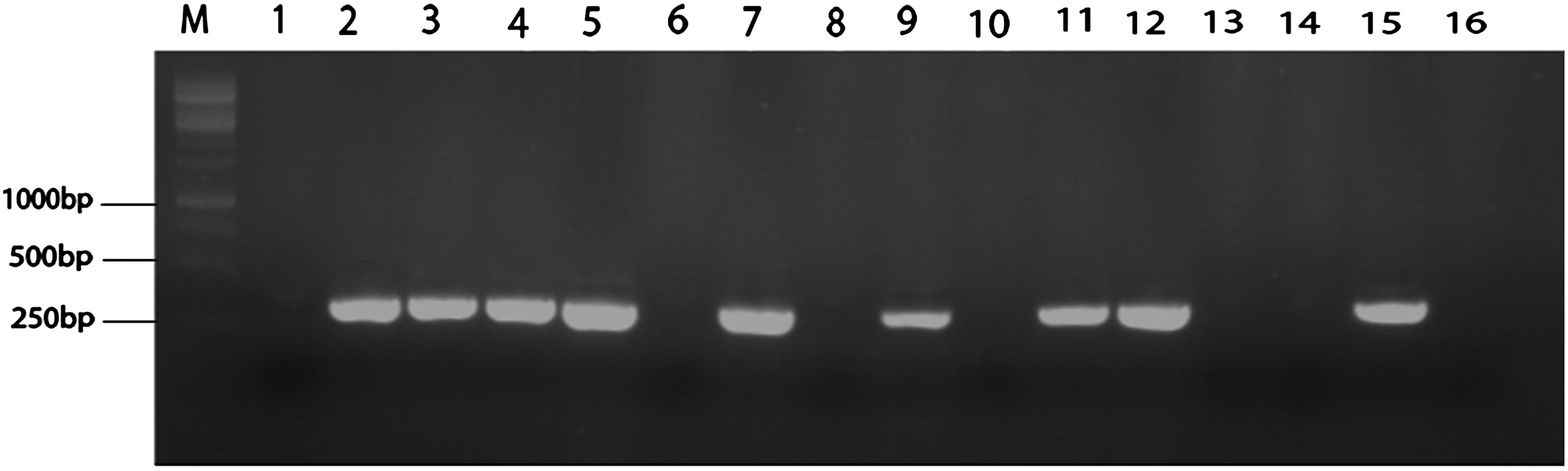
DNA PCR detection of F1 generation of heart tissue. M, 1 kb marker; 1, negative control; 2–16, experimental group.
Western blot detection in F1 generation of heart protein
Western blot detection of the heart protein in F1 mice showed that 7 of 15 mice were positive. The protein was 42 kD (Fig. 12).
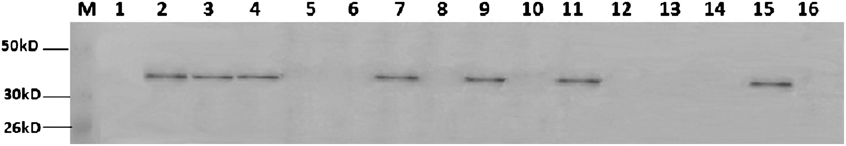
Western blot detection results of F1 generation. M, prestained protein marker; 1, negative control; 2–16, the experimental group.
Discussion
H-FABP is a widely distributed protein among the FABP family (Wang et al., 2002; Yang et al., 2009). Research indicated that there was a high homology of H-FABP in different species: the homology between cattle, sheep, pigs, horses, humans, mice, and chicken is 76.6%–96.8%. In this study, H-FABP was cloned from the heart tissue of the Xuhuai goat for the first time, the length was 402 bp. The gene sequence homology between goat and human, chicken, rat, cow, wild boar, donkey, and zebrafish were between 70% and 93%, and the homology of the amino acid sequence was between 72% and 97%. Nucleotide differences reflected on the protein level were mostly synonymous mutations. It indicated that the gene was highly conservative among species, which suggests that H-FABP is of great significance for the maintenance of normal physiological function (Hao et al., 2008).
H-FABP gene structure consists of four exons and three introns; there is no signal peptide and N-terminal acetylation, suggesting that it is a typical intracellular protein (Gerbens et al., 2001). Crystal X-diffraction analysis of its tertiary structure showed that it contained a domain composed of 10 antiparallel β-chain and two short α-helix formation of a β-sheet barrels (Ye et al., 2003; Li et al., 2006). The main function of this protein is in fat metabolism: it transports fatty acids from the cell membrane to the fatty acid oxidation position and synthesis sites of phospholipids and triglycerides to enter the mitochondrial energy metabolism system for fatty acids oxidative decomposition and ultimately generate adenosine triphosphate (Qiao and Ma, 2009). It plays an important role in regulation of body fat, especially intramuscular fat content. Studies have shown that H-FABP expression and intramuscular fat content are positively related, efficient expression of the protein could significantly improve the body's utilization of fatty acids (Huang et al., 2008).
Xuhuai goat is a landrace species in Xuzhou, Jiangsu province. Currently, most research about the H-FABP gene of Xuhuai goats concerns its polymorphism. The tissue specificity of H-FABP protein expression and the physiological function are not fully understood. In this study, we used Xuhuai goat fetal fibroblasts as host cells transfected with recombinant plasmid that contained the target gene and enhanced fluorescent protein gene for subcellular localization, and analyzed its initial expression at the mRNA level. The result showed the fusion protein was expressed in the cytoplasm in most cells. In a small number of cells, it expressed both in the cytoplasm and the nucleus. This expression may be due to the protein expression system, the protein tag, irradiation or heat stress (Zhou et al., 2011). Overexpression of tagged protein would affect the normal protein localization because EGFP protein expressed both in cytoplasm and nucleus. Western blotting showed that there was a single band of 42 kD in the experimental group, a single band of 27 kD in the empty vector control group, and no band in the blank control group. In cells transfected with recombinant vector by G418 for 14 days, green fluorescence could be observed under fluorescence microscope. Western blot also showed there were fusion proteins expressed. These dataindicated that the target gene integrated into the cell genome and successfully expressed. After testicular injection and intravenous injection of mice (Ding et al., 2008), Xuhuai goat H-FABP e could express transiently and stably. Due to the high homology between mouse and Xuhuai goat H-FABP, the EGFP antibody was selected for western blot. In this study, we cloned Xuhuai goat H-FABP into dissimilar animals, and with highly efficient and stable expression.
Conclusion
This study cloned the Xuhuai goat H-FABP gene and analyzed it bioinformatically. It achieved the recombinant expression of the gene in vitro and explored H-FABP expression in goat fetal fibroblast cells. This study may be the basis for further study of the gene's biological functions and regulation in the process of adipocyte differentiation and lipid metabolism. Transgenic mice was prepared, which will aid in the study of meat quality traits.
Footnotes
Acknowledgments
We are grateful to Pro. Bichun Li, of the College of Animal Science and Technology, Yangzhou University and Lixin Du and Wenguang Cao of the Institute of Animal science, Chinese Academy of Agricultural Sciences.
Disclosure Statement
No competing financial interests exist.