
Abstract
In this study, we report the DNA interaction and cytotoxicity of four dibenzoxanthene compounds
Introduction
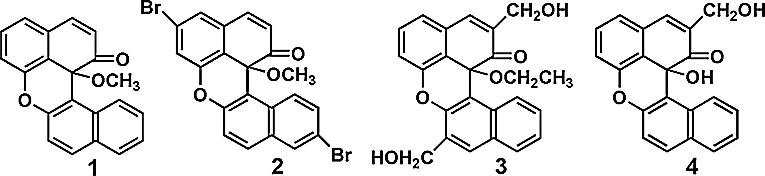
The structures of the compounds
Materials and Methods
Calf thymus DNA (CT DNA) was obtained from the Sino-American Biotechnology Company. Dimethyl sulfoxide (DMSO) was purchased from Sigma. Cell lines of BEL-7402 (hepatocellular), Hela (Human epithelial carcinoma), MG-63 (Human osteosarcoma), and SKBR-3 cells (Human breast cancer) were purchased from American Type Culture Collection. Double-distilled water was used to prepare buffers [5 mM Tris(hydroxymethylaminomethane)-HCl, 50 mM NaCl, pH=7.2]. A solution of CT DNA in the buffer gave a ratio of UV absorbance at 260 and 280 nm of ca. 1.8–1.9:1, indicating that the DNA was sufficiently free of protein (Reichmann et al., 1954). The DNA concentration per nucleotide was determined by absorption spectroscopy using the molar absorption coefficient (6600 M−1 cm−1) at 260 nm (Chaires et al., 1982). UV/Vis spectra were recorded on a Shimadzu UV-3101PC spectrophotometer at room temperature.
Physical measurements
1H NMR and 13C NMR spectra were recorded on a Varian-300 spectrometer. All chemical shifts were given relative to tetramethylsilane. Electrospray mass spectra (ES-MS) was recorded on a LCQ system (Finnigan MAT) using methanol as a mobile phase. Microanalysis was carried out with a Perkin-Elemer 240Q elemental analyzer.
Synthesis of compound 4
To a stirred solution of CuCl2 (0.350 g, 2 mmol) and ethanolamine (0.120 g, 2 mmol) in 15 mL, MeCN was added with 3-hydroxymethyl-2-binaphthol (0.316 g, 0.1 mmol) at 60°C. The mixture was stirred for 15 h, the reaction was quenched with 5% NH3·H2O and the mixture was extracted with EtOAc. The organic extract was washed with water and dried over anhydrous Na2SO4. The solvent was evaporated and the crude product was purified by column chromatography on a neutral alumina (EtOAc-petroleum ether, 1:2 v:v). Compound
DNA-binding studies
The DNA-binding experiments were performed at room temperature. The absorption titrations of the compounds in a buffer were performed using a fixed concentration (5–20 μM) for compounds to which increments of the DNA stock solution were added. Compound–DNA solutions were allowed to incubate for 5 min before the absorption spectra were recorded. The intrinsic binding constants K, based on the absorption titration, were measured by monitoring the changes in absorption at the aromatic band with increasing concentration of DNA using the following equation (Wolf et al., 1987):
where [DNA] is the concentration of DNA in base pairs, ɛa , ɛf , and ɛb correspond to the apparent absorption coefficient Aobsd/[compound], the extinction coefficient for the free compound and the extinction coefficient for a compound in the fully bound form, respectively. In plots of [DNA]/(ɛa −ɛf ) versus [DNA], K b is given by the ratio of slope to the intercept.
Viscosity measurements were carried out using an Ubbelodhe viscometer maintained at a constant temperature at 25.0 (±0.1)°C in a thermostatic bath. DNA samples ∼200 base pairs in average length were prepared by sonication to minimize complexities arising from DNA flexibility (Chaires et al., 1982). Flow time was measured with a digital stopwatch, and each sample was measured three times, and an average flow time was calculated. Relative viscosities for DNA in the presence and absence of compounds were calculated from the relation η=(t−t0)/t0, where t is the observed flow time of the DNA-containing solution and t0 is the flow time of the buffer alone (Satyanarayana et al., 1992, 1993). Data were presented as (η/η0)1/3 versus a binding ratio (Cohen and Eisenberg, 1969), where η is the viscosity of DNA in the presence of compounds and η0 is the viscosity of DNA alone.
Partition coefficient determination
The lipophilicity of the compounds was determined by the shake flask method using the phosphate buffer (0.129 M NaCl, pH=7.4) and n-octanol as the solvent (Graham, 1995). The octanol and buffer solution were presaturated with each other before use. The compound was dissolved in n-octanol. 5 mL of the above solution and 5 mL of the buffer solution were added in 25-mL round-bottomed flask. The mixture was shaken vigorously for 24 h, and then the organic phase and the buffer solution were separated by centrifugation (2500 rpm, 10 min). Each phase was measured by the UV/Vis spectrophotometer and compared with a standard curve to obtain the concentration of compound in two phases. Experiments were performed in triplicate. The partition coefficients P were calculated from the ratio Coct/CPBS, where Coct and CPBS were the concentrations of the compound in the n-octanol and in the phosphate buffer solution, respectively. Equilibration and absorption measurements were made at room temperature (Angeles-Boza et al., 2006).
In vitro cytotoxicity assay
Cytotoxicity of compounds
Flow cytometric analysis
BEL-7402 and SKBR-3 cells were seeded into six-well plates (Costar; Corning Corp.) at a density of 2×10⁵ cells per well and incubated for 24 h. The cells were cultured in RPMI 1640 supplemented with 10% fetal bovine serum and incubated at 37°C and 5% CO2. The medium was removed and replaced with a medium (final DMSO concentration, 1% v/v) containing compounds (25 μM). After incubation for 24 h, the cell layer was trypsinized and washed with cold phosphate-buffered saline (PBS) and fixed with 70% ethanol. About 20 mL of RNAse (0.2 mg/mL) and 20 mL of propidium iodide (0.02 mg/mL) were added to the cell suspensions and incubated at 37°C for 30 min. Then, the samples were analyzed by a FACS Calibur flow cytometer (Becton Dickinson & Co.). The number of cells was 10,000 (Lo et al., 2008).
Scavenger measurements of •OH
The •OH in aqueous media was generated by the Fenton system (Li et al., 2008). The solution of the tested compounds was prepared in DMF. The 4 mL of assay mixture contained following reagents: safranin (28.5 μM), EDTA–Fe(II) (100 μM), H2O2 (44.0 μM), the tested compounds (0.5–3.5 μM) and a phosphate buffer (67 mM, pH=7.4). The assay mixtures were incubated at 37°C for 30 min in a water bath. After which, the absorbance was measured at 520 nm. All the tests were run in triplicate and expressed as the mean. Ai was the absorbance in the presence of the tested compound; A0 was the absorbance in the absence of tested compounds; Ac was the absorbance in the absence of tested compound, EDTA–Fe(II), H2O2. The suppression ratio (ηa) was calculated on the basis of (Ai−A0)/(Ac−A0)×100%.
Results
Electronic absorption spectra studies
Due to the intercalative mode involving a stacking interaction between the aromatic chromophore and the DNA base pairs, the large change in absorbance can be observed, while a compound binds to DNA through intercalation. Figure 2 shows the absorption spectra of compound
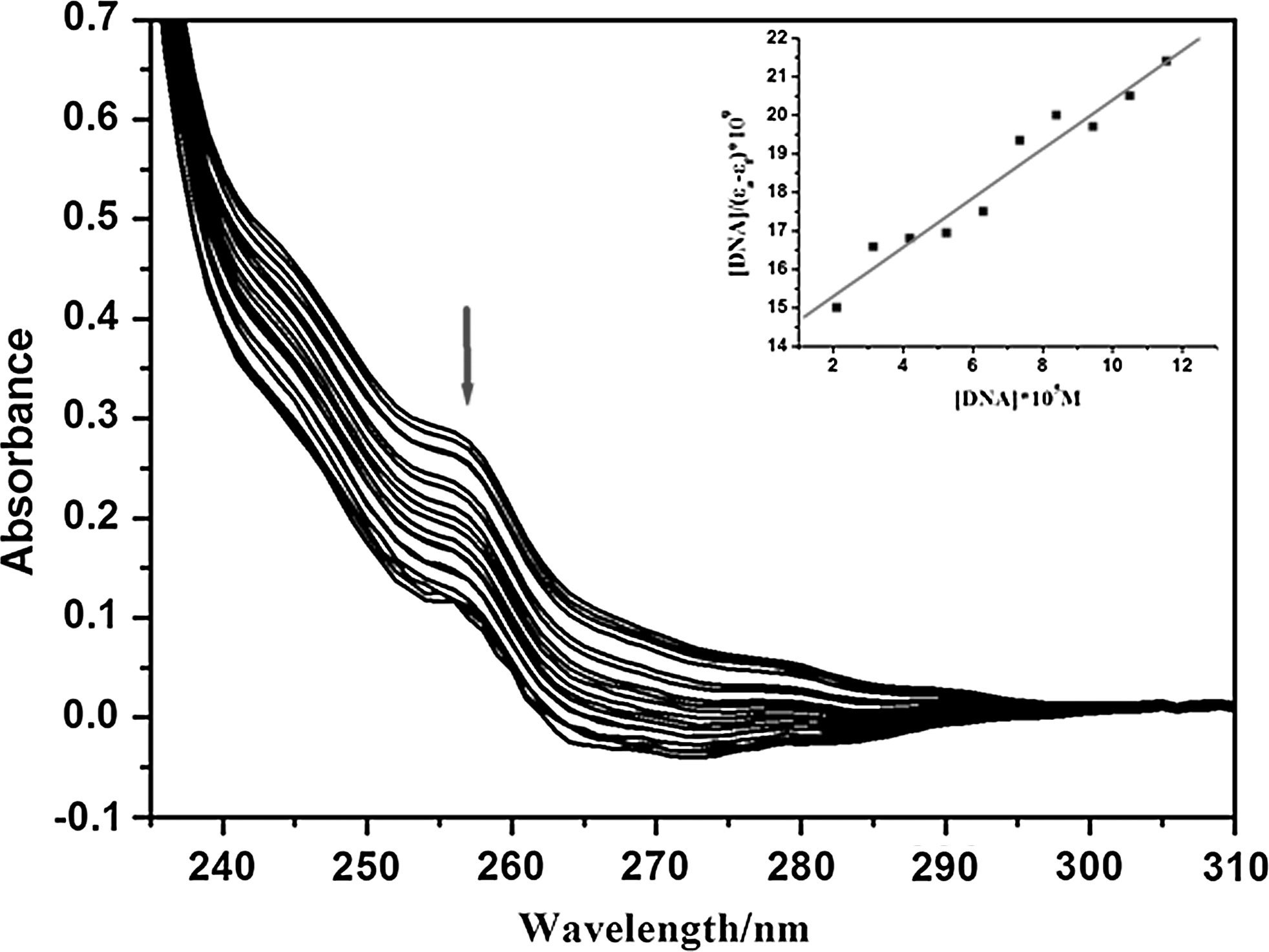
Absorption spectra of the compound
Viscosity measurements
The relative change in viscosity was measured using CT DNA with increasing concentrations of the compounds
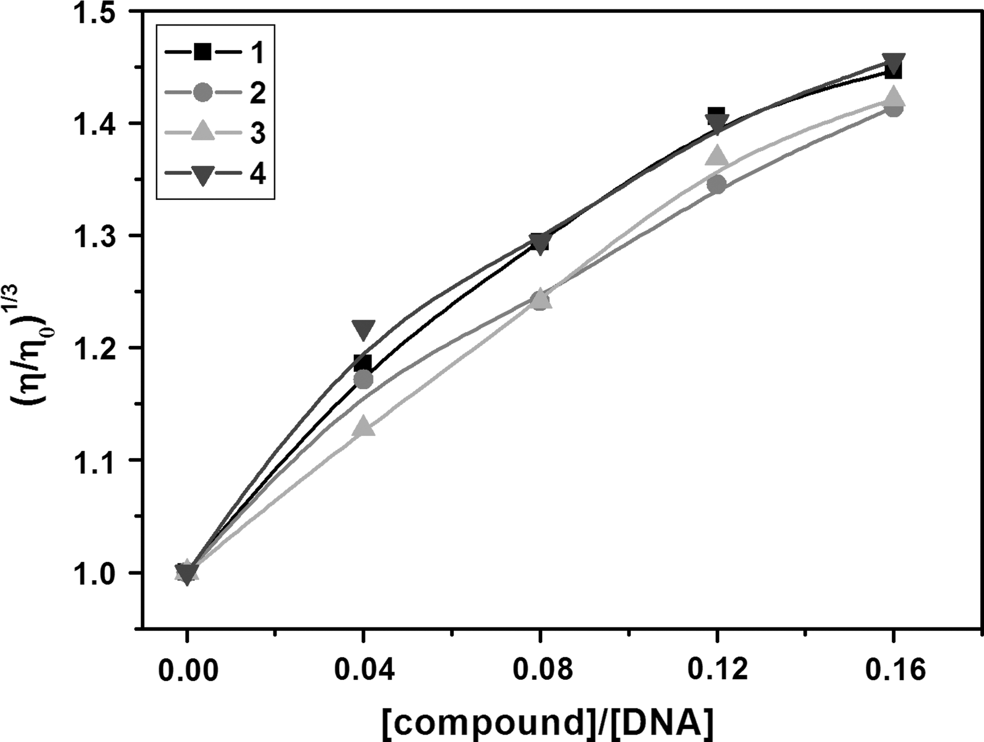
Relative viscosity changes of solutions containing 200 μM CT DNA in the presence of different concentration of compounds
Partition coefficient determination
The partition coefficient, P, is defined as the ratio of the concentration of the compound in octanol and water phases. The logarithm of the partition coefficient, log P, is most commonly used. The shake flask method used during the course of these studies has been shown to work well for molecules with log P values that range from −2 to +4. Octanol is the preferred partitioning solvent for most studies since, with its hydrophobic tail and polar head group, this liquid alcohol serves as a simplified, but well-established, membrane model. The log P of compounds
Partition coefficient P=Coct/CPBS (Coct and CPBS are the compound concentrations in n-octanol and phosphate buffer solution, respectively).
Data from Huang et al.(2011).
Data from Cheng et al. (2009).
In vitro cytotoxicity
The cytotoxicity in vitro for compounds was assessed using the method of MTT reduction. After treatment of BEL-7402, Hela, MG-63, and SKBR-3 cell lines with different concentrations of compounds
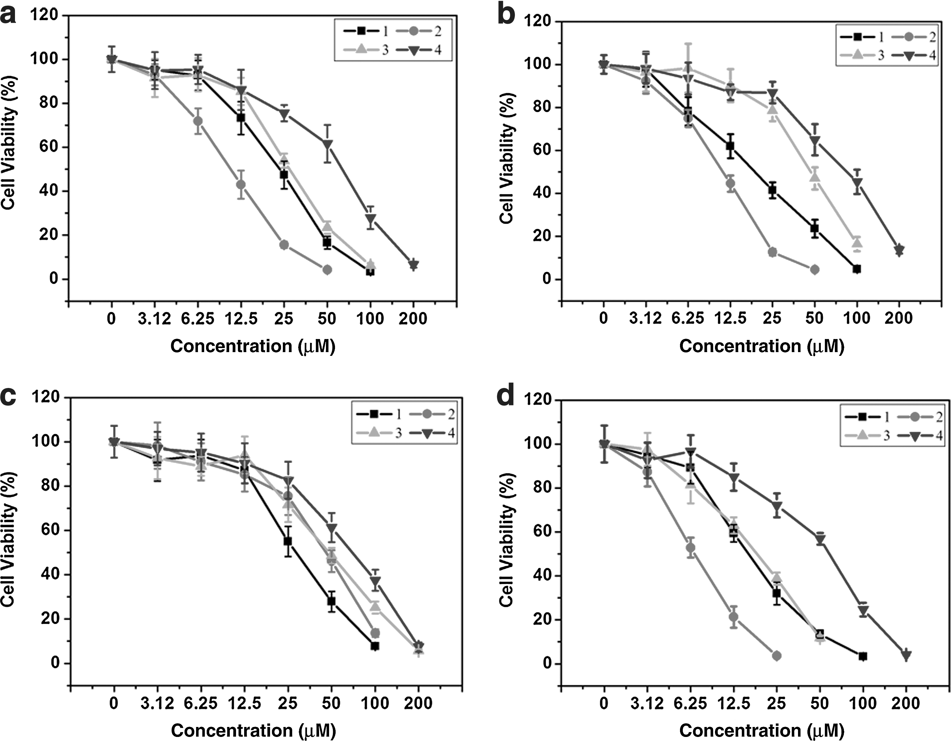
Cell viability of compounds
Flow cytometric analysis
The effect of compounds on cell cycle of BEL-7402 and SKBR-3 cells was investigated by flow cytometry in PI (propidiumiodide)-stained cells after compounds
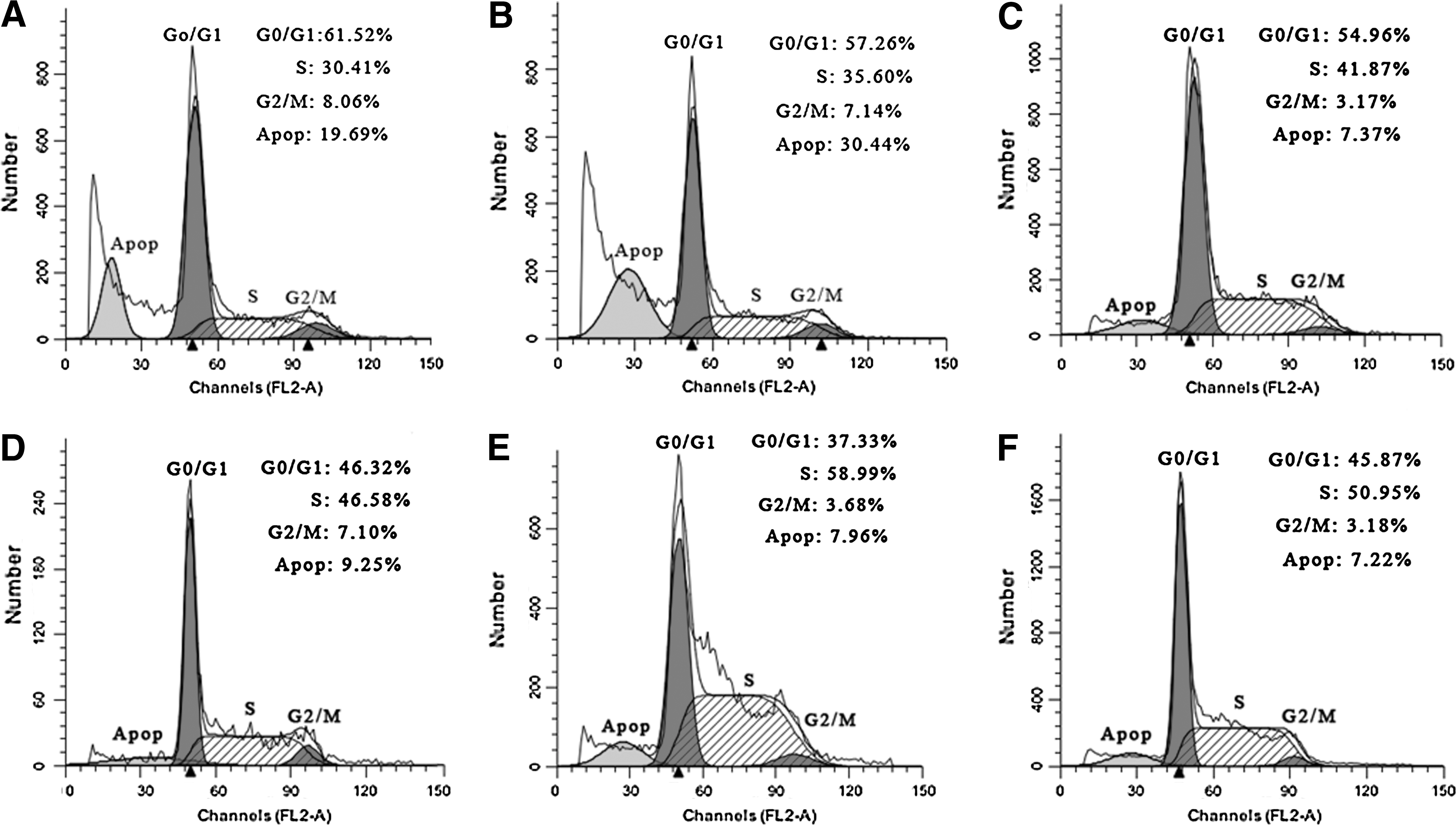
Cell cycle status of the BEL-7402 cell after treatment with compound
Antioxidant activity against •OH
Oxidative damage to DNA has been suggested to contribute to aging and various diseases, including cancer and chronic inflammation. The antioxidant activity of compounds
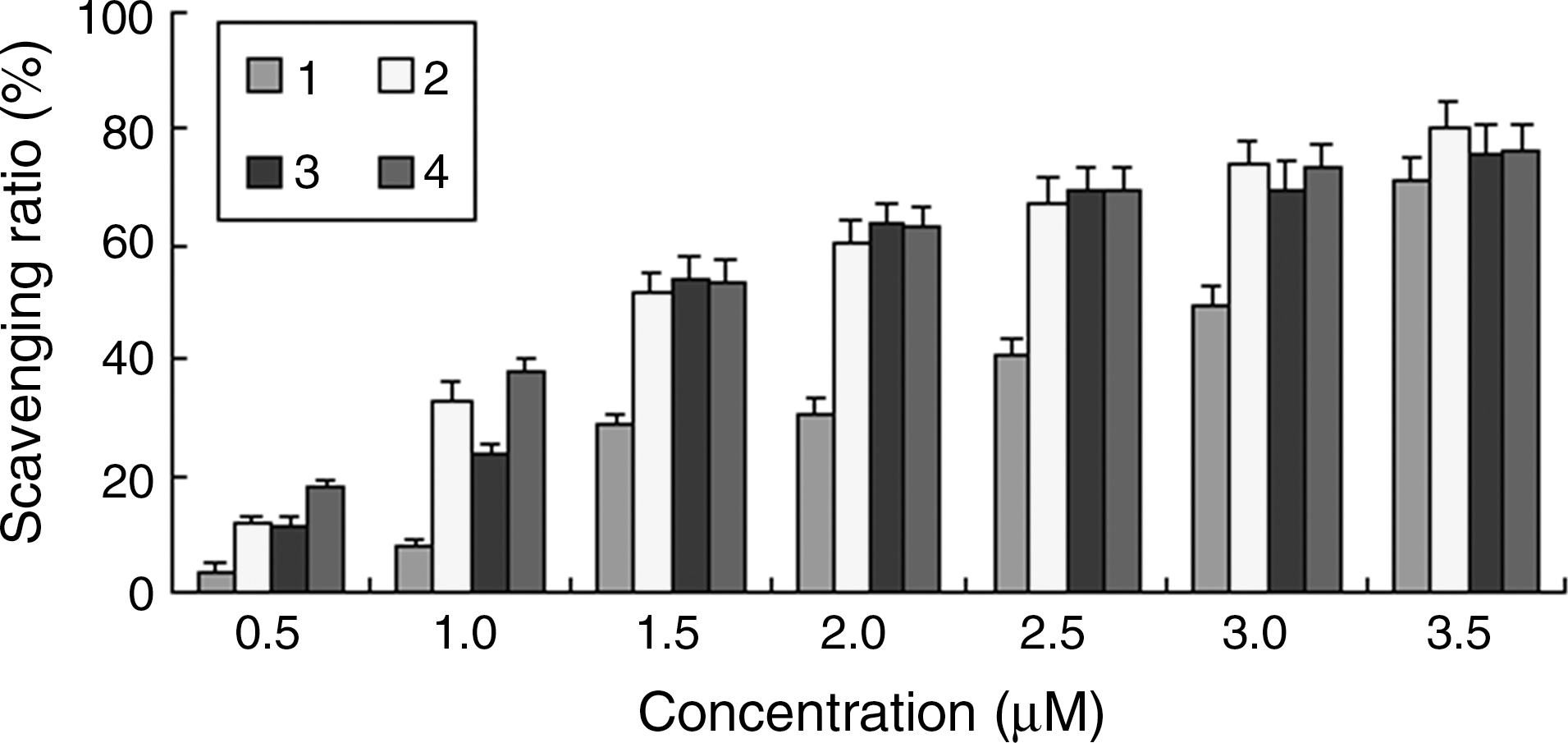
Scavenging effect of the compounds
OH, hydroxyl radical.
Discussion
The band at 256 nm for compound
The viscosity of a DNA solution was sensitive to the addition of organic drugs bound by intercalation. And relative viscosity measurements have proved to be a reliable method for the assignment of the mode of binding compounds to DNA. In general, intercalation of a ligand into DNA is known to cause a significant increase in the viscosity of a DNA solution due to an increase in the separation of the base pairs at the intercalation site and, hence, an increase in the overall DNA molecular length; A partial and/or nonclassical intercalation of ligand could bend (or kink) the DNA helix, reduces its effective length and, concomitantly, its viscosity (Waring, 1965; Satyanarayana et al., 1993). With increasing concentrations of compounds
The logarithm of the octanol–water partition coefficient, log P, is a key physicochemical property for bioactive molecules. It provides valuable information for the overall understanding of the uptake, distribution, biotrasformation, and degradation of drugs (Sangster, 1997). Most bioactive molecules are nowadays designed to have log P values in the range of
The cytotoxicity of compounds was found to be concentration-dependent. The cell viability decreased with increasing the concentrations of compounds
After treatment of the BEL-7402 cell with the compound
Among all reactive oxygen species, the •OH is by far the most potent, and therefore the most dangerous oxygen metabolite; elimination of this radical is one of the major aims of antioxidant administration (Udilova et al., 2003). The antioxidant activity against •OH of compounds
Conclusion
Four dibenzoxanthenes were synthesized and studied on interaction with DNA and cytotoxicity in vitro. The DNA-binding behaviors show that compounds interact with CT-DNA through an intercalative mode. The cytotoxicity assay indicates that compounds
Footnotes
Acknowledgments
This work was supported by the National Nature Science Foundation of China (No 30800227, 31070858) and the Guangdong Pharmaceutical University for financial supports.
Disclosure Statement
No competing financial interests exist.