
Abstract
Cisplatin is a most active drug for the treatment of ovarian cancer; however, acquired cisplatin resistance is easily seen in patients with ovarian cancer. The aim of this study is to clarify the molecular mechanism of cisplatin resistance and try to reverse cisplatin resistance in ovarian cancer lines in vitro and in vivo. First, we used ovarian cancer cell line A2780, and its cisplatin-resistant subline, A2780/DDP as cell model. Cell viability was determined by MTT assay and the IC50 values were observed to increase in a dose- and time-dependent manner. Next, the expression of β-catenin was determined by western blotting analysis, and the results demonstrated that the expression level of β-catenin in A2780/DDP cells was significantly higher than that in A2780 cells (p<0.01). Moreover, we detected the distribution of cytoplasmic and nuclear β-catenin by western blot analysis, which showed that β-catenin was mainly located in nucleus. Compared with A2780 cells, there was no obvious change as the increasing dose of cisplatin in A2780/DDP cells reveal that cisplatin resistance was related to the exrpession of β-catenin. Furthermore, interference with the expression of β-catenin could effectively reverse cisplatin resistance as IC50 was significantly decreased from 123.7 to 42.43 μM in A2780/DDP cells. Additionally, transient interference of β-catenin by siRNA promoted the apoptosis of A2780/DDP cells, for increased apoptosis rates and cleaved caspase-3 levels were detected being treated with cisplatin. Finally, tumorigenicity experiments showed that tumor growth was significantly suppressed in β-catenin shRNA group. The body weight was not significantly changed during the experimental days. In conclusion, all the results showed that cisplatin resistance was partly induced by Wnt/β-catenin signaling pathway. Interfering the expression of β-catenin could reverse cisplatin resistance in vitro and in vivo. Thus, β-catenin could be a potential therapeutic target for the therapy of cisplatin-resistant ovarian cancer.
Introduction
O
The current standard treatment for ovarian cancer is primarily integrated treatment program of cytoreductive surgery in combination of platinum-based chemotherapy (Conteduca et al., 2014). Cisplatin has been the most active drug for the treatment of ovarian cancer for the last 4 decade (Khrunin et al., 2014). However, the majority of patients with ovarian cancers who are resistant to cisplatin will ultimately suffer the disease (Nawrocki et al., 2013). So, the important factor of high recurrence, metastasis, and increased mortality in ovarian cancer patients is probably the decreased sensitivity to cisplatin (Nam et al., 2010). Therefore, it is important to explore the mechanism of drug resistance and to find effective resistance reversal agents in ovarian cancers.
The canonical Wnt signaling plays a pivotal role in intracellular signal transduction. It was reported that wnt/beta-catenin pathway was related to epithelial ovarian cancer, specifically in chemoresistance (Kildal et al., 2005; Arend et al., 2013; Bodnar et al., 2014). The β-catenin is an important adhesion molecule and it is a key regulator in Wnt signal transduction pathway, which play important role in cell–cell adhesion, growth, and differentiation, wound healing, tumorigenesis, metastasis, and others. (Yoshioka et al., 2012). When the levels of β-catenin increase in cytoplasm, it would translocate and accumulate in the nucleus, which activates the expression of TCF/LEF, and promotes downstream target genes, such as c-myc, MMP-7, COX-2, MRP1, cyclin D1, and so on. (Furlong and Morin, 2000; Sillanpaa et al., 2006; Condello et al., 2014). The expression of downstream target genes can lead to abnormal proliferation of tumor cells and affect the sensitivity to chemotherapy drugs (Arend et al., 2014). However, the relationship of drug resistance and the abnormal expression of β-catenin in ovarian cancer cells is not clearly clarified. In this study, we used human ovarian cisplatin-sensitive cancer cell line A2780 and its parental cisplatin-resistant cell lines A2780/DDP as cell models, and clarified the molecular mechanism of cisplatin resistance in ovarian cancer cells. Then, we tried to find an effective way to reverse the cisplatin resistance of ovarian cancer line A2780/DDP in vitro and in vivo. The study would be useful and has important clinical significance for therapy of ovarian cancers.
Materials and Methods
Cells and regents
Human ovarian cancer cell line A2780 and its cisplatin-resistant subline, A2780/DDP was obtained from the Cancer Center of the affiliated hospital of Tongji Medical College, Huazhong University of Science and Technology. The cells were cultured in DMEM medium supplemented with 10% fetal calf serum. The compound cisplatin and MTT agent was purchased from Sigma, Inc. β-catenin shRNA (h) lentiviral particles (sc-29209-V) and control shRNA lentiviral particles A (sc-108080) were obtained from Santa Cruz Biotechnology. The siRNA1 sequences to β-catenin are sense: 5′-UGCUUGGUUCACCAGUGGAUU-3′ and reverse: 5′-AAUCCACUGGUGAACCAAGCA-3′. The siRNA2 sequences to β-catenin are: sense: 5′-AGCUGAUAUUGAUGGACAG-3′ and reverse: 5′-CUGUCCAUCAAUAUCAGCU-3′. The negative control siRNA sequences are: sense 5′-UUCUCCGAACGUGUCACGU-3′ and reverse 5′-ACGUGACACGUUCGGAGAA-3′.
MTT assay
Cell proliferation is tested by MTT assay as described (Bernas and Dobrucki, 2002; Sylvester, 2011). Generally, the human ovarian cancer cells were plated into 96-well plates. Then, the cells were cultured for 8 h and treated with increasing concentrations of cisplatin for 24, 48, and 72 h, respectively. Four hours before testing, MTT agent was added into the medium. At last, the purple crystals were dissolved with DMSO, and the 96-well plates were read on a microplate reader at a test wavelength of 490 nm and a reference wavelength of 570 nm.
Flow cytometric analysis
The apoptosis rate of human ovarian cancer cells was tested by annexin V-FITC/PI staining analysis according to the kit protocols (Santa Cruz Biotechnology). Briefly, the ovarian cancer cells (3×10⁶ cells/well) were transfected with siRNA1 and siRNA2 specific to β-catenin. Then, the cells were treated with 40 μM of cisplatin for 48 h. They were washed in PBS buffer and resuspended in binding buffer with HEPES–NaOH 10 mM pH 7.4, 25 mM CaCl2 and 144 mM NaCl. The staining dye of Annexin V (0.1 μg/μL) and PI (0.05 μg/μL) was added and incubated in dark for 30 min on ice and cells were then subjected to FACS analysis.
Western blotting analysis
The ovarian cancer cells A2780 and A2780/DDP were plated into 10 cm dishes. After adherence of the cells, they were treated with increasing concentrations of cisplatin for 48 h. The whole cell extracts were prepared and the proteins were separated by PAGE as previously described (Schulman et al., 2000; Nishitani et al., 2006; Peng et al., 2010). The cytoplasmic and nuclear expression levels of β-catenin were detected. Here, α-tublin was used as the marker in cytoplasm and lamin B1 was used as the internal reference in nucleus.
Statistical analysis
All data were analyzed by SPSS software. The results were shown as the mean value±standard error of the mean. All the experiments were performed thrice in duplicates. p value<0.01 means that there is a significant difference.
Results
The antitumor effects of cisplatin on human ovarian cancer cell line A2780 and A2780/DDP
To examine the effects of cisplatin on cellular proliferation, MTT assay was performed to test the inhibitory effects of cisplatin on ovarian cancer cells. Here, we used the human ovarian cancer cell line A2780 and its cisplatin-resistant subline, A2780/DDP as the cell models. As shown in Figure 1, cisplatin had an increasing antitumor effect on human ovarian cancer cell line A2780 and A2780/DDP in a time- and dose-dependent manner. The IC50 values of cisplatin on sensitive cell line A2780 was 105.1, 17.47, and 8.3 μM, being treated with different concentrations of cisplatin for 24, 48, and 72 h, respectively. And, the IC50 values were 431.8, 123.7, and 98.6 μM on cisplatin-resistant cell line A2780/DDP, being treated for 24, 48, and 72 h. We chose 48 h as the appropriate time to treat A2780 cells and A2780/DDP cells. The ratio of IC50 value was approximately seven-fold in A2780/DDP cells compared with A2780 cells suggesting A2780/DDP had higher resistance to the chemotherapy drug cisplatin.

The antitumor effects of cisplatin on human ovarian cancer cell line A2780 and A2780/DDP. The proliferation of human ovarian cancer cell line A2780 and A2780/DDP was determined by MTT assay. The cells were exposed to the increasing concentrations of cisplatin for 24, 48, and 72 h, respectively. Data were collected from at least three independent experiments conducted on different individuals and IC50s were calculated.
The expression of β-catenin increases in A2780/DDP cells
To detect whether the cisplatin resistance in A2780/DDP cells is associated with Wnt/β-catenin pathway, the expression levels of β-catenin were detected in A2780 and A2780/DDP cells by western blotting analysis. As shown in Figure 2, the expression level of β-catenin was much higher in A2780/DDP cells than that in A2780 cells (**p<0.01).
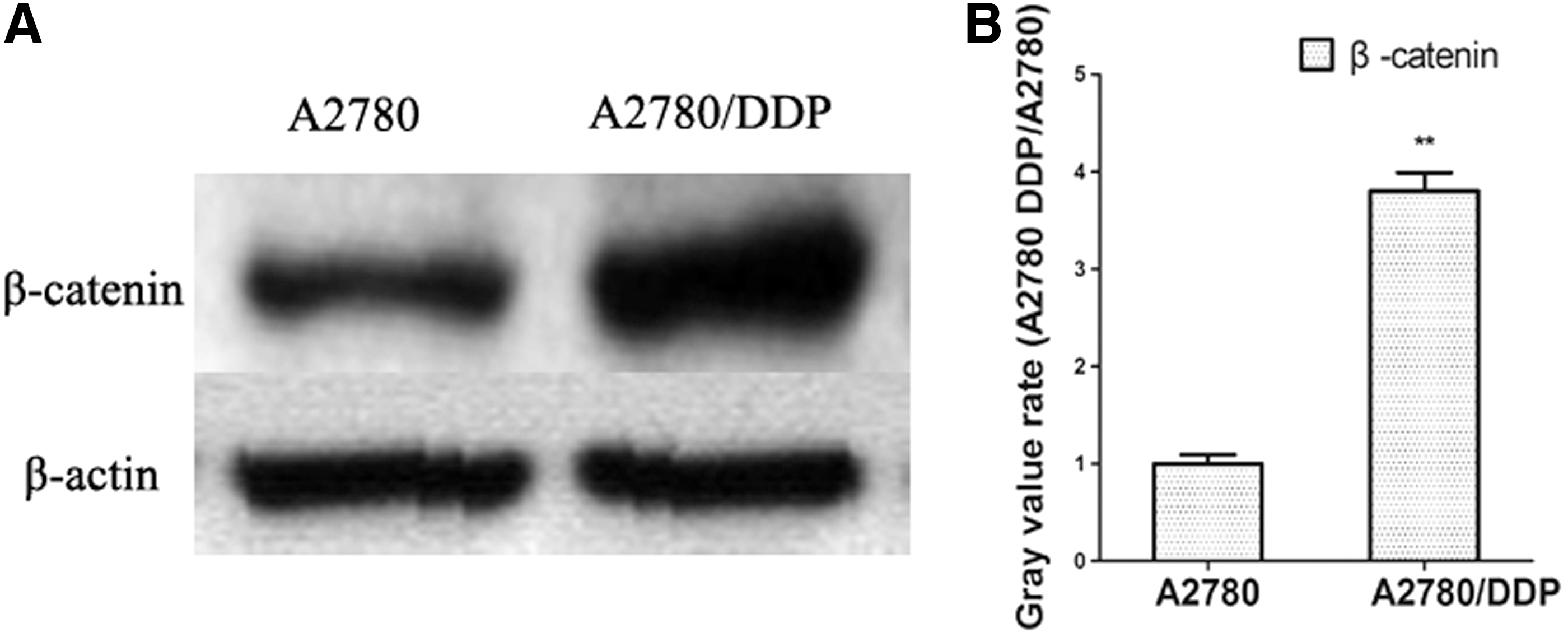
The expression of β-catenin increases in A2780/DDP cells compared with that of A2780 cells.
The β-catenin is principally located in nucleus in A2780/DDP cells
Next, the expression levels of β-catenin in A2780 cells and A2780/DDP cells were detected by western blot analysis. As shown in Figure 3, the results revealed that β-catenin levels in cytoplasm or nucleus of A2780/DDP cells were both higher than that of A2780 cells (**p<0.01), suggesting that β-catenin was highly activated and located in the nucleus in A2780/DDP cells. It could be speculated that the abnormal activation of β-catenin may be associated with cisplatin resistance. Here, the gray value of cytoplasmic β-catenin expression in A2780 cells was used as control.

The β-catenin is principally located in nucleus in A2780/DDP cells.
β-catenin expression is tested in ovarian cancer cells A2780 and A2780/DDP
To explore how cisplatin affects the β-catenin levels in A2780 and A2780/DDP cells, the ovarian cancer cells were treated with increasing concentrations of cisplatin for 48 h. As shown in Figure 4, in cisplatin-sensitive ovarian cancer cell line A2780, the expression levels of β-catenin were significantly decreased with increasing concentrations of cisplatin. However, there were no obvious changes on the β-catenin levels of A2780/DDP cells. All the data revealed that the expression levels of β-catenin in A2780 cells were suppressed by higher concentration of cisplatin (40 μM), but the β-catenin levels were not affected by the increasing concentrations of cisplatin in A2780/DDP cells, suggesting that the resistence of cisplatin in A2780/DDP cells was due to abnormal activation of β-catenin.
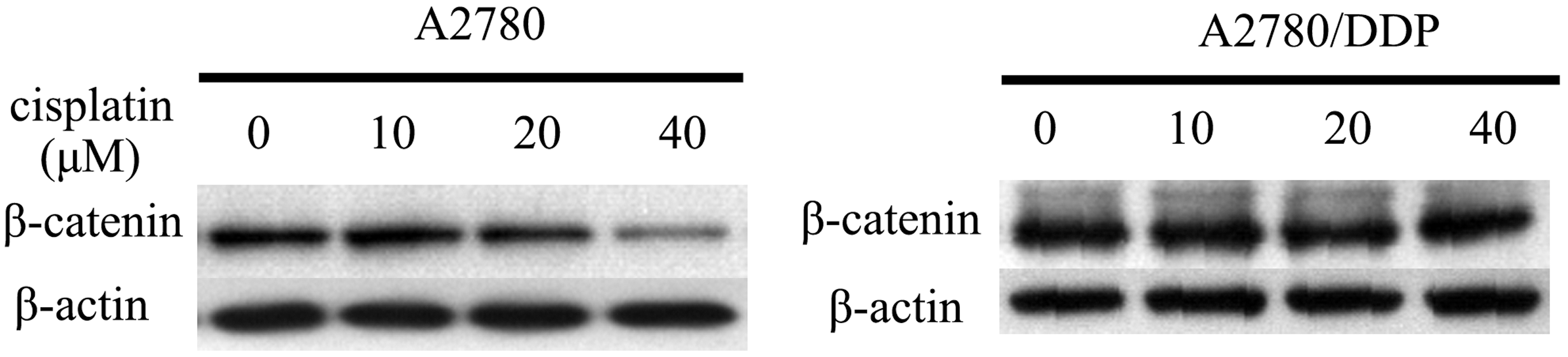
β-catenin expression is tested in ovarian cancer cells A2780 and A2780/DDP. The ovarian cancer cells (2×10⁶ cells/well) were plated into 48-well plate. After 8 h, the cells were treated with increasing concentrations of cisplatin of 10, 20, and 40 μM, respectively. The cells were treated with cisplatin for 48 h, and cell lysates were prepared for western blotting analysis. The expression of β-catenin was detected in A2780 cells and A2780/DDP cells.
The expression of β-catenin is interferenced in ovarian cancer cell line A2780/DDP by specific siRNA to β-catenin
It was demonstrated that the resistence to cisplatin in A2780/DDP cells was related to abnormal activation of nuclear β-catenin. We designed two pairs of siRNA specific to β-catenin and the two pairs of specific siRNA were used to knock down β-catenin expression in A2780/DDP cells. As shown in Figure 5, the silencing effects were determined by western blot and real time PCR. The result showed that both the siRNA specific to β-catenin could effectively interfere the expression of β-catenin. Here, the untreated cells and the cells transfected by negative control siRNA or transfection reagent lipofectamin were used as controls.

The expression of β-catenin is interferenced in ovarian cancer cell line A2780/DDP by specific siRNA to β-catenin. A2780/DDP cells were plated into 48-well plate. After 8 h, the cells were transfected with siRNA1 and siRNA2 specific to β-catenin. Then, 48 h later, the expression of β-catenin was determined by western blotting analysis
The IC50 value of cisplatin is detected in β-catenin-knocking down ovarian cancer cell line A2780/DDP for 48 h
We successfully interfered the expression of β-catenin in cisplatin-resistant cell line A2780/DDP cells. Next, MTT assay was performed to detect the variation of IC50 values to cisplatin in β-catenin-knocking down ovarian cancer cell line A2780/DDP. The cells were treated with increasing concentrations of cisplatin for 48 h. All of samples were collected in duplicates and the test was repeated again. The untreated cells were used as negative controls. As shown in Figure 6A, the IC 50 values in siRNA1 and siRNA2 interfering A2780/DDP cells were calculated as 42.43 and 73 μM for 48 h. The ratio of IC50 value was downregulated for approximately 2.9-fold and 1.7-fold in β-catenin-knocking down A2780/DDP cells compared with that of A2780/DDP cells, which demonstrated that interferencing the expression of β-catenin in A2780/DDP could downregulate the resistance to chemotherapy drug cisplatin in A2780/DDP cells.
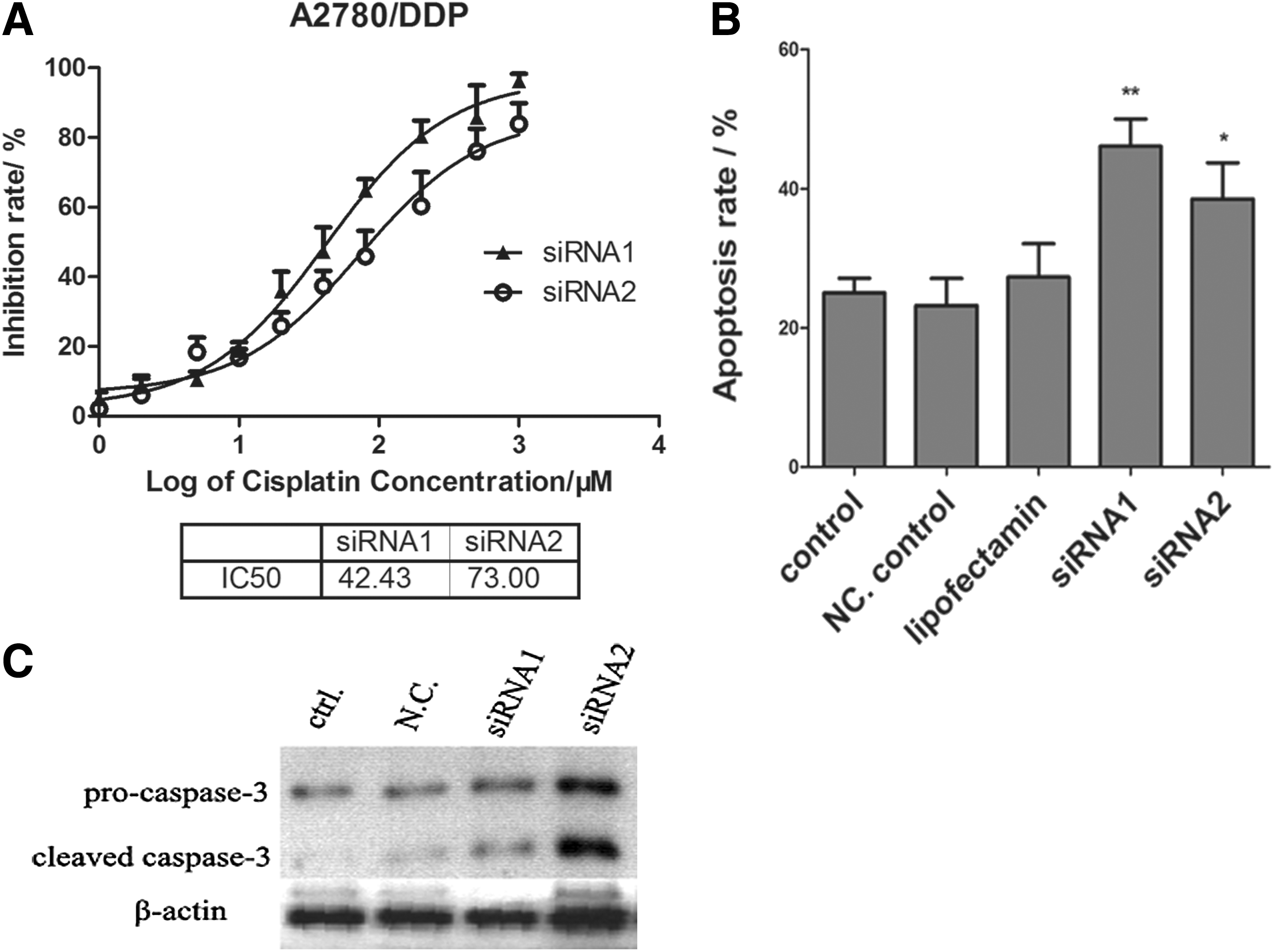
Interferencing β-catenin expression induces cell apoptosis of cisplatin-resistant ovarian cancer cell line A2780/DDP cells.
Interferencing β-catenin expression induces cell apoptosis of cisplatin-resistant ovarian cancer cell line A2780/DDP cells
Then, the cell apoptosis rates were detected by AnnexinV-FITC/PI dual staining analysis. The results demonstrated that cell apoptosis rates increased in A2780/DDP cells transfected by siRNA1 and siRNA2 specific to β-catenin (Fig. 6B). Additionally, the expression of caspase-3 was also detected by western blotting analysis. As shown in Figure 6C, caspase-3 was activated in siRNA1 and siRNA2 transfected cells, although the expression levels of pro-caspase-3 were not obviously changed. All the data demonstrated that interferencing the expression of β-catenin could effectively induce cell apoptosis of cisplatin-resistant ovarian cancer cell line A2780/DDP cells.
Interfering the expression of β-catenin inhibits tumor growth of A2780/DDP cells in nude mice model
To detect the in vivo antitumor effects of interfering the expression of β-catenin in A2780/DDP, a nude mouse xenograft model was constructed. We first constructed the stable cell line of β-catenin interferencing A2780/DDP cell line using specific lentivirus system. The expression of β-catenin was successfully interferenced in A2780/DDP cells by western blot analysis (Fig. 7A, B). Then, 2×10⁵ tumor cells were subcutaneously injected into the axillary region of nude mice. Eight days later, the tumors were successfully planted in animals injected by ovarian cells A2780/DDP or A2780/DDP transfected with negative control shRNA. All the animals were injected with the chemotherapy drug cisplatin every 3 days. As shown in Figure 7C, the tumor volume in β-catenin shRNA-transfected group was much smaller compared with the controls (**p<0.01), which showed that tumors grew the slowest in β-catenin shRNA-transfected group.
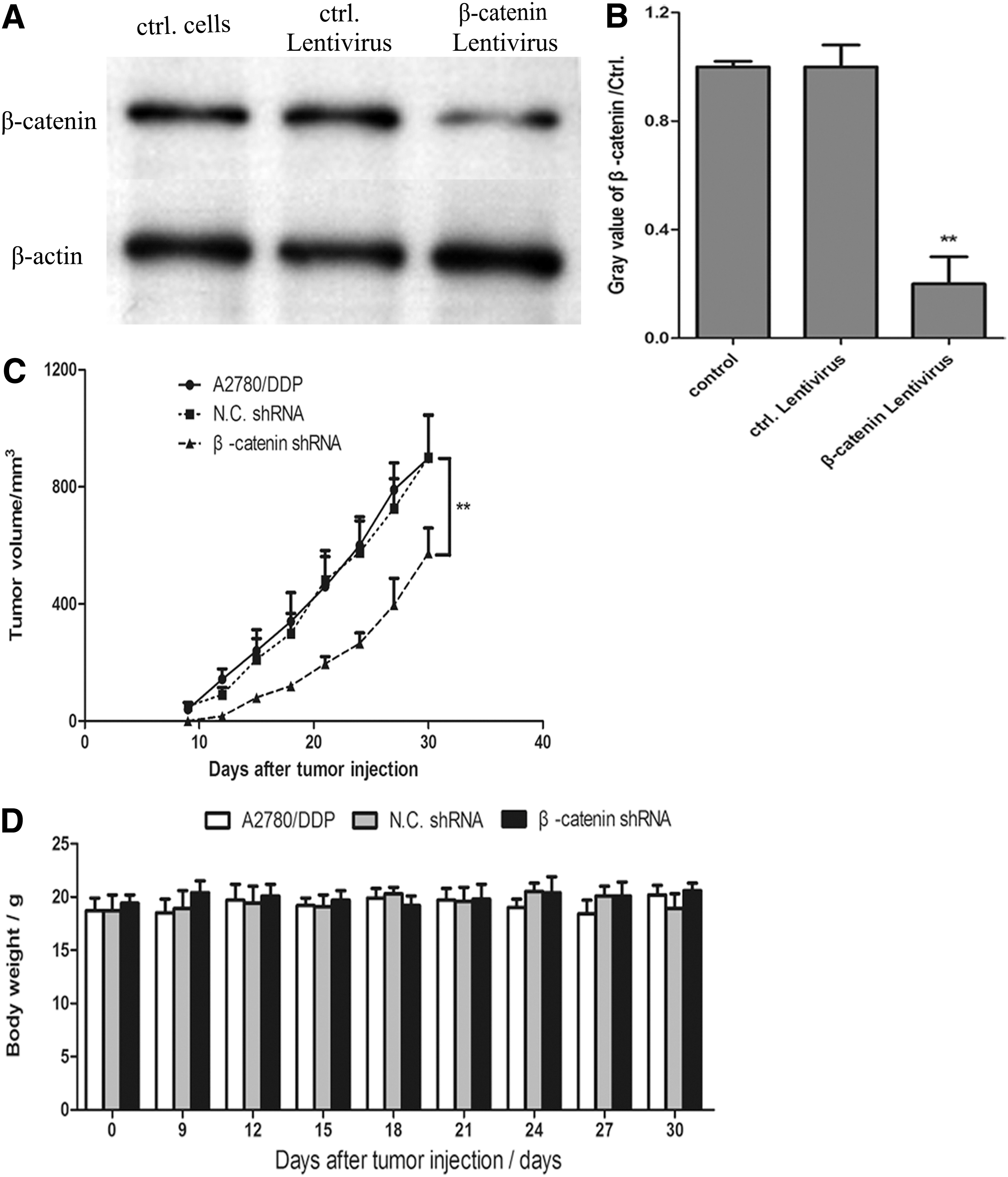
Interfering the expression of β-catenin inhibits tumor growth of A2780/DDP cells in nude mice model.
The mice had normal drinking and eating, without diarrhea and retarded activity. Body weights were recorded every 3 days after tumor injection. As shown in Figure 7D, there was no statistical difference among groups, suggesting no significant side effects in mice treated with the dose of cisplatin.
Discussion
Currently, chemotherapy remains a primary treatment for ovarian cancers and platinum-based chemotherapy plays an irreplaceable role in the treatment of ovarian cancers (Bicaku et al., 2012; Chambers et al., 2012). However, it does not always achieve fully satisfactory outcome due to platinum drug resistance in ovarian cancer patients (Baekelandt et al., 2001; Surowiak et al., 2007). Thus, it is crucial to find a new strategy to reverse drug resistance in ovarian cancer patients (Wang et al., 2014). In this study, we used RNA interference technology to knock down the expression of β-catenin and explored the reversal of cisplatin resistance in cisplatin-resistant ovarian cancer cells.
First, we chose cisplatin-sensitive ovarian cancer cell line A2780, and its cisplatin-resistant subline, A2780/DDP as cell models. Exposure of human ovarian tumor cell line A2780 to increasing concentrations of cisplatin led to development of cell lines that exhibited increasing degrees of drug resistance. The ratio of IC50 value of A2780/DDP cells was approximately seven-fold to that of the cisplatin-sensitive A2780 cells, which revealed A2780/DDP had much higher resistance to cisplatin. Thus, the cell model was perfect to be used to explore the drug resistance to the chemotherapy drug cisplatin.
In ovarian endometrioid adenocarcinoma patients, mutations of CTNNB1 that encodes β-catenin are usually accompanied by abnormal expression of β-catenin protein in the nucleus (Palacios and Gamallo, 1998; Schwartz et al., 2003; Wu et al., 2014). We demonstrated that β-catenin expression was highly expressed in cisplatin-resistant ovarian cancer cell line A2780/DDP compared with that of the cisplatin-sensitive ovarian cancer cell line A2780. Next, RNA interference technique was used to investigate the effect of silencing the expression of β-catenin in the cisplatin-resistant ovarian cancer cell line. Our results demonstrated that inhibition of β-catenin expression significantly decreased cellular proliferation and increased cell death of A2780/DDP cells.
To test the effect of interferencing β-catenin on tumor growth in vivo, we constructed a xenograft mouse model by lentiviral delivery system. To be consistent with in vitro results, inhibition of β-catenin expression in combination with cisplatin treatment, a synergistic antitumor activity can be readily achieved in a nude mouse xenograft model. Additionally, the body weights of the mice in every group were not obviously changed suggesting there was no overt toxicity under these experimental conditions.
In conclusion, synergistic antitumor activity can be successfully achieved by combining cisplatin therapy with β-catenin interference. It would possibly provide a new clue for the treatment of drug-resistant ovarian cancer.
Footnotes
Disclosure Statement
No competing financial interests exist.