
Abstract
Laminar shear stress is considered to improve endothelial cell (EC) function. However, the underlying mechanism is unclear. Autophagy has been found to protect cell survival under stress. In this study, the effect of laminar shear stress on EC autophagy and its potential mechanism were explored. The autophagic markers, Beclin 1 and LC3 II, in human umbilical vascular endothelial cells increased after laminar shear stress treatment. Meanwhile, the autophagic substrate, p62, decreased. The protein level of Rab4 increased under laminar shear stress. When pretreated with Rab4 siRNA, the increased levels of Beclin 1 and LC3 II were attenuated and p62 levels significantly increased. In addition, the MCP level and the adhesion of monocytes were also obviously increased by Rab4 siRNA. Laminar shear stress upregulated Rab4 expression, which contributed to improved EC autophagy and function.
Introduction
A
Studies showed that autophagy exists in the progress of atherosclerosis (Li et al., 2015a, b; Magné et al., 2015). Autophagy, which is a conserved process that uses double-membrane vesicles to deliver cytoplasmic contents to lysosomes for degradation, enhances the clearance of toxic, cytoplasmic, aggregate-prone proteins, and infectious agents, thereby supporting cell survival and inhibiting inflammation (Nussenzweig et al., 2015). A recent study showed that EC autophagy is regulated by laminar shear stress and has a protective role in EC function (Guo et al., 2014).
Rab GTPases are implicated in endosome-to-plasma membrane recycling and autophagy regulation. Rab4, a member of small GTPases, has been reported to drive the assembly of carrier vesicles emanating from the early endosomal compartment (D'Souza et al., 2014). In the present study, we hypothesized that Rab4 expression may be upregulated by laminar shear stress and regulates EC autophagy under shear stress, which is beneficial for EC function.
Materials and Methods
Cell culture and shear stress exposure
A human umbilical vascular endothelial cell (HUVEC) line was obtained from the China Center for Type Culture Collection. Cells were cultured in Dulbecco's modified Eagle's medium (DMEM) supplemented with 10% fetal bovine serum (Invitrogen), 100 μg/mL penicillin, and 100 μg/mL streptomycin at 37°C in a 5% CO2 atmosphere.
In fluid shear stress experiments, cells were treated with 0 dyne/cm2 (static cultured) or 15 dyne/cm2 (laminar shear stress) for 1 h. Briefly, confluent endothelial monolayers were grown onto a glass slide precoated with fibronectin and connected with a parallel plate flow chamber. The perfusion culture medium consisted of serum-free DMEM containing antibiotics (100 IU/L penicillin, 100 mg/L streptomycin). The shear stress (τw) acting on the cultured EC monolayer was determined by τw = 6μQ/bh 2, where μ is fluid viscosity, Q is the flow rate, b is the chamber width, and h is the flow path height.
RNA interference
HUVECs were plated in 24-well culture plates at 1 × 10⁵ cells/well. After reaching 70% confluence, the cells were transfected into Rab4 siRNA oligonucleotides (specific for Rab4A, 5′-GGACCTGGATGCAGATCGT-3′) or a nonrelated scrambled siRNA (5′-CCUACGCCACCAAUUUCGU-3′) with Lipofectamine™ 2000 reagent according to the manufacturer's instructions. The medium was changed after 24 h, and cells that were cultured until confluence of controls were used in the following experiments. Knockdown of Rab4 by siRNA was confirmed by Western blot.
Western blots
Cells were rinsed twice with phosphate-buffered saline (PBS), and then lysed with assay buffer. Equal amounts of whole proteins were separated by sodium dodecyl sulfate–polyacrylamide gel electrophoresis and transferred onto polyvinylidene fluoride membranes. After incubation with anti-Beclin1 (1:500 diluted), anti-LC3 (1:500 diluted), anti-Rab4 (1:200 diluted), anti-MCP1 (1:500 diluted), or anti-β-actin (1:100 diluted) antibodies overnight at 4°C, the corresponding secondary antibody was applied for 2 h. Enhanced chemiluminescence reagents were used to detect the targeted antigen. The abundance of the targeted protein was analyzed using Labwork image analysis software. All experiments were performed in triplicate.
Immunofluorescence staining
After treatment, ECs were fixed in 95% ethanol for 30 min at 4°C and washed twice with PBS. After blocking with bovine serum albumin for 30 min, the cells were incubated with anti-LC3 II antibody overnight at 4°C. The corresponding Cy3-labeled second antibody was subsequently incubated for 30 min. The cells were rinsed with PBS and examined under a fluorescence microscope (IX70; Olympus).
Monocyte adhesion assay
HUVECs (5.0 × 10⁴ cells/mL) were cultured in 24-well plates for 12 h and divided into four different groups, namely the static cultured group, 15 dyne/cm2 group, Rab4 siRNA +15 dyne/cm2 group, and 3-methyladenine (3-MA) +15 dyne/cm2 group. After treatment, the HUVECs were incubated with 2 × 10⁴ THP-1 cells for 1 h, and plates were washed by PBS. The number of adherent cells was counted in at least three different locations for each sample.
Statistical analysis
Data are expressed as the mean ± standard error, with n representing the number of biological replicates using different shear experiments. Student's unpaired t-test was used to establish significance between groups, and one-way analysis of variance was applied for multiple comparisons. p < 0.05 was considered statistically significant.
Results
Laminar shear stress promoted HUVEC autophagy
Given that previous results showed that autophagy plays a protective role in atherosclerosis and is regulated by shear stress (Ding et al., 2014; Li et al., 2015a, b), we proposed that EC autophagy may be improved by laminar shear stress. To examine this hypothesis, we detected the expression of the autophagy marker, Beclin 1, and the transformation of LC3 from type I to type II. Results showed that laminar shear stress increased Beclin 1 (Fig. 1A, B) and LC3 II expression (Fig. 1A, C). In addition, immunofluorescence staining also showed that laminar shear stress significantly increased LC3 II protein abundance (Fig. 1E, F). The autophagy substrate, p62, decreased in laminar shear stress-treated HUVECs (Fig. 1A, D). Taken together, these results indicated that EC autophagy was strengthened under laminar shear stress.
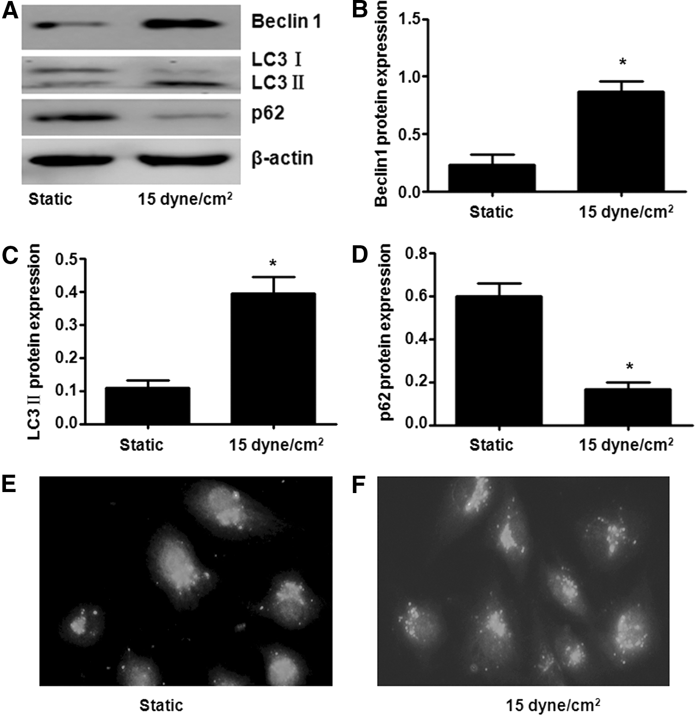
Effect of laminar shear stress on endothelial cell autophagy. Laminar shear stress increased Beclin 1
Laminar shear stress increased HUVEC autophagy flux
To determine if laminar shear stress increases autophagic flux, HUVECs were cotreated with bafilomycin A1. Results showed that laminar shear stress increased LC3 II even in the presence of bafilomycin A1, which suggested that laminar shear stress improved HUVEC autophagic flux (Fig. 2).
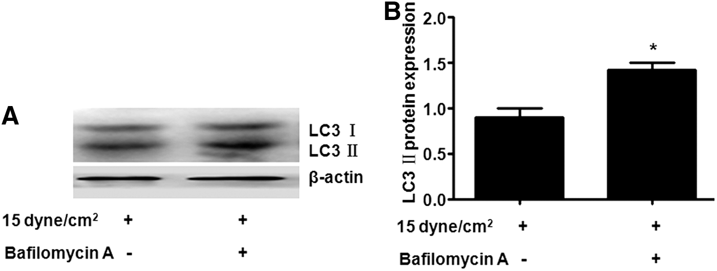
Effect of laminar shear stress on human umbilical vascular endothelial cell (HUVEC) autophagy flux. The conversion of LC3 protein in HUVECs was detected by Western blot after treatment with laminar shear stress in the presence or absence of bafilomycin A1
Laminar shear stress upregulated HUVEC Rab4 expression
We explored the mechanism of laminar shear stress on improved HUVEC autophagy. In this study, we detected the expression of Rab4 under laminar shear stress. Results showed that Rab4 protein expression increased after laminar shear stress treatment (Fig. 3).

Effect of laminar shear stress on HUVEC Rab4 expression. After treatment with laminar shear stress, Rab4 protein expression was increased
Rab4 siRNA attenuated HUVEC autophagy under laminar shear stress
To explore the function of Rab4 in HUVEC autophagy under laminar shear stress, we used Rab4 siRNA. As shown in Figure 4A and B, Rab4 siRNA evidently decreased HUVEC Rab4 protein expression. After treatment with Rab4 siRNA, the expression of Beclin1 (Fig. 4C, D) and the transformation of LC3 I to LC3 II were inhibited (Fig. 4C, E). Meanwhile, immunofluorescence staining showed that the protein content of LC3 II decreased (Fig. 4G, H), whereas the p62 level increased. Thus, laminar shear stress-induced EC autophagy was inhibited by Rab4 siRNA, suggesting that Rab4 was involved in the regulation of HUVEC autophagy induced by laminar shear stress.
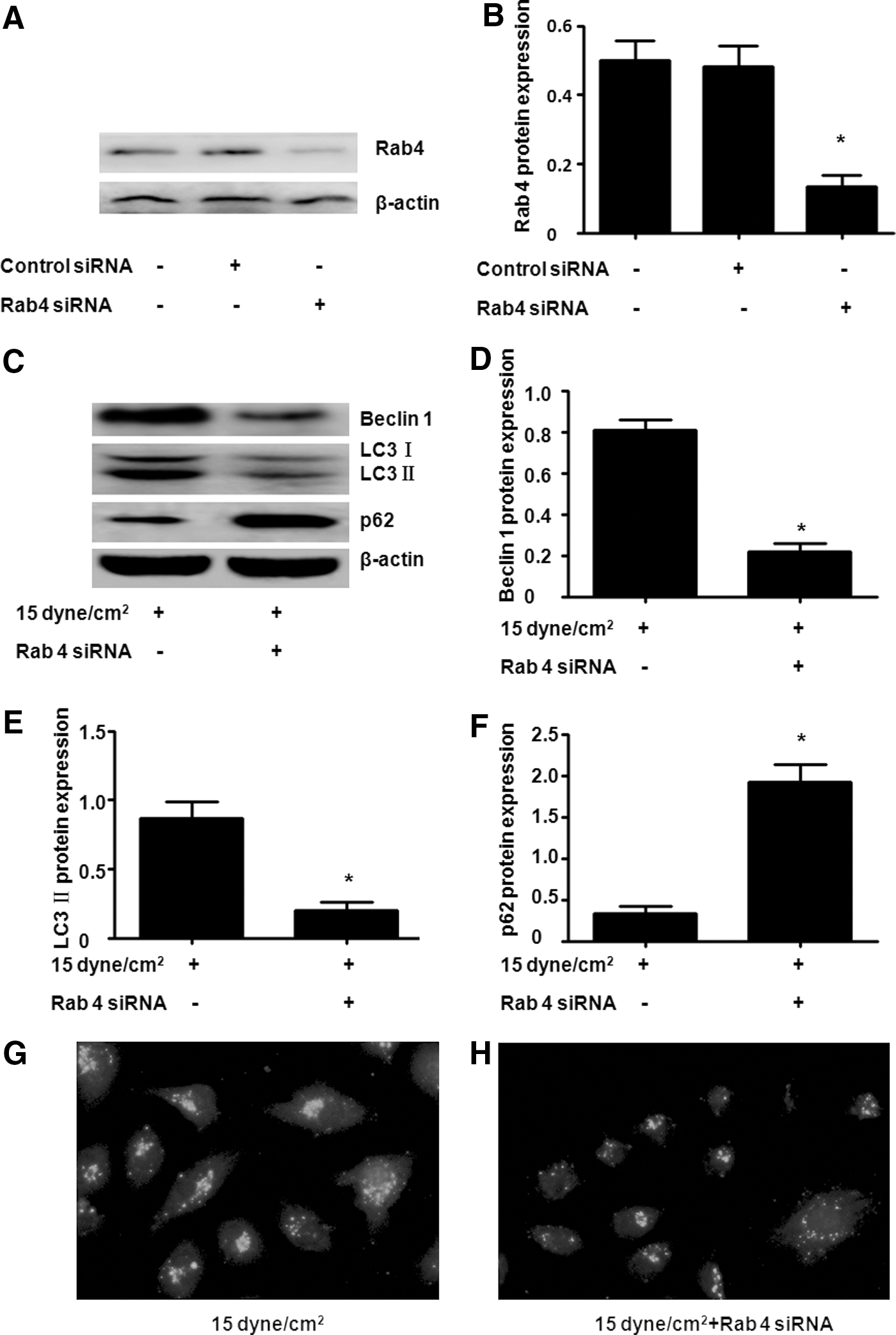
Effect of Rab4 siRNA on HUVEC autophagy. Rab4 siRNA inhibited HUVEC Rab4 protein expression
Autophagy inhibition increased monocyte adhesion to HUVECs
Numerous studies showed that laminar shear stress is antiatherogenic through inhibiting inflammation and apoptosis, as well as maintaining cell shape and vessel tone (Woo et al., 2011; Rennier and Ji, 2013; Kwak et al., 2014; Chen et al., 2015; Kanthi et al., 2015). The effect of autophagy on the adhesion of monocytes to HUVECs was observed. First, we detected the effect of 3-MA (an autophagy inhibitor) and Rab4 siRNA on HUVEC MCP 1 protein expression. As shown in Figure 5A, B, laminar shear stress significantly inhibited HUVEC MCP1 expression. However, both 3-MA and Rab4 siRNA upregulated HUVEC MCP1 expression even under laminar shear stress conditions. In agreement with the increase in MCP-1, 3-MA and Rab4 siRNA evidently increased the adhesion of monocytes with ECs (Fig. 5C).
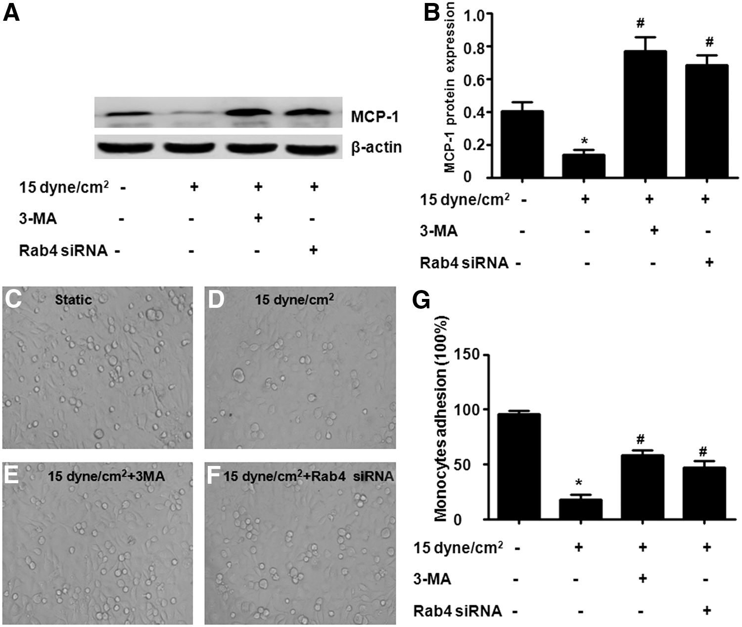
Effects of autophagy on the adhesion of monocytes to HUVECs. Laminar shear stress evidently inhibited HUVEC MCP 1 protein expression, whereas both 3-methyladenine (3-MA) and Rab4 siRNA upregulated HUVEC MCP1 expression
Discussion
Laminar shear stress was established to perform a protective role in vascular EC function. This study showed that laminar shear stress increased EC autophagy and autophagy flux, as well as upregulated Rab4 expression. Rab4 siRNA decreased autophagy and increased the adhesion of monocytes in laminar shear stress-treated HUVECs. These results indicate that Rab4 is involved in EC autophagy, which contributed to EC inflammatory response under shear stress.
Biomechanics performs a vital function in vascular biology, affecting the cells of arteries and veins under both physiological and pathological conditions. Laminar shear stress exerts its antiatherogenic effects through inhibition of inflammation and apoptosis and maintaining cell shape and vessel tone (Baeyens et al., 2014). A recent study showed that autophagy dysregulation may be a central mechanism that elicits endothelial dysfunction by risk factors, such as oxidized low-density lipoprotein (Guo et al., 2013). Autophagy is a cell survival mechanism that involves the degradation of damaged intracellular components and protection of cells from apoptosis (Chen et al., 2014; Shan et al., 2014). Autophagy protects the heart by maintaining contractile function as autophagosome dysfunction is associated with increased apoptosis, mitochondrial injury, intracellular Ca2+ dysregulation, and cardiac dysfunction (Xu et al., 2013). In this study, results showed that laminar shear stress increased the expression of the HUVEC autophagic marker, Beclin 1, and the ratio of LC3 II/LC3 I and decreased the level of the autophagy substrate, p62. Moreover, the ratio of LC3 II/LC3 I was increased in the presence of bafilomycin A1. These results showed that laminar shear stress increased HUVEC autophagy and autophagic flux.
Increasing evidence showed that GTPases frequently act in autophagy. For example, the early endosomal marker, Rab11, is required for autophagosome formation by mediating vesicular transport from the endoplasmic reticulum to the expanding phagophore, whereas the late endosomal Rab7 directs the maturation of autophagosomes by guiding the trafficking of cargos along microtubules to participate in the fusion step with lysosomes (Longatti et al., 2012; Hyttinen et al., 2013). Recent studies showed that Rab4, the master regulator of receptor recycling from endocytic compartments to the plasma membrane, performs an autophagy function by promoting the formation of LC3+ autophagosomes and regulates EC migration, proliferation, and angiogenesis (Jopling et al., 2014; Talaber et al., 2014). In this study, results showed that the HUVEC Rab4 protein level increased under laminar shear stress. Rab4 siRNA downregulated HUVEC Beclin 1 expression and the transformation of LC3 I to LC3 II while increasing the p62 level. Those results showed that this small GTPase may mediate the improved HUVEC autophagy under shear stress.
The literature suggested that the loss of autophagy may play a central mechanism in endothelial homeostasis (Du et al., 2012; Soto-Pantoja et al., 2012; Kim et al., 2013). The loss of autophagy promotes endothelial reactive oxygen species generation and inflammatory cytokine production, thus promoting the ongoing chronic inflammatory process (Lavandero et al., 2015). Previous studies showed that Rab4 is a selective regulator of autophagy, thus contributing to lysosomal degradation of proteins such as the membrane receptors, CD4 and CD3ζ, and mitophagy initiator, Drp1 (Nagy et al., 2006; Fernandez et al., 2009; Caza et al., 2014). The present study showed that Rab4 siRNA and autophagy inhibitor, 3-MA, upregulated the HUVEC MCP-1 level. These data showed that Rab4 might also increase MCP-1 degradation through the autophagy pathway. MCP-1 is a major chemokine and plays critical roles in the initiation of arteriogenesis. Under hemodynamic forces, MCP-1 is induced in ECs and activates monocytes, which facilitates their capture by adhesion molecules and their transmigration through the endothelium into tissues. (Lin et al., 2014). Along with this result, HUVECs exposed to 3-MA or Rab4 siRNA significantly increased THP-1 monocyte adhesion, suggesting that the function of laminar shear stress anti-inflammation, at least partially, contributed to the increased EC autophagy.
In conclusion, EC autophagy was upregulated and improved by laminar shear stress, which was regulated by Rab4. Moreover, improved autophagy contributed to EC inflammation inhibition, which is induced by laminar shear stress.
Conclusion
Laminar shear stress upregulated Rab4 expression, which contributed to improved EC autophagy and function.
Footnotes
Disclosure Statement
No competing financial interests exist.