
Abstract
Apoptosis of renal proximal tubular epithelial cells (PTECs) plays a vital role in the pathogenesis and progression of diabetic kidney disease (DKD). Calcium dobesilate is a vascular protective compound used for treatment of diabetic retinopathy and chronic venous insufficiency. The aim of this study was to determine whether calcium dobesilate can protect PTECs from glucose-induced apoptosis and the potential mechanism of this effect. It is indicated that high glucose promoted abnormal apoptosis of HK2 cells, which was inhibited by treatment of calcium dobesilate, while Bim expression decreased in response to calcium dobesilate in high-glucose-treated HK2 cells. These findings confirmed the therapeutic effects of calcium dobesilate on DKD and emphasized the importance of it as a potentially crucial drug in treatment of DKD.
Introduction
A
Hodiernal therapies focus on controlling blood pressure and glucose, in which angiotensin-converting enzyme inhibitors and angiotensin receptor blockers are recommended as the standard drugs (Abdel-Rahman et al., 2012). Nevertheless, deterioration of renal function cannot be fully blocked or reversed through these therapies (Lovell, 2000). Hence, it has become an imperative and significant task to develop new medicament or new pharmacologic actions of current drugs to forestall the occurrence and progression of DKD.
Calcium dobesilate (calcium dihydroxy-2,5-benzenesulfonate), a vascular protective compound, has been affirmed to be the optimal choice in efficient fibroblast growth factor inhibitors. It is widely used for treating diabetic retinopathy and chronic venous insufficiency (Allain et al., 2004; Ribeiro et al., 2006). In recent years, it has been demonstrated in many clinical trials that treatment with calcium dobesilate had significant effects on DKD in decrement of urinary albumin excretion rates (Menon et al., 2012; Xiaoqian Zhang, 2013).
Although a large quantity of investigations focused on the role of glomerulopathy, increasing evidences have shown that tubulopathy has surpassed glomerulopathy (Tang and Lai, 2012). Renal proximal tubular epithelial cells (PTECs) played a vital role in DKD pathogenesis, which participated in the procedure of progression from microalbuminuria to overt proteinuria. Some investigations even pointed out that PTEC apoptosis was the principal characteristic of DKD (Vallon, 2011; Barzilay et al., 2013).
Previous studies indicated that calcium dobesilate effectively inhibited cell apoptosis in venous wall of primary varicose veins by regulating the expression of Bcl-2 and p53 (Iriz et al., 2008). However, the exact pathway of calcium dobesilate improving renal function in DKD is still under investigation. This study is designed to answer this question.
Materials and Methods
Cell culture and treatment
HK2 cells of proximal tubule epithelial cell line (American Type Cell Collection, Rockville, MD) were incubated in RPMI 1640 medium containing 10% FBS, 11.1 mM glucose, and 100 units/mL penicillin–streptomycin (Sigma, St. Louis, MO) at 37°C, 5% CO2, and 95% humidity. The cells were then cultured in different medium for 24 and 72 h, respectively. Cells in the experimental group were cotreated with 30 mM glucose and calcium dobesilate (20 μM) for indicated times, and those in the control group were incubated in medium containing 30 mM glucose only for indicated times.
Antibodies, reagents, and calcium dobesilate
Antibodies were obtained from the following sources: anti-Bim (Mono) from Abcam (Cambridge, MA), anti-β-actin from Sigma, and all secondary antibodies (polyclonal) were from Jackson ImmunoResearch Laboratories, Inc. (West Grove, PA). Unless indicated above, other reagents were purchased from Sigma. Calcium dobesilate was purchased from Shanghai Zhaohui Pharmaceutical Company; 20 μM calcium dobesilate, which is equivalent to the maximum plasma concentration (8 μg/mL) after 500 mg oral administration,13 was used to treat HK2 cells in vitro.
Quantitative real time-PCR
The mRNA expression of Bim induced by high glucose (30 mM) and calcium dobesilate was measured by quantitative real time (RT)-PCR. HK2 cells were treated with 30 mM glucose or 30 mM glucose + calcium dobesilate for indicated times. Subsequently, cells were washed with phosphate-buffered saline (PBS) before RNA extraction. Total RNA was extracted with TRNzol-A+ RNA isolation reagent (TIANGEN) according to the manufacturer's instructions. Reverse transcription was performed with 1 μg of total RNA and RevertAid First-Strand cDKDA Synthesis Kit (Fermentas). Primer sequences used were as follows: Bim forward primer: 5′-ATTACCAAGCAGCCGAAGAC-3′ and reverse primer: 5′-TCCGCAAAGAACCTGTCAAT-3′; β-actin forward primer:5′-TGACGTGGACATCCGCAAAG-3′ and reverse primer: 5′- CTG GAA GGT GGA CAG CGA GGT-3′. Quantitative RT-PCR was performed in a cycler (MyiQ2; Bio-Rad, Inc.) using SYBR green (Roche). The mRNA expression levels of Bim were normalized by β-actin. Experiments were conducted in triplicate and repeated three times.
Assessment of apoptosis
Annexin V-FITC apoptosis detection kit (FAK011) was used to assess apoptosis. The inhibitive effect of calcium dobesilate on HK2 cell apoptosis induced by high concentration of glucose was detected qualitatively by flow cytometry with Annexin V-FITC/PI double staining. Briefly, the cells were cultured in six-well plates overnight and then exposed to 30 mM glucose (control group) or 30 mM glucose + calcium dobesilate (experimental group), respectively, for indicated times. Then, the cells were washed with ice-cold PBS, detached by trypsinization procedure, and centrifuged at 1000 rpm (4°C) for 5 min, according to the manufacturer's instructions of Annexin V-FITC apoptosis detection kit. After being resuspended with 1 × binding buffer (195 μL), the cells were stained with Annexin V-FITC (5 μL) and propidium iodide (10 μL) at room temperature for 13 min while shielded from light. The apoptosis rates of cell samples were acquired using a flow cytometer (BD FACSAriaTM II Cell Sorter).
Western blot
Western blotting was used to measure protein levels of Bim in samples. Summarily, equivalent protein samples were added into gel and separated with sodium dodecyl sulfate–polyacrylamide gel electrophoresis (SDS-PAGE) and then transferred to PVDF membranes (Bio-Rad, Inc.). The membranes were incubated in blocking buffer (0.2 mM Tris, 137 mM NaCl, 5% fat-free milk, and 0.1% Tween 20) for 1 h and probed with specific primary antibodies at 4°C overnight. Subsequently, the membranes were rinsed with TBST buffer (0.1% Tween 20, 0.2 mM Tris, and 137 mM NaCl) and incubated with HRP-conjugated secondary antibody (1:5000) at room temperature for 1 h. Finally, the protein level of Bim was evaluated by chemiluminescence detection.
Statistical analyses
All statistical analyses were performed using SPSS (Statistical Product and Service Solutions) 19.0 software (from IBM). Student's t-test was used to assess significance of data within two groups. Data are presented as mean ± SEM and significance was set at p < 0.05.
Results
Apoptosis is induced in response to high glucose in HK2 cells
To examine the effect of high glucose on HK2 cells, the cells were treated with glucose of different concentrations for different periods of time. Apoptosis of HK2 cells was detected by Annexin V-FITC/PI double staining.
A significant increase in cell apoptosis rate was observed in the cells treated with 30 mM glucose compared with those with 5.5 mM glucose (Fig. 1) (p < 0.05).

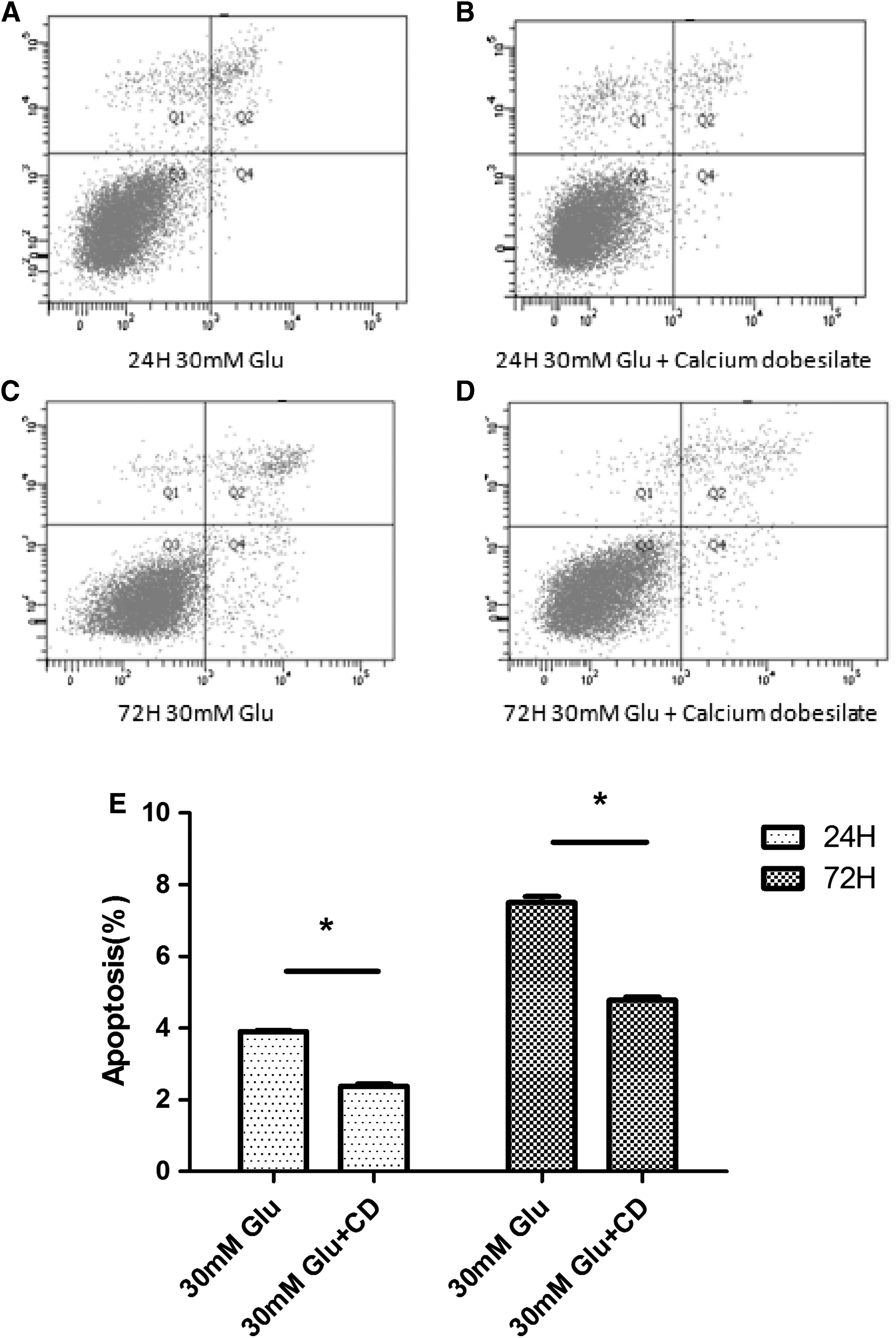
The apoptotic status of HK2 cells after indicated treatment with different medium was determined by Annexin V-FITC binding assay. The Q4 (Annexin V-FITC+/PI−) and Q2 (Annexin V-FITC+/PI+) were considered as early stage and late stage of apoptotic cells, respectively. The percent of cell apoptosis was quantified by Q2+Q4. In Figure
Apoptosis of HK2 cells induced by high glucose in cotreated group was inhibited by calcium dobesilate
To investigate the effect of calcium dobesilate on HK2 cells under high-glucose condition, apoptosis of cells treated with high glucose and high glucose + calcium dobesilate for different periods of time was detected by Annexin V-FITC/PI double staining. As is shown in Figure 2, it is observed that the apoptosis rate of the calcium dobesilate-treated group was significantly decreased in comparison with that of control group (30 mM glucose) (p < 0.05).
Bim expression was decreased in response to calcium dobesilate in high-glucose-treated HK2 cells
To investigate the mechanism of calcium dobesilate inhibiting HK2 cell apoptosis, Bim expression at both protein and mRNA levels was determined. HK2 cells were incubated with high glucose or high glucose + calcium dobesilate for 24 and 72 h. The variation of Bim expression was detected by western blotting to verify that Bim was involved in apoptosis of HK2 cells.
The results are shown in Figure 3. Compared with control group, the expression of Bim protein reduced remarkably in the calcium dobesilate group (p < 0.05).
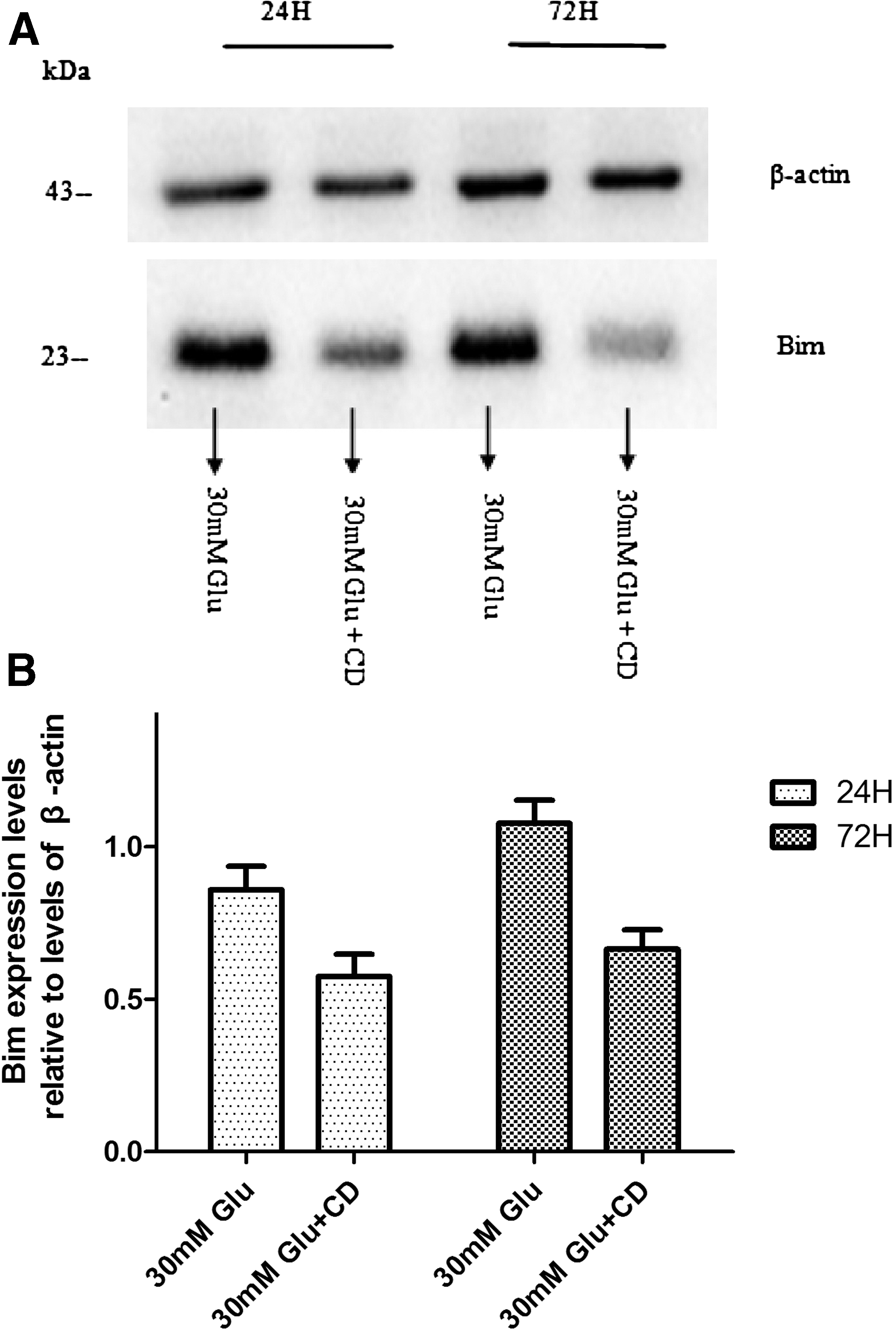
Bim protein expression levels relative to levels of β-actin are quantitated by immunoblotting, and optical density values are expressed as a ratio between the Bim protein and β-actin.
To determine whether upregulation of Bim expression was attributed to enhanced transcription, Bim mRNA was also analyzed by RT-PCR. A significant decrease of Bim mRNA in the calcium dobesilate group was detected as well (*p < 0.05) (Fig. 4). It was observed that Bim mRNA was notably decreased in the calcium dobesilate group (*p < 0.05) (Fig. 4).

Total RNA was extracted from HK2 cell after treatment with 30 mM glucose + calcium dobesilate or control (30 mM glucose only) for 24 and 72 h, respectively. Relative mRNA levels of Bim in HK2 cells after indicated treatment analyzed by real-time RT-PCR, and β-actin was used as an internal control. Incubation in 30 mM glucose + calcium dobesilate resulted in significantly decreased Bim mRNA levels compared with cells incubated in medium containing 30 mM glucose only. Experiments were performed in triplicate and repeated three times. Data are shown as the mean ± SEM. n = 3 for each group, *p < 0.05, compared with control; Student's t-test.
Discussion
DM is a cosmopolitan health problem that affects more than 590 million people globally and leads to target organ dysfunction and impairment (Boyle et al., 2010; Guariguata et al., 2014). Among all the complications of DM, DKD is one of the most important complications (Lovell, 2000; Abdel-Rahman et al., 2012). Despite accumulating comprehension of DKD, current therapeutic methods such as control of blood pressure and blood glucose, as well as treatment with renin–angiotensin–aldosterone system inhibitors, have failed to prevent the progress of DKD (Awad et al., 2015).
Based on pathogenesis and progression (Mogensen, 1999), DKD is divided into five phases: acute renal hypertrophy hyperfunction, normoalbuminuria, incipient diabetic nephropathy also known as early stage of diabetic nephropathy, proteinuria also known as clinical overt diabetic nephropathy, and ESRD. Progression from early-stage to clinical overt diabetic nephropathy is marked by the transformation from microalbuminuria to overt proteinuria.
Although prevalent investigations have illustrated the materiality of glomerular lesion in the progress of DKD, it is the extent of tubulointerstitial injury that conclusively determines the rate of attrition of renal function (Tang and Lai, 2012). Under physiological conditions, renal tubules reabsorb albumin from crude urine, in which 71% albumin is reabsorbed by PTECs (Tojo and Kinugasa, 2012). Tojo et al. (2001) found that tubulopathy occurred earlier than glomerulopathy in DKD rats. Similarly, it has been proved that microalbuminuria occurred earlier than the glomerulopathy in clinical trials (Magri and Fava, 2011; Barzilay et al., 2013). Further research indicated that apoptosis of PTECs was the main feature of DKD (Vallon, 2011; Tang and Lai, 2012). To this end, the renal PTEC is progressively implicated in pathogenesis and progression of DKD (Tang and Lai, 2012). Thus, preventing PTECs from apoptosis may ameliorate DKD (Bonventre, 2012).
Our previous study showed that proapoptosis protein Bim (Bcl-2-interacting mediator), which is only expressed in PTECs specifically in renal system, played a vital role in inducing the apoptosis of PTECs. Bim belongs to Bcl-2 protein family, which regulates apoptosis by balancing activity of pro- and antiapoptotic members. Bim is a proapoptotic protein, which has only one Bcl-2 homology (BH3) domain. High glucose significantly increased the expression of Bim and further accelerated apoptosis of PTECs.
Bim is a proapoptosis protein belonging to Bcl-2 family (O'Connor et al., 1998), which contains a series of evolutionarily related proteins that are closely connected with cell apoptosis, including proapoptotic members (Bim, Bax, BAD, Bak, and Bok) and antiapoptotic members (Bcl-2, Bcl-xl, and Bcl-w) (Muchmore et al., 1996). As a proapoptotic regulator, Bim can trigger cell apoptosis not merely by interfering antiapoptotic function of Bcl-2 but also by activating proapoptotic elements Bax/Bak in mitochondrial membrane to trigger mitochondria-related programmed cell death (Kuwana et al., 2005). Compared with other members in Bcl-2 family, Bim is expressed in renal tubule cells uniquely in the renal system, which makes it highlighted in apoptosis of PTECs (O'Reilly et al., 2000).
Calcium dobesilate (calcium dihydroxy-2, 5-benzenesulfonate) is a small-molecule drug, which has been widely used in microvascular complications of DM. Previous investigations focused on its anti-inflammatory effect and amelioration of microvessel permeability (Allain et al., 2004; Ribeiro et al., 2006; Menon et al., 2012; Xiaoqian Zhang, 2013). Investigations have indicated that calcium dobesilate could reduce the apoptosis of cells by reducing the level of reactive oxygen species (ROS) (Iriz et al., 2008; Zheng et al., 2013). Zheng et al. (2013) reported that calcium dobesilate of different doses could protect the lens of rats against galactose damage in a dose-independent way, but the exact pathway was still unclear. Iriz et al. (2008) confirmed that calcium dobesilate could reduce apoptosis of venous wall in primary varicose veins and put forward that apoptosis regulation factors of Bcl-2 family were probably involved in this progress. Antiapoptosis effect of calcium dobesilate in treating DKD and the specific pathway are still under discussion and investigation.
In this study, we affirmed that calcium dobesilate protects against PTEC apoptosis induced by high glucose. Besides the antioxidant effect, the potential mechanism was through downregulating the inordinate expression of Bim in PTECs.
Our study indicated that the expression of Bim increased significantly under high glucose, which led to excess apoptosis of PTECs. The elevated expression of Bim under high glucose could be reduced by calcium dobesilate, and the apoptosis of PTECs was in turn decreased. In consideration of the materiality of PTECs in the early stage of DKD as well as the transition of microalbuminuria to overt proteinuria, this effect of calcium dobesilate on PTECs may promise its expanding usage in treating DKD and ameliorating renal injury, especially in the early stage, to postpone the progress from microalbuminuria to overt proteinuria, even reverse it to normality.
Besides cell apoptosis, there are many other mechanisms that participated in DKD onset and progression. Hyperglycemia-induced renal hyperfiltration and renal injury, oxidative stress, PKC-induced production of cytokines, chemokines, and different inflammatory signals also have been postulated as playing an important role in DKD course (Bhattacharjee et al., 2016). Soo-Hyun Park found that coincubation of PTECs and high glucose increase both ROS and TGF β-1 levels, which inhibit the cellular proliferation of PTECs (Soo-Hyun Park, et al., 2001). Additionally, in previous researches, Verzola had indicated that incubation of PTECs with high glucose activated cellular death through production of ROS (Verzola et al., 2004). These two researches demonstrated two vital molecular factors in DKD: ROS and TGF-β1.
As correlation studies have demonstrated, Bim is one of the downstream effectors of ROS. Deng et al. (2015) found that the ROS/JNK pathway is involved in Bim upregulation and Bim mediated the human hepatoma-derived cell l apoptosis. Costa et al. (2016) discovered in human respiratory epithelial cells that ROS involved in apoptosis through increasing Bim level. In addition, Bim also is a downstream effecter of TGF β-1. Weiner et al. (2014) indicated that in intestinal adenomas, oncogenic mutations regulate Bim-mediated apoptosis induced by TGF β-1. Based on this information, high glucose induces apoptosis of PTECs through activation of ROS, TGF β-1, and Bim, and calcium dobesilate reduces the apoptosis of PTECs through decrease of ROS, TGF β-1, and Bim. However, whether calcium dobesilate could affect TGF β-1 directly needs to be verified in the future.
Although the protective function of calcium dobesilate to PTECs has been proved, further study is needed to investigate its detailed pathway of functioning.
Conclusion
In this study, it was indicated that calcium dobesilate has protective function in the kidney against PTEC apoptosis induced by high glucose. Besides the antioxidant effect, the potential mechanism was through downregulating the inordinate expression of Bim, which is a vital factor of cell apoptosis expressed only in PTECs in the kidney.
Footnotes
Acknowledgments
This work was funded by National Natural Science Foundation of China Grants (81670757, 81570742), Grant for the development of science and technology of Shandong Province (NO.2010GSF10228, 2012GGH11862, 2014GSF118118), Shandong Provincial Natural Science Foundation of China Grants (No. ZR2016HQ26, Y2008C73, ZR2010HM044), Shandong Provincial Science and Technology Development Program, China (2009GGB14001), Grant for Excellent Young and Middle-aged Scientists of Shandong Province (No. 2004BS02016), and Grant for the development of science and technology of JiNan City (201602172). We want to thank Merck Serono Co., Ltd, for their help and sponsorship.
Disclosure Statement
No competing financial interests exist.