
Abstract
Klotho is originally discovered as an anti-aging gene and recently identified as a tumor suppressor in various human cancers. Drug resistance is a major obstacle to affect the treatment of chemotherapy. In the present study, we explore the role of klotho on drug resistance in human lung cancers and investigate the mechanism of klotho on drug resistance in lung cancer cells. First, we detected a panel of six human lung cancer cell lines, including H460, SK-MES-1, cisplatin (DDP)-resistant A549/DDP, its parental subline A549, docetaxel (DTX)-resistant SPC-A-1/DTX, and SPC-A-1 by western blotting analysis. The results showed that klotho level was significantly decreased in chemotherapeutic drug-resistant lung cancer cells. Next, klotho was overexpressed in drug-resistant cancer cell lines and the results showed that overexpression of klotho significantly inhibited cell proliferation of A549/DDP and SPC-A-1/DTX. Conversely, knockdown of the expression of klotho significantly promoted cell growth of lung cancer cells. Furthermore, overexpression of klotho had synergistic effects with cisplatin to inhibit the proliferation of drug-resistant lung cancer cells in a dose- and time-dependent manner. The molecular mechanism was explored by western blotting analysis and the results revealed that the levels of beclin 1 and LC3-II were obviously increased, suggesting cell autophagy enhanced in drug-resistant cancer cells. Importantly, overexpression of klotho would inhibit cell autophagy in A549/DDP cells. All the results demonstrated that the levels of klotho were significantly decreased, which was accompanied by the increased cell autophagy in drug-resistant lung cancer cells. Overexpression of klotho would inhibit cell autophagy in drug-resistant lung cancers, which may probably contribute to reverse drug resistance in lung cancer cells.
Introduction
L
Klotho was first discovered by Kuro-o et al. (1997) involving in the suppression of several aging phenotypes. A defect in klotho gene expression in the mouse leads to a syndrome that resembles human aging, including a short lifespan, infertility, arteriosclerosis, skin atrophy, osteoporosis, and emphysema. Next, it has been found that klotho expression is associated with epithelial ovarian cancer risk and progression, and klotho serves as an independent marker for ovarian cancer prognosis (Lu et al., 2008). Gradually, much attention has been focused on the role of klotho in tumorigenesis, progression, and prognosis, including breast cancer (Rubinek et al., 2012), gastric cancer (Wang et al., 2011a), lung cancer (Wang et al., 2011b; Chen et al., 2012), colorectal cancer (Pan et al., 2011; Liu et al., 2015), pancreatic cancer (Abramovitz et al., 2011), and so on. It has been reported that klotho inhibits growth and promotes apoptosis in human lung cancer cell line A549 (Chen et al., 2010) and also inhibits the capacity of cell migration and invasion in cervical cancer cells (Chang et al., 2012). He has found that miR-199a-5p acts as an oncogene in gastric cancer, which functions by targeting and negative regulation of klotho (He et al., 2014). It works as a tumor suppressor and a modulator involving in IGF-1 and FGF pathways (Wolf et al., 2008; Abramovitz et al., 2011), transforming growth factor-beta1 (TGF-beta1) signaling pathway (Doi et al., 2011), IGF1R-mediated PI3K/AKT pathway (Li et al., 2014), and Wnt/beta-catenin signaling pathway (Sun et al., 2015).
Wang et al. (2013) found that klotho knocking-down cells showed enhanced resistance to chemotherapy by PI3k/Akt pathway. However, the role of klotho in drug resistance has not been clearly clarified till now. In the present study, we used two pairs of cell lines as cell model, including cisplatin (DDP)-resistant A549/DDP and its parental subline A549, docetaxel (DTX)-resistant SPC-A-1/DTX, and SPC-A-1. We further investigated the molecular mechanism of klotho in drug-resistant lung cancer cells. Klotho may be used as a new target for gene therapy of drug-resistant lung cancers and it would give new clues for the chemotherapy for lung cancer patients.
Materials and Methods
Cell lines and agents
The lung cancer cell lines NCI-H460 [H460] (ATCC® HTB-177™) and SK-MES-1 (ATCC HTB-58™) were purchased from American Type Culture Collection (ATCC, Rockville, MD). The cells were cultured in DMEM medium (GIBCO, Gaithersburg, MD) supplemented with 10% fetal bovine serum in 37°C with 5% CO2 atmosphere. A549 cells and A549/DDP cells were purchased from Cell Bank of Chinese Academy of Medical Sciences (Beijing, China). SPC-A-1 was obtained from MeiLian ShengWu Corporation (Shanghai, China). SPC-A-1/DTX cells were selected and kept by our laboratory. Docetaxel (Cat. No. 01885) was obtained from Sigma-Aldrich with the purity more than 97.0%. Cisplatin (Cat. No. 479306) was also purchased from Sigma-Aldrich Corporation and the purity was more than 99.9%. MTT agent (Cat. No. M2128) was purchased from Sigma-Aldrich Corporation and stock concentration was 5 mg/mL. The autophagy inhibitor, 3-Methyladenine (3-MA, Cat. No. M9281), was obtained from Sigma-Aldrich Corporation. Lipofectamine 2000 (Cat. No. 11668-019) was obtained from Invitrogen. The myc-tagged klotho expression vector (pCMV6-klotho) and the control vector (pCMV6) were purchased from OriGene Corporation (Rockville, MD).
Cell transfection
The lung cancer cells were plated at a density of 3 × 10⁴/well in a six-well plate. The cells were cultured before transfection to achieve more than 70% confluence. The cells were transfected according to the manufacturer's instructions. Briefly, for transfection, 1 μL of pCMV6-klotho plasmid or pCMV6 vector were added into 100 μL Opti-MEM (Invitrogen, Carlsbad), and 2.5 μL of Lipofectamine 2000 (Invitrogen, Carlsbad) were added to 100 μL Opti-MEM I (Invitrogen, Carlsbad). Five minutes later, both of the liquids were mixed gently and cultured for 15 min at room temperature. The plate was incubated at 37°C for 48–72 h, respectively, and used in the experiments described below.
MTT assay
Cell viability was determined by MTT assay. Briefly, human lung cancer A549 cells and A549/DDP cells (SPC-A-1 cells and SPC-A-1/DTX cells) were plated into 96-well plate. After 6 h, they were treated with increasing concentrations of chemotherapeutic drug cisplatin (or docetaxel) for 24 h. In another treatment, the A549/DDP cells or SPC-A-1/DTX cells transfected with pCMV6-klotho and pCMV6 vector were treated with increasing concentrations of cisplatin (10, 20, 40, and 80 μM) for 24 h. Four hours before test, 10 μL of MTT agent were added into the medium. Finally, the purple crystals were dissolved with DMSO. The data were read on a microplate reader at a test wavelength of 490 nm and a reference wavelength of 570 nm.
Antibodies
Anti-klotho antibody (Cat. No. ab18131) was purchased from Abcam Corporation. Mouse monoclonal anti-β-actin antibody (Cat. No. TA310155) was obtained from OriGene Corporation (Beijing, China). Anti-beclin 1 antibody (Cat. No. 11306-1-AP) was a rabbit polyclonal IgG and was purchased from Proteintech Corporation (Wuhan, China). LC3 Antibody (LC3-I and LC3-II) was a rabbit polyclonal IgG (Cat. No. 14600-1-AP) obtained from Proteintech Corporation (Wuhan, China). Goat anti-rabbit IgG-HRP (sc-2004; Santa Cruz) and goat anti-mouse IgG-HRP (sc-2005; Santa Cruz) were both obtained from Santa Cruz Corporation.
Western blotting assay
The cells in different groups were lysed with RIPA buffer, including Tris 50 mmol/L, NP-40 1%, NaCl 150 mmol/L, EDTA 1 mmol, SDS 0.1%, and SDC 0.25%. The whole proteins were separated by SDS-PAGE at constant voltage of 80 v for 15 min and 120 v for 60 min. Then, the whole proteins were transferred electrophoretically into a nitrocellulose membrane at constant current of 400 mA for 2 h. The membrane was blocked with 5% nonfat milk in TBST (Tris-HCl 50 mmol/L, NaCl 150 mmol/L, and 0.1% Tween) for 30 min at room temperature. Then, the membranes were incubated with primary antibodies at 4°C overnight and secondary antibodies for 30 min at room temperature. Each time was washed with TBST buffer for 5 min. The bands were detected using an enhanced chemiluminescence western blotting detection system according to the kit protocol.
Statistical analysis
All the results were analyzed by SPSS 20.0 software. The data for MTT assay were repeated twice and each sample was given three replicates. The data are shown as mean value ± standard deviation.
Result
The expression level of klotho is decreased in drug-resistant lung cancer cells
To identify the role of klotho on chemotherapy of lung cancers, we detected the expression levels of klotho in a panel of lung cancer cell lines by western blotting analysis. As shown in Figure 1, the level of klotho was obviously decreased in A549/DDP cells compared to that in A549 cells. Simultaneously, the western blotting result also showed that klotho was obviously decreased in SPC-A-1/DTX cells compared to that in SPC-A-1 cells. Moreover, the expression of klotho was observed in the other lung cancer cell lines, such as H460 cells and SK-MES-1 cells. All the data suggested that the levels of klotho were obviously decreased in chemotherapeutic drug-resistant lung cancer cells.
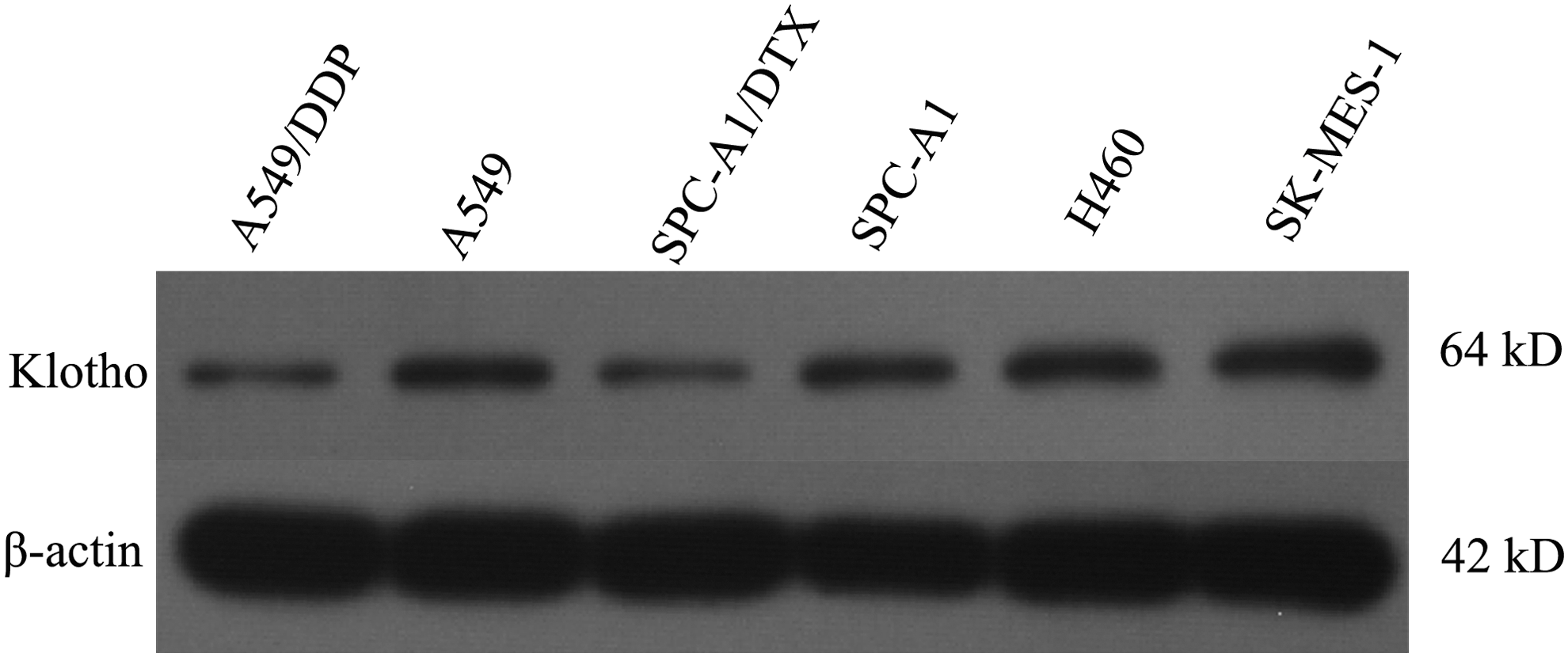
The expression level of klotho is decreased in drug-resistant lung cancer cells. A panel of lung cancer cell lines was plated into 48-well plate and cultured for 24 h. The levels of klotho were detected by western blotting analysis. In this study, β-actin was used as the internal reference gene.
Overexpression of klotho inhibits cell proliferation of A549/DDP and SPC-A1/DTX
To detect whether the levels of klotho affected the cell proliferation of lung cancer cells, functional assay was performed by MTT assay. We had found that the levels of klotho were relatively lower in drug-resistant lung cancer cells A549/DDP and SPC-A1/DTX cells (**p < 0.01, compared with that of A549 cells or SPC-A1 cells). Next, A549/DDP and SPC-A1/DTX cells were transfected with pCMV6-klotho and control plasmid for 48, 72, and 96 h, and cell viability was determined by MTT assay. As shown in Figure 2, the cell proliferation was significantly suppressed in pCMV6-klotho transfected cells compared with pCMV6 vector transfected cells (**p < 0.01). The results obviously demonstrated that overexpression of klotho would inhibit cell proliferation of chemotherapeutic drug-resistant cancer cells.

Overexpression of klotho inhibits cell proliferation of A549/DDP and SPC-A1/DTX. The drug-resistant lung cancer cell lines A549/DDP cells
Transfection with pCMV6-klotho sensitizes the effects of cisplatin in A549/DDP cells
Next, we tested whether klotho expression could sensitize the effects of chemotherapeutic drug cisplatin; MTT assay was performed to determine the cell viability in the experiment. A549/DDP cells were treated with different concentrations of cisplatin for 24-h, including 10, 20, 40, and 80 μM, respectively. As shown in Figure 3A, the inhibition rate was significantly increased in pCMV6-klotho transfected cells than that in pCMV6 vector transfected cells (**p < 0.01). Furthermore, A549/DDP cells were transfected with pCMV6-klotho or pCMV6 vector and treated with 40 μM of cisplatin for 24, 48, and 72 h, respectively. The results revealed that the proliferative activity was significantly inhibited in pCMV6-klotho transfected—A549/DDP cells, compared with the pCMV6 plasmid transfected cells (Fig. 3B). All the data demonstrated that transfection with klotho in A549/DDP cells significantly suppressed the cell proliferation and sensitized the effects of chemotherapeutic drug cisplatin.
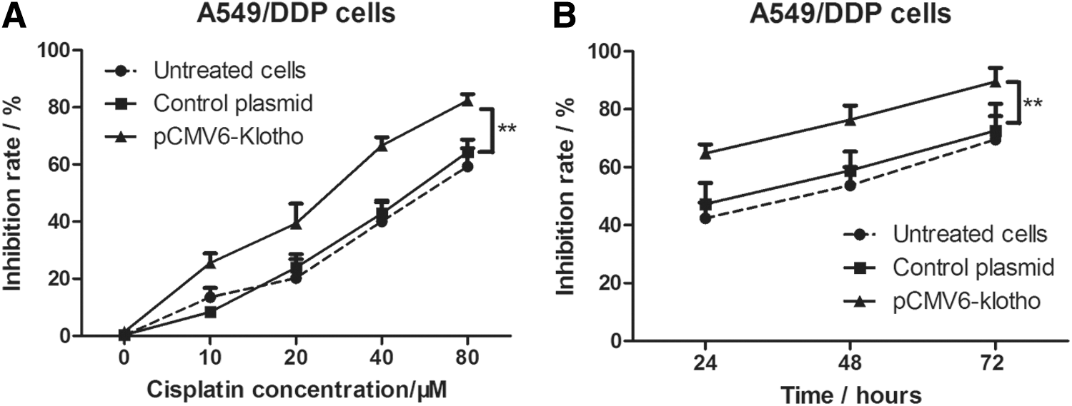
Transfection with pCMV6-klotho sensitizes the effects of cisplatin in A549/DDP cells.
Knockdown of the expression of klotho increases cell proliferation of A549 and H460 cells
Next, klotho was knocked down by shRNA specific to klotho in A549 cells and H460 cells and the effects of klotho were further to be explored on the cell proliferation of lung cancer cells. Briefly, the cells were transfected with klotho- shRNA and control shRNA for 48, 72, and 96 h. Cell viability in each group was determined by MTT assay. As shown in Figure 4, OD490 values were significantly increased after 72 and 96 h, suggesting that knockdown of the expression of klotho promoted the cell proliferation of lung cancer cells.
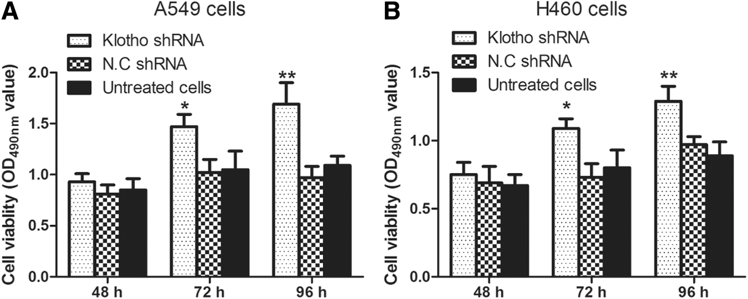
Knockdown of the expression of klotho increases the cell proliferation of A549 and H460 cells. The lung cancer cells A549
The levels of Beclin1 and LC3-II are upregulated in drug-resistant lung cancer cells
Cell autophagy is a new type of programmed cell death, and the role of cell autophagy in cancer progression is still not clarified. To determine whether cell autophagy was involved in chemotherapeutic drug resistance in lung cancer cells, we detected the levels of related proteins in cell autophagy signaling pathway, such as beclin 1, LC3-I, and LC3-II. As shown in Figure 5, the levels of beclin 1 and LC3-II were obviously increased in drug-resistant lung cancer cells, including A549/DDP cells and SPC-A1/DTX cells; however, the level of LC3-I was not obviously changed between A549 cells and A549/DDP cells, or SPC-A1 cells and SPC-A1/DTX cells. The results demonstrated that cell autophagy was increased in chemotherapeutic drug-resistant lung cancer cells.
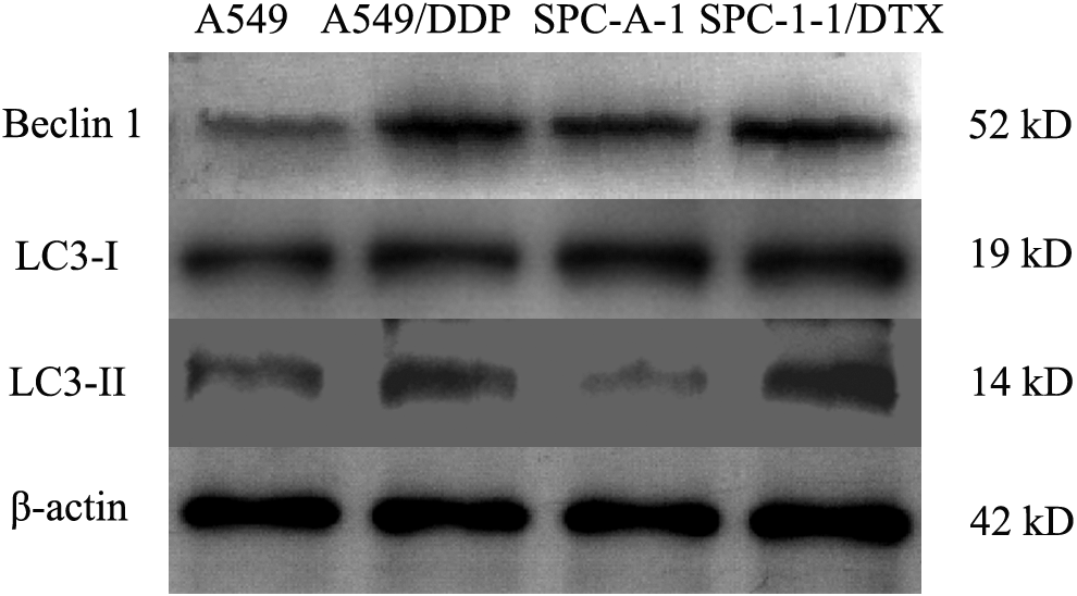
The levels of Beclin1 and LC3-II are upregulated in drug-resistant lung cancer cells. The lung cancer cells, A549 cells, A549/DDP cells, SPC-A1 cells, and SPC-A1/DTX cells, were plated into 48-well plate and cultured for 24 h. The cell lysates were prepared by RIPA buffer. The levels of beclin 1, LC3-I, and LC3-II were determined by western blotting analysis. In this study, β-actin was used as the internal reference.
Overexpression of klotho decreases the levels of beclin 1 and LC3-II in A549/DDP cells
To further clarify whether cell autophagy was mediated by klotho in DDP-resistant lung cancer cells, we overexpressed klotho in A549/DDP cells and detected the levels of proteins involved in cell autophagy in A549/DDP cells. As shown in Figure 6, the A549/DDP cells were treated with the inhibitor of cell autophagy, 3-MA, for 24 h and the levels of beclin 1 and LC3-II were obviously decreased. Moreover, the cells were transfected with klotho for 24 h and the results demonstrated that the levels of beclin 1 and LC3-II were significantly decreased compared with control plasmid-transfected A549/DDP cells. Interestingly, A549/DDP cells were transfected with pCMV6-klotho plasmid or control plasmid and treated with 10 mM of 3-MA for 24 and 48 h, and MTT assay results demonstrated that cell viability was significantly decreased in 3-MA or pCMV6-klotho plasmid-transfected cells compared with untreated controls. All the results demonstrated that overexpression of klotho could decrease the cell autophagy in cisplatin-resistant lung cancer cells, which would probably contribute to increasing the drug sensitivity in lung cancer cells.
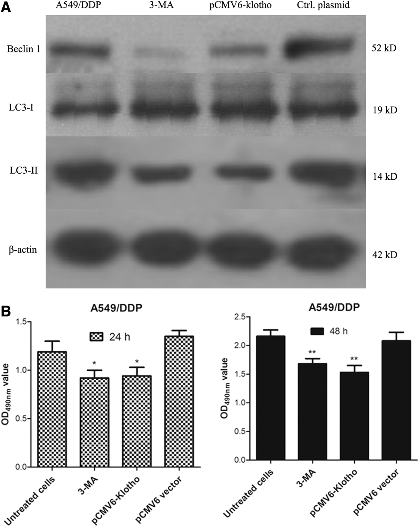
Overexpression of klotho decreases the levels of beclin 1 and LC3-II in A549/DDP cells.
Discussion
Klotho is first discovered as an antiaging gene and knockout of klotho would promote rapid aging in mice (Kuro-o et al., 1997). Gradually, it has been found that klotho possesses a wide range of biological effects not only in anti-aging but also involving in calcium and phosphorus metabolism, anti-oxidation, anti-apoptosis, and the protection of organs, which have broad application prospects (Torres et al., 2009; Oswiecimska et al., 2015; Salanova Villanueva et al., 2016). Primary and acquired drug resistance is a big obstacle in the chemotherapy of human lung cancers, which severely affects the outcome and prognosis of lung cancer patients (Kim, 2016; Tetsu et al., 2016). In the present study, we investigated the role of klotho on drug resistance in human lung cancer cells and further investigated the molecular mechanism of klotho in drug resistance of lung cancer cells.
First, we identified the role of klotho in DDP- or DTX-sensitive and DDP- or DTX-resistant lung cancer cell lines by western blotting analysis. We observed that the expression of klotho was significantly decreased in drug-resistant lung cancer cells than that in drug-sensitive lung cancer cells. The results demonstrated that chemotherapeutic drug-resistant lung cancer cells had lower levels of klotho compared with drug sensitive cells. We speculated that klotho may contribute to sensitization of chemotherapeutic drug-resistant cancer cells.
Next, klotho was overexpressed in the drug-resistant lung cancer cell lines A549/DDP cells and SPC-A1/DTX cells. MTT results demonstrated that increased klotho protein would suppress cell proliferation of A549/DDP cells and SPC-A1/DTX cells. Moreover, overexpression of klotho also sensitized the effects of chemotherapeutic drug in A549/DDP cells in a time- and dose-dependent manner. Conversely, klotho was knocked down by shRNA specific to klotho in A549 cells and H460 cells and the results obviously revealed that knockdown of klotho increased the cell proliferation of A549 cells and H460 cells.
Furthermore, the molecular mechanism of klotho on the antidrug resistance in lung cancer cells was investigated. Development of acquired chemoresistance severely limits the efficiency of chemotherapy drugs. It is a persistent clinical problem in cancer therapy (Huang et al., 2016). Enormous evidences are clarifying different roles of autophagy in inducing chemoresistance. Recently, more and more researchers found that upregulation of autophagy promoted chemoresistance in various human cancers, such as leukemia cells (Yang et al., 2012; Kong et al., 2015), gastric cancer (Ge et al., 2014), cervical cancer (Zhang et al., 2013), colon cancer (Wu et al., 2013), neuroblastoma cancer (Wang et al., 2015), prostate cancer (Chang et al., 2014), and so on. However, the role of autophagy in lung cancer was not clearly clarified. It will be critical to reveal the roles of autophagy in chemoresistance and the involved molecular mechanisms for the molecular therapy of human lung cancers. In the present study, cell autophagy was observed in A549/DDP cells and SPC-A1/DTX cells, and the levels of beclin 1 and LC3-II were obviously higher in drug-resistant lung cancer than that in drug-sensitive lung cancer cells. Interestingly, in pCMV6-klotho transfected lung cancer cells, the cell autophagy was significantly inhibited, as well as 3-MA treated lung cancer cells. Thus, klotho may play an important role to reverse chemotherapeutic drug resistance in the therapy of lung cancers.
Footnotes
Acknowledgment
This work was supported by a grant from the Scientific and Technological Research Project of Shaanxi Province (No. 2015KW-038).
Disclosure Statement
No competing financial interests exist.