
Abstract
The aim of this study was to determine the role of DNA methylation of the platelet-derived growth factor-D (PDGFD) gene promoter in the development of intracranial aneurysms (IAs) and brain arteriovenous malformations (BAVMs). A total of 70 patients with IAs or BAVMs and 26 control individuals were enrolled for this study. The PDGFD level in the plasma was determined using enzyme-linked immunosorbent assay. DNA methylation levels of seven cytosine-phosphate-guanine (CpG) dinucleotides present in the PDGFD gene promoter were measured using bisulfite pyrosequencing technology. The plasma PDGFD levels in IA and BAVM were significantly lower than those in the control group (p = 0.0008 and 0.002, respectively). CpG1 methylation levels of the PDGFD gene promoter were significantly higher in IA patients (4.63 ± 0.35, p = 0.017) than in the control group (3.36 ± 0.35). CpG1 methylation of the PDGFD gene promoter in BAVM patients (6.00 ± 0.86, p = 0.003) was also significantly higher than that in the control group, although these differences were seen in both male and female patients (p = 2.81E-04 and p = 0.017, respectively). In addition, CpG1 methylation of the PDGFD promoter was associated with apolipoprotein E (APOE) levels in IA patients (p = 0.013). In conclusion, our study has demonstrated significant correlations between DNA methylation of the PDGFD gene promoter and the risk of developing either IA or BAVM. Furthermore, PDGFD gene promoter CpG1 methylation shows a significant correlation with APOE in IAs. Further functional studies on these relationships and correlations are warranted.
Introduction
C
DNA methylation, which occurs at the cytosine-phosphate-guanine sequences (CpGs), is a unique chemical modification in the human genome (Chen et al., 2016). DNA methylation of a promoter is correlated with transcriptional silencing of protein-coding genes, thus regulating its protein functions in the human body (Caranci et al., 2013). Aberrant DNA methylations have been extensively studied in the pathogenesis of multiple diseases (Mendizabal and Yi, 2016). For example, DNA hypermethylation of promoter-associated CpG islands (CGIs) in tumor suppressor genes was found to cause carcinogenesis (Moarii et al., 2015). DNA methylation of the PLA2G7 gene promoter may play a role in the molecular mechanisms underlying the pathophysiology of cardiovascular diseases (Jiang et al., 2013).
The platelet-derived growth factor-D (PDGFD) is the fourth member of the PDGF family and was first identified by Eriksson's group in 2001 (Bergsten et al., 2001). Emerging evidence had implicated that the PDGFD gene was mainly expressed in endothelial cells and adventitial connective layers of vessels (Chen et al., 2005). The PDGFD protein was predominantly expressed in artery, often localized to vascular bifurcations (Gladh et al., 2016). PDGFD was considered as the specific agonistic ligand for PDGF beta-receptor, and its activation could induce downstream signaling through PDGF-β-receptor (Bergsten et al., 2001). Through binding to PDGF-β-receptor, PDGFD played an important function in the vasculature (Uutela et al., 2001). It emerged as a vascular modulator during pathological conditions. PDGFD could induce vascular smooth muscle cell migration and proliferation by stabilizing newly formed vessels and neointimal hyperplasia after vessel injury (Chen et al., 2005). In the experimental mice model, Yang et al.'s (2016) study certified that PDGFD/PDGFR-β played an important role in the proinflammatory response of secondary brain injury after intracerebral hemorrhage. Gladh et al.'s (2016) study suggested that PDGFD might have a role in regulation of arterial blood pressure and described a possible role for PDGFD in mural cell physiology and regulation of vascular homeostasis. PDGFD polymorphisms were also shown to increase cardiovascular mortality risk in elders (Alehagen et al., 2016). Studies demonstrated that PDGFD variation was associated with higher risk for ischemic stroke in a Chinese population (Han et al., 2008). Furthermore, the PDGFD polymorphism rs974819 displayed a suggestive correlation with coronary stenosis index in women with coronary artery disease (Dechamethakun et al., 2014). Based on these results, we predict that the PDGFD gene is associated with cerebral artery disease. The only epigenetic study on this topic indicated that DNA methylation levels of the PDGFD gene promoter are significantly associated with cumulative arsenic exposure in urothelial carcinoma patients (Yang et al., 2014). The objective of our research is to determine the role of DNA methylation of the PDGFD gene promoter in the development of IAs and BAVMs.
Materials and Methods
Samples and clinical data
A total of 70 cerebrovascular patients (48 patients with IAs and 22 patients with BAVMs) and 26 patients with cerebral trauma (used as controls) were included in this study. All of the individuals were enrolled between September 2013 and December 2014 at the Department of Neurosurgery, Ningbo First Hospital. All patients were diagnosed using magnetic resonance imaging and angiography according to the standardized definition (Arngrim et al., 2016). Subjects who had any history of cardiovascular, severe liver, or kidney diseases were excluded. Plasma levels of several biochemical factors and the clinical characteristics of the patients are presented in Table 1. The detection methods for the biochemical factors used were the same as those described in our previous study (Huang et al., 2015). The Ethics Committee of Ningbo First Hospital approved this study, and written informed consent was obtained from all the participants.
Data presented are mean ± SE, p-value less than or equal to 0.017 is in bold.
p-value after log10 transformation.
A total of 28 IA patients, 28 BAVM patients, and 28 controls were recruited for the PDGFD expression analysis.
IA, intracranial aneurysm; BAVM, brain arteriovenous malformation; TG, triglyceride; TC, total cholesterol; HDL, high-density lipoprotein; LDL, low-density lipoprotein; APOA, apolipoprotein A; APOB, apolipoprotein B; APOE, apolipoprotein E; CK-MB, creatine kinase isoenzyme; hs-CRP, hypersensitive c-reactive protein; PDGFD, platelet-derived growth factor-D.
Phenotypes collection
Blood samples were collected from the participants in a fasting state. Plasma levels of triglyceride, total cholesterol, high-density lipoprotein cholesterol (HDL), low-density lipoprotein cholesterol, apolipoprotein A-I, apolipoprotein B, apolipoprotein E (APOE), creatine kinase (CK), creatine kinase isoenzyme (CK-MB), and hypersensitive c-reactive protein were measured using the automatic biochemical analyzers [14]. A total of 84 gender–age matched individuals including 28 patients with IAs, 28 patients with BAVMs, and 28 controls were collected for the protein expression analysis. The PDGFD level in the plasma was determined using human PDGFD enzyme-linked immunosorbent assay kit (QiaoDu Biotechnology) according to the manufacturer's instructions.
Pyrosequencing assay
The magnetic bead isolation method was used to extract DNA from peripheral blood samples. The concentrations of the extracted DNA were measured by the NanoDrop 2000 spectrophotometer (Thermo Fisher Scientific, Inc.). The Epi Tech Bisulfite kit (Qiagen; CAT. 59104) was used to measure methylation levels through the conversion of unmethylated cytosine to uracil. DNA methylation was measured using the sodium bisulphite DNA conversion coupled with pyrosequencing (Wang et al., 2016). Pyrosequencing was performed on the PyroMark Q96 ID System. The primers were designed by the PyroMark Assay Design software. The forward primer sequence for the CGI region on the PDGFD gene promoter was 5′- TGGGTATGTTTAGTGGTAGAGT -3′, the reverse primer sequence was 5′- biotin- AAACAACAACCTTCTCCAACCC -3′, and the sequence of the sequencing primer was 5′- TGGGAAATTGGGGAG - 3′.
Statistical analyses
SPSS statistical software (version 16.0) was used to determine the correlation between the DNA methylation of the PDGFD gene promoter and IA, BAVM, and various biochemical factors. Biochemical indicator values that deviated from normality were corrected through a logarithmic transformation. A more conservative nonparametric approach was used to statistically test the data that could not be normalized. The correlation of DNA methylation with biochemical indicators was performed using both SPSS and R statistical software. A two-tailed p < 0.05 value was considered to be significant. Bonferroni's adjustment was applied to the significance thresholds for the multiple testing corrections. Given the three groups tested, the p value 0.05/3 = 0.017 was considered a corrected significance threshold.
Results
The clinical characteristics of the tested subjects are presented in Table 1. CK-MB levels were statistically different between IAs and controls (p = 0.014), BAVMs and controls (p = 0.001), and among the three groups (p = 0.004). The plasma PDGFD concentration was significantly different among three groups (Table 1, IAs vs. BAVMs vs. controls = 13.47 ± 0.64 vs. 13.92 ± 0.66 vs. 18.83 ± 1.36, p = 0.002). The plasma PDGFD levels in IAs and BAVMs were significantly lower than those in the control group (Fig. 1, p = 0.0008 and 0.002, respectively).
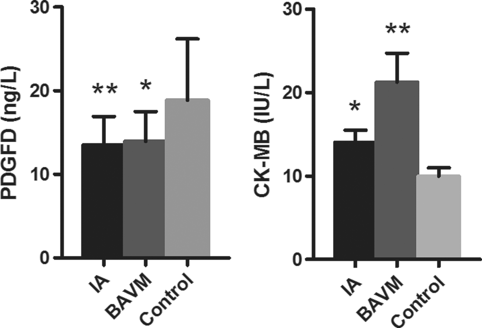
Comparison of PDGFD, CK-MB, and hs-CRP levels among the three groups. *p ≤ 0.017, **p ≤ 0.001. CK-MB, creatine kinase isoenzyme; hs-CRP, hypersensitive c-reactive protein; PDGFD, platelet-derived growth factor-D.
The CGI (GRCh/hg38, chr11: 104163809-104164330) region of the PDGFD is located in the gene promoter (Fig. 2). The percentages of DNA methylation found on the seven CpG sites on a 134-base pair (bp) polymerase chain reaction product were obtained using the bisulfite pyrosequencing assay. There was no significant correlation in DNA methylation levels among the seven CpGs (Fig. 2, r = 0.076–0.828). Therefore, we used the DNA methylation percentage of each CpG and the mean DNA methylation level to compare the differences between cases and controls, between male and female patients, and the relationship between PDGFD methylation and biochemical indicators.

Correlation among the seven CpGs in PDGFD gene promoter.
As shown in Figure 3, significant differences were observed between cerebrovascular and control groups in CpG1 methylation levels only. IA patients had significantly higher methylation levels (4.63 ± 0.35) than control subjects (3.36 ± 0.35, p = 0.017). BAVM patients (6.00 ± 0.86) had even higher levels than controls (3.36 ± 0.35, p = 0.003). There were no differences in DNA methylation levels between male and female patients in the case of IA, BAVM, and control subjects (Table 2, p = 0.926, 0.763, and 0.539, respectively). Both male and female patients with BAVM had higher CpG1 methylation levels than control subjects (Table 2, male patient: p = 2.81E-04, female patient: p = 0.017).
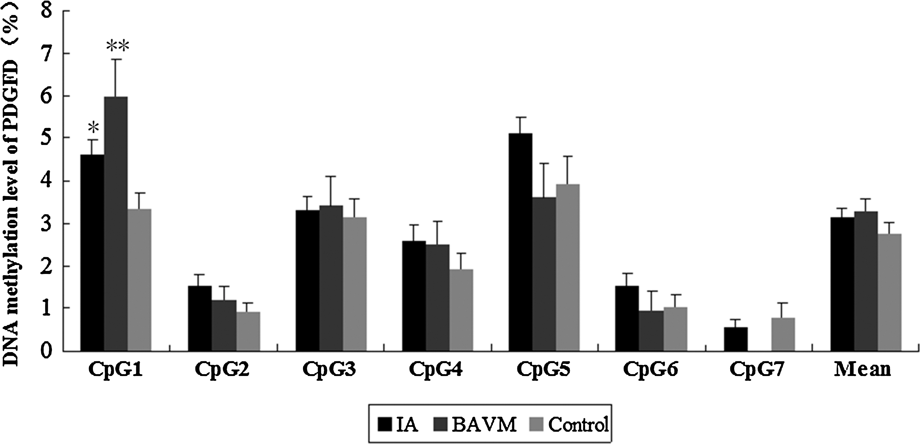
DNA methylation differences of seven CpG dinucleotides analyzed within the PDGFD. *p IA VS Control = 0.017, **p BAVM VS Control = 0.003.
Data presented are mean ± SE, p-value less than or equal to 0.017 is in bold.
Next, we explored the correlation between biochemical indicators and DNA methylation levels. Significant correlations were observed between APOE and CpG1 methylation levels in IA patients (Fig. 4a, p = 0.013). CK-MB was negatively correlated with CpG1 methylation levels in all individuals (Fig. 4b, p = 0.031), but no correlations were found between each group. Furthermore, HDL was negatively correlated with CpG1 methylation in IAs (Fig. 4c, p = 0.044).
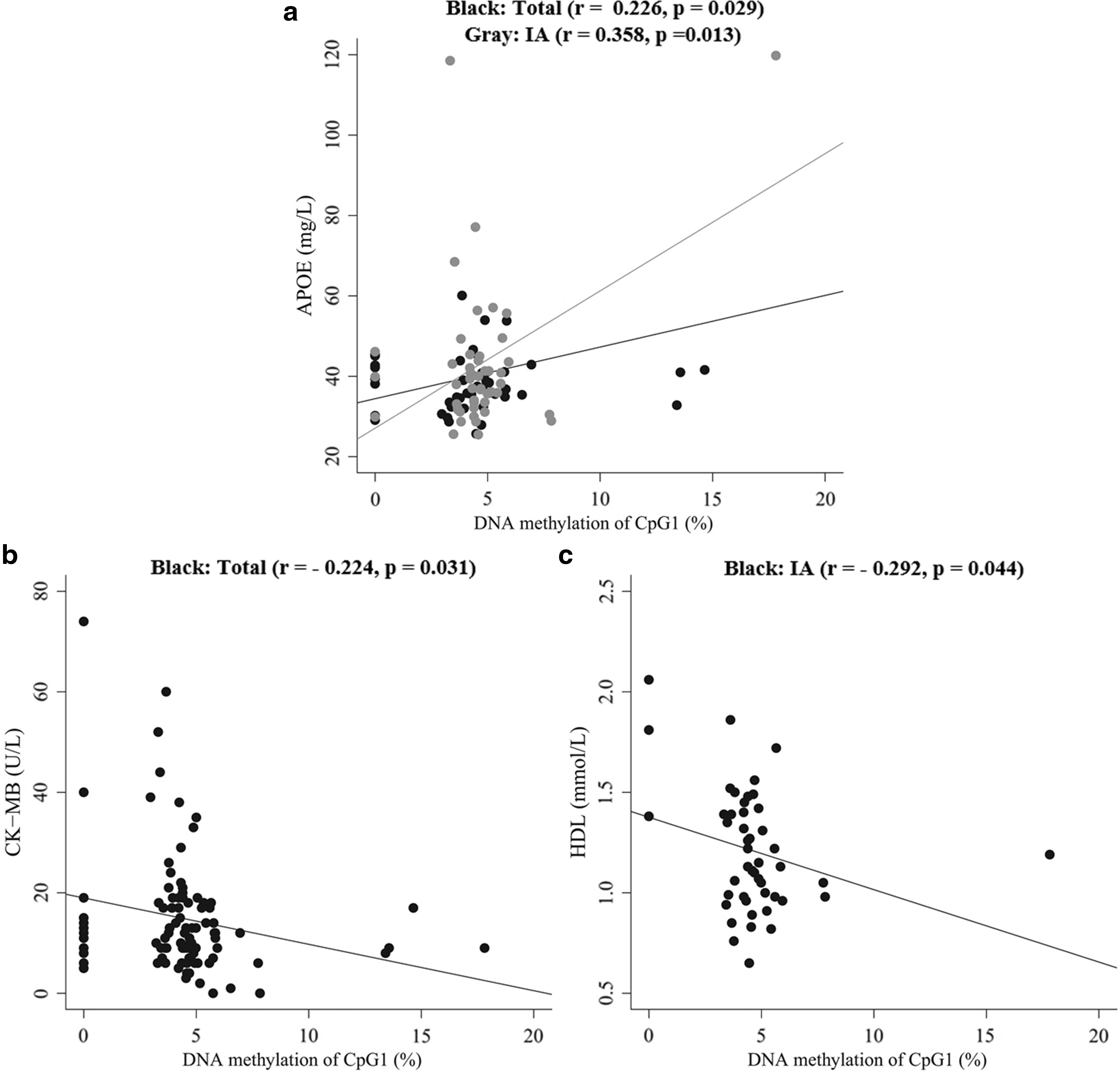
Pearson correlation between PDGFD gene DNA methylation and APOE, CK-MB, and HDL.
Discussion
In recent years, genetic research on cerebrovascular disease has focused on genes involved in vascular smooth muscle cell proliferation and differentiation, blood pressure regulation, and vascular inflammation (Bendjilali et al., 2014). Recent studies reported that genetic variations in angiogenesis and inflammation processes could play an important role in vascular development and differentiation and in arteriovenous specifications (Sturiale et al., 2013, 2014). Susceptible chromosomal loci for IAs are as follows: 1p34.3-p36.13, 7q11, 8q11.23-q12.1, 9p21, 19q13, and Xp22 (Ruigrok and Rinkel, 2008; Mohan et al., 2015); the associated genetic variants are located in genes such as CDKN2B-AS1, ELN, COL1A2, SOX17, and ADAMTS15 (Ruigrok and Rinkel, 2008; Alg et al., 2013; Yan et al., 2015). Two aneurysm-associated loci, SOX-17 and RBBP8, are also possibly associated with BAVMs (Alg et al., 2013; Kremer et al., 2015). Moreover, associated genes including IL6, MMP, and APOE were identified in both IAs and BAVMs (Fontanella et al., 2012; Huai et al., 2013; Sturiale et al., 2013). In the previous study, we had found that gender modulated the interaction between nitric oxide synthase 1 adaptor (NOS1AP) promoter DNA methylation in IA and BAVM patients. And the results also confirmed that regular tobacco smoking could increase NOS1AP methylation in humans (Wang et al., 2016). To our knowledge, this study is the first to report the correlation between DNA methylation of the PDGFD promoter and the risk of developing either BAVM or IA.
The disulfide-linked homodimer, PDGFD, is activated by proteolysis and becomes a specific agonist for PDGF-beta-receptor (Bergsten et al., 2001). This binding specificity suggested that the PDGFD could be important for the development and pathophysiology of several organs. The PDGFD gene is predominantly expressed in fibroblastic adventitial cells and is active in cultured endothelial cells (Uutela et al., 2001). PDGFD secreted by glioma cells is capable of promoting the migration of stem cells toward glioma cells (Gondi et al., 2010). A PDGFD variation was found to be significantly correlated with a higher risk of developing cardiovascular and cerebrovascular disease (Han et al., 2008; Zhou et al., 2012). Homocysteine is an independent risk factor for vascular diseases. Han et al.'s (2014) study suggested that aberrant DNA methylation of the PDGF gene in homocysteine could mediate vascular smooth muscle cell proliferation in atherosclerosis. The only available research on DNA methylation of the PDGFD gene promoter suggested a correlation with cumulative arsenic exposure in patients with urothelial carcinomas, results that were derived from genome-wide DNA methylation profiles (Yang et al., 2014). In this study, we found that the plasma PDGFD levels in IA and BAVM were significantly lower than those in the control group. Both IA and BAVM patients had significantly higher DNA methylation levels of the PDGFD gene promoter than the control group. CpG1 methylation of the PDGFD gene promoter was significantly correlated with the risk of developing BAVM, in both male and female patients. However, a similar correlation was only found in male IA patients. Therefore, there may be interplay between DNA methylation of the PDGFD gene promoter and gender in IA. This significant correlation could provide new information regarding epigenetic regulation of the PDGFD gene promoter in both BAVM and IA.
On analysis of clinical data, CK-MB, a biomarker for detecting myocardial injury (Ozdemir et al., 2011), caught our attention. Increased CK-MB levels often indicate acute myocardial infarction (Jaruvongvanich et al., 2015) and cerebrovascular disease (Manea et al., 2015). The cerebral trauma control group had higher CK-MB levels than IA and BAVM patients, which may have been caused by an acute craniocerebral trauma in the recent past. Analysis of clinical data and DNA methylation of the PDGFD gene promoter showed a negative correlation between CK-MB and CpG1 methylation levels in all participants. APOE is a major cholesterol carrier that plays an important role in maintaining lipid homeostasis in both the periphery and the brain (Zhong et al., 2016). HDL is a biomarker in various arterial diseases, including cardiovascular (Cho et al., 2016) and cerebrovascular events (Dong et al., 2015). APOE and HDL were recognized for their importance in lipoprotein metabolism and cerebrovascular disease. Recent studies demonstrated that the HDL particle profile is associated with DNA methylation of the ABCA1 gene promoter in coronary heart disease (Guay et al., 2012). The plasma APOE level was positively correlated with the gene promoter DNA methylation (Marioni et al., 2015). Summarizing, our results showed that CpG1 methylation of the PDGFD gene promoter has a significant correlation with APOE in IA patients. This observation can possibly be explained by the interaction between epigenetic and clinical data.
There are some limitations of our results that have to be examined with caution. First, we explored only a portion of the CGI; seven CpGs were selected for analysis and no correlation was found between them. Therefore, our findings involving CpG1 may not represent all the CGIs of the PDGFD promoter gene. Second, the small sample size is also a limitation. Our findings need to be validated in a larger number of samples and in other ethnic populations. Finally, we cannot exclude the possibility that variations in administered drugs and diets could affect our findings regarding the DNA methylation levels of the PDGFD gene promoter. Our results have not been adjusted by multiple factors, including hypertension, smoking habits, and diabetes, all of which can influence DNA methylation in subjects.
In conclusion, our study has proved that there are significant correlations between DNA methylation levels of the PDGFD gene promoter and the risk for developing IAs and BAVMs. In addition, CpG1 methylation of the PDGFD gene promoter shows a significant correlation with APOE in IAs. Further functional studies about these relationships and correlations are warranted.
Footnotes
Acknowledgments
This study was supported by grants from the Ningbo People of Science and Technology Projects (2015C50005), Ningbo Natural Science Foundation (2015A610232 and 2016A610154), Zhejiang Traditional Chinese Medicine Science and Technology Projects (2015ZB100), Ningbo Youth and Doctor Foundation, Ningbo High Level Innovative Talents Program, the National Key Research and Development Plan (2016YFC0106106), and Zhejiang Provincial Natural Science Foundation of China (LQ17H090002).
Disclosure Statement
The authors declare no conflict of interest.