
Abstract
Glioblastoma multiforme is a type of central nervous system tumor with extremely poor prognosis. Previously, hydrogen peroxide (H2O2), which promotes the oxidative stress response, has been reported to induce the apoptosis of glioma cells. Recently, secreted frizzled-related protein 1 (SFRP1) has been shown to be associated with various types of malignant tumors and with H2O2-induced oxidative stress in cardiomyocytes by negatively regulating the Wnt signaling pathway. This study aimed to explore SFRP1 expression and its roles in H2O2-induced apoptosis in human glioma cells. We found that the SFRP1 level was decreased in several human glioma cell lines, including U87, U251, and SW1783 cells. In U251 cells, SFRP1 could function as a cancer suppressor gene, and the growth of U251 cells could be inhibited not only by H2O2 but also by the overexpression of SFRP1. Furthermore, we demonstrated that H2O2-induced SFRP1 gene demethylation partially contributed to H2O2-induced U251 cell apoptosis, which was verified by studies using an SFRP inhibitor (WAY-316606). Our research identified that H2O2-induced SFRP1 gene demethylation contributes to H2O2-induced apoptosis in human U251 glioma cells.
Introduction
G
The canonical Wnt/β-catenin pathway plays a crucial role in modulating multiple cellular processes, such as cell survival, proliferation, differentiation, and oncogenesis (Clevers and Nusse, 2012). Secreted frizzled-related protein 1 (SFRP1), mapped to chromosome 8p12-p11.1, has become a research focus in recent years, as it can negatively regulate Wnt signaling (Jones and Jomary, 2002; Garcia-Hoyos et al., 2004). SFRP1 possesses a cysteine-rich domain that binds and then sequesters Wnts away from active receptor complexes (Jones and Jomary, 2002; Garcia-Hoyos et al., 2004). Besides, SFRP1 can antagonize Wnt signaling by forming a Wnt signaling inhibitory complex with frizzled receptors (Wu et al., 2013). In recent years, downregulation or inactivation of SFRP1 has been reported in various types of malignant tumors, including prostate, gastric, breast, ovarian, and bladder cancers (Kang et al., 2014). Using a Kaplan–Meier analysis, one study revealed that GBM patients with positive SFRP1 expression had a significantly longer overall survival time relative to those with negative SFRP1 expression (Chang et al., 2016). Hence, SFRP1 may possess significant prognostic value for GBM patients. However, the exact role of SFRP1 in GBM is still unclear.
Reactive oxygen species (ROS) are implicated in the pathogenesis of numerous diseases such as neurodegenerative disorders (Lee et al., 2001), brain tumors (Chen et al., 2006; Hsieh et al., 2010), and heart failure (Hafstad et al., 2013). Accumulation of ROS such as hydrogen peroxide (H2O2) can lead to oxidative stress, which can induce various defense mechanisms or cell apoptosis (Zhang et al., 2009). SFRP1 has been found to confer protective effects on cardiomyocytes during oxidative stress (Tao et al., 2015, 2016). For instance, Tao et al. (2015) demonstrated that the SFRP1 gene protects cardiomyocytes from H2O2-induced oxidative stress through restraining the Wnt/frizzled pathway. Previous work has also shown that H2O2 can cause cell apoptosis in glioma cells (Lee et al., 2001; Zhang et al., 2009). However, whether SFRP1 also confers protective effects on H2O2-treated glioma cells remains undetermined.
The purpose of this study was to explore SFRP1 expression and its role in H2O2-induced apoptosis in human glioma cells. We discovered that SFRP1 expression is decreased in glioma cells and functions as a cancer suppressor gene in U251 cells. H2O2-induced U251 cell apoptosis is also enhanced in cells overexpressing SFRP1. We further discovered that H2O2-induced SFRP1 gene demethylation led to the accumulation of SFRP1 in U251 cells, which partially contributed to H2O2-induced U251 cell apoptosis. Our study identified that H2O2-induced SFRP1 demethylation contributes to H2O2-induced apoptosis in human U251 glioma cells.
Materials and Methods
Cell culture
Primary human astrocytes (HA1800) were purchased from ScienCell. Human GBM cell lines (U87, U251, and SW1783) were purchased from American Type Culture Collection (ATCC). All cell lines were cultured in Dulbecco's modified Eagle medium (Invitrogen) supplemented with 10% fetal bovine serum (Sigma-Aldrich), 100 U/mL penicillin (Sigma), and 100 μg/mL streptomycin (Sigma) (Wang et al., 2015; Liu et al., 2016). Cells were maintained at 37°C with 5% CO2 in a humidified atmosphere.
Reverse transcription–polymerase chain reaction
Total RNA was extracted from HA1800, U87, U251, or SW1783 cells using TRIzol reagent (Invitrogen). After quantitation, total RNA (4 μg) from each sample was reverse transcribed and amplified with the Super-Script One-Step Reverse transcription–polymerase chain reaction (RT-PCR) system (Invitrogen). Real-time PCRs were carried out on the Applied Biosystems Prism 7500 Fast Sequence Detection System (Applied Biosystems). PCR parameters were as follows: 95°C for 5 min, followed by 35 cycles of 95°C for 30 s, 56°C for 30 s, and 72°C for 45 s. The following gene-specific primers (synthesized by Shanghai Sangon Biological Engineering and Technology Service, China) were used: forward: 5′-GGC CCA TCT ACC CGT GTC G-3′ and reverse: 5′-GAT GGC CTC AGA TTT CAA CTC GT-3′ for SFRP1; forward: 5′-CTC CTT AAT GTC ACG CAG GAT TTC-3′ and reverse: 5′-GTG GGG CGC CCC AGG CAC CA-3′ for β-actin. The relative SFRP1 mRNA levels were calculated using the 2−ΔΔCt method. Data were normalized to β-actin.
Western blot assay
Cells were lysed with RIPA buffer (Radio-Immunoprecipitation Assay, Sigma). The protein concentration was determined using the BCA assay (Bicinchoninic Acid, Sigma). For each sample, 40 μg of proteins was separated by sodium dodecyl sulfate-polyacrylamide gel electrophoresis and transferred onto polyvinylidene fluoride membranes (Sigma). The membranes were blocked with 0.5% skim milk powder (Sigma) and incubated with the primary antibodies of target proteins overnight at 4°C. After washing, membranes were incubated with horseradish peroxidase (HRP)-conjugated secondary antibodies at room temperature for 1.5 h. The reactive bands were observed by enhanced chemiluminescence (Thermo-Pierce) according to the manufacturer's protocols. The bands were analyzed by using Image-Pro 6.0 software and normalized to β-actin. The following antibodies (purchased from Abcam) were used: monoclonal rabbit antihuman SFRP1 antibody (Cat. #ab126613; dilution: 1:2500), polyclonal rabbit antihuman active-caspase 3 antibody (Cat. #ab2302; dilution: 1:1500), monoclonal rabbit antihuman Bcl-2 antibody (Cat. #ab32124; dilution: 1:2500), polyclonal rabbit antihuman β-actin antibody (Cat. #ab8227; dilution: 1:2000), and HRP-goat antirabbit immunoglobulin G second antibody (Cat. #ab6721; dilution: 1: 4000).
Construction of SFRP1 overexpressing vector
For SFRP1 overexpression, the pcDNA3.1/V5-HisA-SFRP1 (pSFRP1-V5) vector was constructed as described previously (Fukui et al., 2005). The full-length SFRP1 cDNA was subcloned using pryobest DNA polymerase (TaKaRa Biotechnology Co. Ltd.) and inserted into a pcDNA3.1/V5-HisA vector (Invitrogen). The full length SPFR1-containing vector (1 μg/well) was transfected into U251 cells (96-well plates, 2 × 10⁴ cells/well) using Lipofectamine 2000 (Invitrogen) to produce the “pSFRP1-V5 vector group.” Cells transfected with empty pcDNA3.1/V5-HisA vector were labeled the “empty vector group” and cells without any transfection were used as a “control group.” After transfection for 48 h, SFRP1 levels were verified by RT-PCR and Western blot.
Cell viability assays
The viability of U251 cells was determined using the MTT assay. In brief, cells (2 × 10⁴ cells/well) were cultured in a 96-well plate in 200 μL complete medium to allow confluence. Cells were then treated with or without 1 mM H2O2 (Sigma) for 12 h (Zhang et al., 2009). After treatment, cell viability was examined in the cultures at 24, 48, 72, and 96 h. At each time point, MTT reagent (10 μL, 5 mg/mL in phosphate-buffered saline; Sigma) was added to the corresponding wells and incubated for 4 h. The microtiter plate was centrifuged at 300 g for 5 min at 4°C. After removing the MTT solution, the formazan crystals were dissolved in 150 μL dimethylsulfoxide (Sigma) and the absorbance was recorded at 570 nm using a spectrophotometer (Bio-Rad Laboratories). When WAY-316606 [an inhibitor of SFRP1 (Moore et al., 2008; Komatsu and Warden, 2010; Ohishi et al., 2014); Sigma] was introduced, cell viability was retested at 72 h post-treatment by parallel MTT assays. Before MTT assay, cells containing empty vector were treated with or without 2.0 μM WAY-316606 for 12 h. And also, cells containing empty vector were allowed to receive 1 mM H2O2 treatment or 1 mM H2O2 + 2.0 μM WAY-316606 treatment for 12 h.
Apoptosis assays
After cells were treated with or without 1 mM H2O2 for 12 h, cell apoptosis was analyzed at 72 h post-treatment by flow cytometry. The Annexin V-FITC Apoptosis Detection Kit (Sigma) was used to stain the cells. In brief, cells were washed with Dulbecco's phosphate buffered saline (DPBS) and resuspended in 1 × binding buffer at a final concentration of 1 × 10⁶ cells per ml. Then, 5 μL Annexin V-fluorescein isothiocyanate conjugate and 10 μL propidium iodide solution were added to 500 μL of cell suspension in a test tube. The tubes were incubated at room temperature for 10 min and kept in the dark. Cells were then analyzed using an FACS analyzer (BD Biosciences). When WAY-316606 was introduced, cell apoptosis was retested at 72 h post-treatment.
Methylation-specific polymerase chain reaction
SFRP1 promoter methylation was assessed with methylation-specific polymerase chain reaction (MS-PCR) (Shih et al., 2006; Majchrzak-Celińska et al., 2016). DNA was isolated from U251 cells and subjected to bisulfite methylation modification by applying the EZ DNA Methylation Kit (Zymo Research) as per the manufacturer's protocols. The MS-PCR was performed in a MyCycler Thermal Cycler with Gradient (Bio-Rad), using Gold Taq DNA polymerase (PE Applied Biosystems). The SFRP1 primer sequences used were 5′-TGT AGT TTT CGG AGT TAG TGT CGC GC-3′ (forward) and 5′-CCT ACG ATC GAA AAC GAC GCG AAC G-3′ (reverse) for methylated DNA, and 5′-GTT TTG TAG TTT TTG GAG TTA GTG TTG TGT-3′ (forward) and 5′-CTC AAC CTA CAA TCA AAA ACA ACA CAA ACA-3′ (reverse) for unmethylated DNA. The reaction parameters were as follows: polymerase activation at 92°C for 10 min, 35 cycles of 92°C for 30 s, annealing at 60°C for 30 s, and extension at 72°C for 30 s, then final elongation at 72°C for 10 min and hold at 4°C. The products of MS-PCR were separated on a 3% agarose gel and then stained with Simply Safe reagent (EURx). The bands were observed under UV light.
Statistical analysis
All experiments were performed in triplicate in three independent experiments. Statistical analyses were performed using the SPSS 16.0 software (SPSS). Data were expressed as mean ± standard deviation and analyzed using a Student's t-test. The symbols *p < 0.05 or **p < 0.01 represent a statistically significant difference.
Results
SFRP1 is markedly decreased in human glioma cells
To explore the expression of SFRP1 in GBM, normal human astrocytes (HA1800) and glioma cells (U87, U251, and SW1783) were prepared as described in “Materials and Methods”. Through RT-PCR, we discovered that the relative SFRP1 mRNA levels in U87 (*p < 0.05), U251 (**p < 0.01), and SW1783 (*p < 0.05) cells were markedly lower than those in HA1800 cells (Fig. 1A). Similar results were observed at the protein level (Fig. 1B). These results indicate that the level of SFRP1 is decreased in glioma cells, hinting that alterations in SFRP1 expression may be associated with the growth of GBM. Based on these results, U251 cells were chosen for use in the subsequent experiments. To explore the roles of SFRP1 in GBM, SFRP1 was overexpressed in U251 cells (Fig. 1C, D).
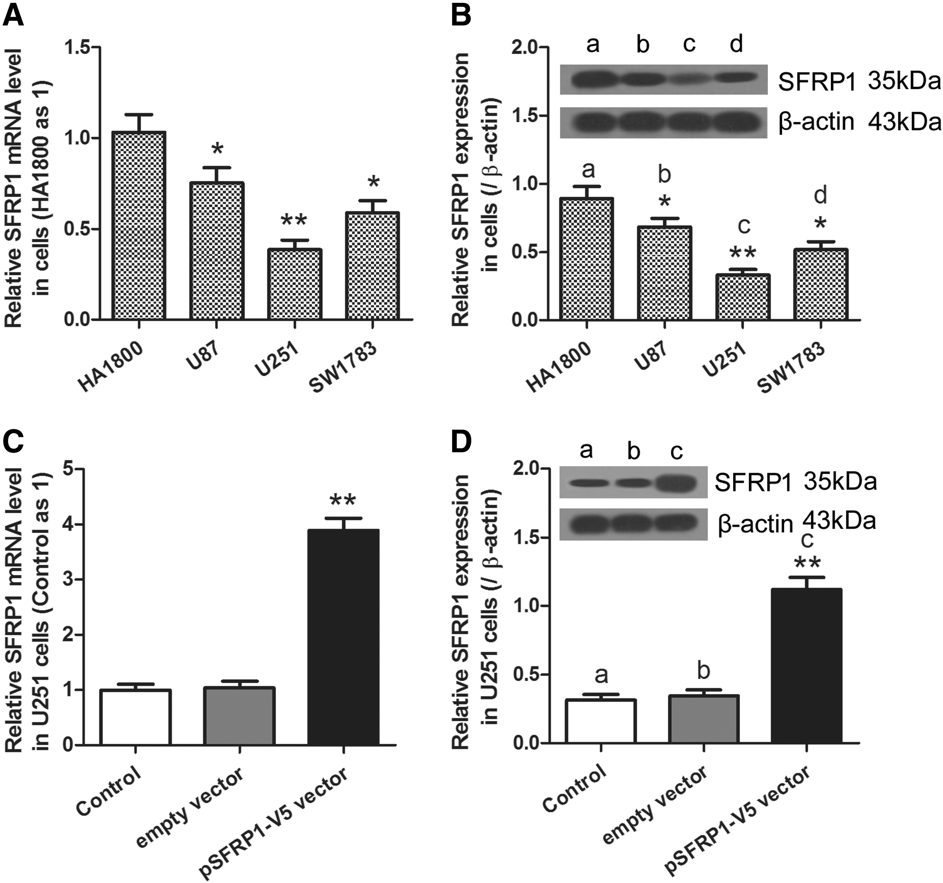
Expression of SFRP1 in normal human astrocytes and human glioma cells. Cultured cells were subjected to RT-PCR and Western blot assays.
H2O2-induced U251 cell apoptosis is enhanced in SFRP1 overexpressing cells
The human U251 cell line was chosen to explore the role of SFRP1 in H2O2-induced cell apoptosis. Cultured U251 cells (pSFRP1-V5 vector and empty vector groups) were treated with or without H2O2 as described in “Materials and Methods”. Cell viability, apoptosis, and apoptosis-related protein levels were then examined. When compared with cell viability in H2O2-untreated cells containing the empty vector, we observed that cell viability was significantly decreased in H2O2-untreated cells containing the pSFRP1-V5 vector or in H2O2-treated cells containing the empty vector in a time-dependent manner. This decrease was sharply enhanced in H2O2-treated cells containing pSFRP1-V5 vector (Fig. 2A).

U251 cell viability, apoptosis, and apoptosis-related protein levels.
In contrast, when compared with cell apoptosis in H2O2-untreated cells containing the empty vector, cell apoptosis was dramatically increased in H2O2-untreated cells containing the pSFRP1-V5 vector or in H2O2-treated cells containing the empty vector. This increase was sharply enhanced in H2O2-treated cells containing pSFRP1-V5 vector (Fig. 2B, C). In addition, similar results for active-caspase 3 expression and opposite results for Bcl-2 expression were observed (Fig. 2D). These results suggest that both H2O2 and SFRP1 overexpression contribute to the decline in cell viability and enhanced apoptosis in U251 cells and that H2O2-induced U251 cell apoptosis can be further aggravated in SFRP1-overexpressing cells.
H2O2-induced SFRP1 gene demethylation contributes to the accumulation of SFRP1 in U251 cells
It has been reported that SFRP1 methylation predicts shorter survival of glioma patients (Majchrzak-Celińska et al., 2016). Oxidative stress has also been shown to induce DNA demethylation (Gu et al., 2013; Wu and Ni, 2015). Hence, we suspected that the methylation status of SFRP1 may be altered by H2O2. Through RT-PCR assays, we first discovered that mRNA levels of SFRP1 in cells containing the empty vector or cells containing the pSFRP1-V5 vector were significantly increased after treatment with H2O2 (Fig. 3A). MS-PCR was then performed to determine the methylation status of the SFRP1 gene. As shown in Figure 3B, in cells in the empty vector group, we found that the level of unmethylated SFRP1 gene was markedly increased after H2O2 treatment. Similar results were observed in cells containing the pSFRP1-V5 vector (Fig. 3B). These results suggest that H2O2 can induce SFRP1 gene demethylation in U251 cells, which contributes to the accumulation of SFRP1.
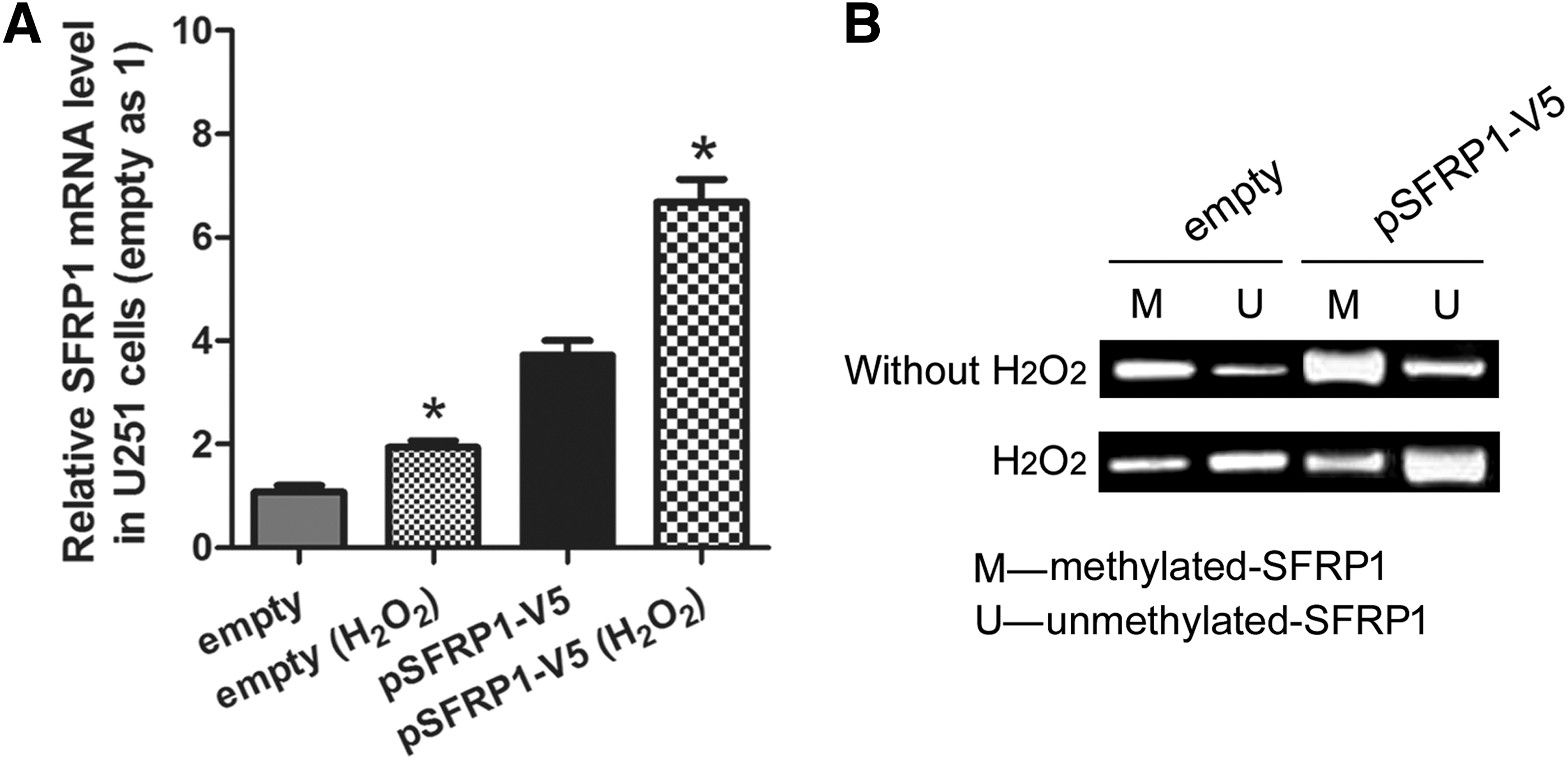
mRNA levels of methylated or unmethylated SFRP1 gene in U251 cells. Cultured U251 cells were treated with or without H2O2 and then subjected to RT-PCR or methylation-specific polymerase chain reaction assays.
H2O2-induced U251 cell growth inhibition can be partially attenuated by WAY-316606
As overexpressing SFRP1 contributes to U251 cell apoptosis, we inferred that H2O2-induced accumulation of SFRP1 partially contributes to H2O2-induced U251 cell apoptosis. To verify this inference, WAY-316606, an inhibitor of SFRP1, was introduced as described in “Materials and Methods”. The results of the MTT assay (Fig. 4A) demonstrated that WAY-316606 treatment resulted in a slight, nonsignificant increase in cell viability in the H2O2-untreated empty group, but led to a prominent increase in cell viability in the H2O2-treated empty group (*p < 0.05). Flow cytometry analysis (Fig. 4B) showed that WAY-316606 treatment resulted in a slight, nonsignificant decrease in cell apoptosis in the H2O2-untreated empty group, but led to a significant decrease in cell apoptosis in the H2O2-treated empty group (*p < 0.05). These results suggest that H2O2-induced U251 cell growth inhibition can be partially attenuated by WAY-316606, which can be explained by the demethylation effect of H2O2 on the SFRP1 gene in U251 cells and the inhibition effect of SFRP1 on U251 cell growth. Hence, we concluded that H2O2-induced accumulation of SFRP1 partially contributes to H2O2-induced U251 cell apoptosis.
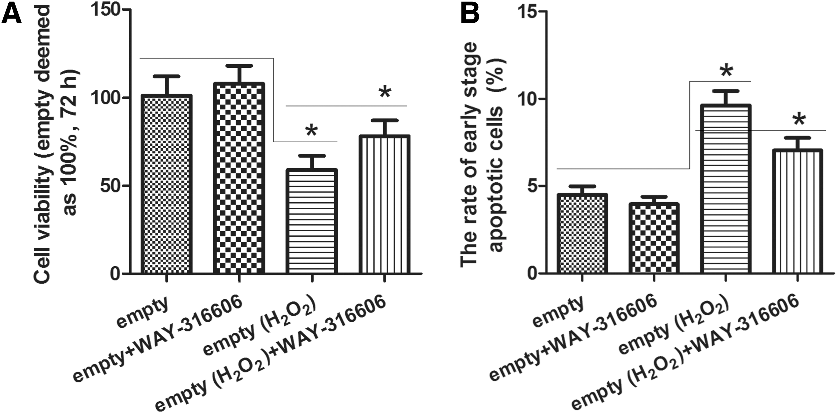
Detection of U251 cell viability and apoptosis in empty group.
Discussion
SFRPs are a family of secreted proteins that can modulate the Wnt signaling cascades (Esteve and Bovolenta, 2010). In this study, the SFRP1 level was found to decline in human glioma cells as compared with that in normal human astrocytes, which is consistent with its expression in other cancers, including those of the stomach, kidney, small intestine, parathyroid, pancreas, adrenal gland, endometrium, gall bladder, and testis (Dahl et al., 2007). In addition, Chang et al. (2016) reported that SFRP1 was expressed in 77.5% (31 of 40 samples) of normal brain tissues and just 33.63% (38 of 113 samples) of GBM tissue samples; they pointed out that the downregulation of SFRP1 protein may be important features of GBM. SFRP1 has been recognized as a candidate tumor suppressor in multiple human malignancies, including colorectal cancer (Caldwell et al., 2004), breast cancer (Klopocki et al., 2004), and hepatocellular carcinoma (Shih et al., 2007). As predicted, in our study, SFRP1 also functions as a cancer suppressor molecule in human glioma, which is a supplement and extension of our knowledge to its role in human cancers.
Previously, studies have demonstrated that H2O2 treatment can lead to the apoptosis of glioma cells (Lee et al., 2001). As SFRP1 has been reported to play a protective effect in H2O2-induced cardiomyocyte apoptosis (Tao et al., 2015), we continued to focus on the role of SFRP1 in H2O2-induced glioma cell apoptosis. Through a series of experiments, we discovered that overexpression of SFRP1 can aggravate H2O2-induced growth inhibition in U251 cells, which is different from its role in H2O2-induced cardiomyocytes apoptosis. This difference may be associated with the microenvironment of cancer cells and needs to be investigated in future studies. Besides, Delic et al. (2013) reported on a novel molecular miR-328-dependent mechanism that through SFRP1 inhibition and Wnt activation contributes to the infiltrative glioma phenotype at already early stages of glioma progression, which also hints the cancer suppressor roles of SFRP1 in glioma. Meanwhile, in glioma stem cells (GSCs), expressional and functional analysis of SFRP1-treated GSCs revealed that SFRP1 halts cell cycling and induces apoptosis (Kierulf-Vieira et al., 2016).
The frequent loss of SFRP1 expression in multiple human solid tumors, such as hepatocellular carcinoma (Shih et al., 2006), breast cancer (Veeck et al., 2006), and nonsmall-cell lung cancer (Fukui et al., 2005), is associated with aberrant methylation of the SFRP1 gene promoter (Dahl et al., 2007). Majchrzak-Celińska et al. (2016) also demonstrated that many genes that inhibit the Wnt pathway, including SFRP1, SFRP2, SOX17, and PPP2R2B, are methylated in human gliomas and SFRP1 methylation predicts shorter survival. As H2O2-induced oxidative stress has been shown to lead to DNA demethylation (Gu et al., 2013; Wu and Ni, 2015), we examined whether the methylation status of SFRP1 could be influenced by H2O2 in U251 cells. Through MS-PCR, we observed that H2O2 induced SFRP1 gene demethylation in U251 cells, which led to the accumulation of SFRP1. After introducing the SFRP1 inhibitor (WAY-316606), we demonstrated that H2O2-induced accumulation of SFRP1 partially contributes to the decline in cell viability and increase in apoptosis induced by H2O2 treatment in these cells.
In conclusion, this study was the first to demonstrate the downregulation of SFRP1 in glioma cell lines. The discovery of SFRP1's anticancer effects on glioma cells furthers our understanding of the important role this gene plays in human cancers. Furthermore, we demonstrated that both H2O2 and SFRP1 overexpression contribute to the decline in viability increase in apoptosis of U251 cells. In addition, H2O2-induced SFRP1 gene demethylation partially contributes to H2O2-induced apoptosis in human U251 glioma cells. Since that hypoxia also leads to the oxidative stress response as with H2O2, our findings hint that the appropriate regional hypoxia of GBM tissues may be an effective strategy to control the progress of GBM, which needs to be investigated in our future researches.
Footnotes
Disclosure Statement
No competing financial interests exist.