
Abstract
Oral squamous cell carcinoma (OSCC) is characterized by rapid local migration and invasion. This study was aimed at clarifying the effect of miR-654-5p on progression of OSCC. miR-654-5p promoted proliferation, metastasis, and chemoresistance of OSCC in vitro and in vivo. Consistently, miR-654-5p was upregulated in late-stage OSCC and was correlated with poor prognosis of OSCC patients. Furthermore, miR-654-5p was mechanistically verified to target Grb-2-related adaptor protein (GRAP), accompanied by the activation of Ras/MAPK signaling and the facilitation of epithelial-mesenchymal transition in OSCC cells. GRAP was downregulated in T1–2 stage versus T3–4 stage head and neck squamous cell carcinoma (HNSC) and was negatively correlated with tumor-node-metastases (TNM) stage in HNSC patients based on The Cancer Genome Atlas (TCGA) analysis. In addition, GRAP was positively correlated with good prognosis in HNSC patients. Our findings suggest that the miR-654-5p/GRAP/Ras/Erk signaling pathway in OSCC cells might contribute to the underlying mechanism through which miR-654-5p participates in the regulation of OSCC progression. miR-654-5p, as a potential biomarker for the clinical diagnosis and prognosis of OSCC, may be an effective anticancer target for the treatment of OSCC.
Introduction
A
microRNAs (miRNAs or miRs) play important roles in oncogenesis and cancer progression (Nana-Sinkam and Croce, 2014; Rupaimoole et al., 2016; Karatas et al., 2017). Furthermore, miRNAs regulate various biological functions, including development and differentiation, proliferation, apoptosis, metastasis, metabolism, and immune response, and are also involved in the development of chemoresistance in cancers (Gorenchtein et al., 2012; Ayers and Vandesompele, 2017; Caliskan et al., 2017). Therefore, therapies that target miRNAs may aid in overcoming chemoresistance in OSCC.
MAPKs, also known as extracellular signal-regulated kinases (ERKs), are involved in multiple cellular processes, such as proliferation, transcription regulation, differentiation, and development. The specific components of the MAPK signaling cascade vary greatly among different stimuli, but the pathway usually includes adaptors (e.g., Shc, GRB2, Crk, and so on), a guanine nucleotide exchange factor (e.g., SOS, C3G, and so on), and small GTP-binding proteins (e.g., Ras, Rap1), which activate the core unit of the cascade composed of a MAPKKK (Raf), a MAPKK (MEK1/2), and MAPK (Erk) (Keyse, 2008; De Luca et al., 2012; Roskoski, 2012). The activation of the MAPK signaling pathway could promote cancer cell proliferation, metastasis, and resistance to chemotherapy (Si et al., 2017; Tao et al., 2017).
In the present study, we constructed cisplatin-resistant Tca-8113 and CAL-27 cell lines (named Tca-8113/CDDP and CAL-27/CDDP) and performed microarray analysis in CAL-27/CDDP cells and wild-type CAL-27 cells, which identified miRNA-654-5p as a potential onco-miRNA in the regulation of chemoresistance in OSCC. The specific effect of miR-654-5p on OSCC is yet to be uncovered. Therefore, we examined the function of miR-654-5p in OSCC and the molecular mechanisms underlying its tumor-promoting ability. We showed that miR-654-5p was positively correlated with poor patient prognosis, as well as enhanced proliferation, metastasis, and chemoresistance of OSCC. Mechanistically, miR-654-5p targeted Grb-2-related adaptor protein (GRAP) to activate Ras/MAPK signaling and facilitate epithelial–mesenchymal transition (EMT), positively impacting cancer progression.
Materials and Methods
Cell culture
Human Tca-8113 and CAL-27 OSCC cell lines were purchased from the Shanghai Cell Bank of the Chinese Academy of Sciences and were cultured in DMEM supplemented with 10% (v/v) FBS in a humidified atmosphere of 5% CO2 at 37°C.
Lentivirus infection
Lentiviral particles encoding shRNA targeting miR-654-5p (sh-miR-654-5p) and their control sequences (NC) were constructed by Neuron Biotech Co., Ltd., Shanghai, China. Lentivirus was then packed, amplified, and transfected into cells using Polybrene reagent (Sigma-Aldrich, St. Louis, MO).
MTT assay
Cell proliferation and drug sensitivity tests were investigated by MTT assay. The exponentially growing cells were dissociated and seeded into 96-well plates at a density of 8 × 10³ cells/well for cell proliferation test and 1.5 × 10⁴ cells/well for drug sensitivity test and incubated overnight to allow for cell adherence. Cell viability was measured using 3-(4,5-dimethylthiazol-2-yl)-2,5-diphenyl tetrazolium bromide (MTT) as previously described (Lin et al., 2015).
EdU incorporation assay
EdU incorporation was performed using Apollo567 Imaging Kit (RiboBio Co., Ltd, Guangzhou, China) according to the manufacturer's protocol. Briefly, cells were incubated with 5-ethynyl-2′-deoxyuridine (EdU) and were fixed with paraformaldehyde. After permeabilization, cells were stained with Apollo fluorescent dyes and 4′,6-diamidino-2-phenylindole (DAPI). The EdU-positive cells were then counted under a microscope.
Migration and invasion assays
The migration and invasion assays were conducted using a 24-well 8.0 μm Transwell chamber (Corning Incorporated, Corning, NY). 1 × 10⁵ cells in 200 μL serum-free medium were seeded in the upper chamber, and 600 μL of 10% FBS culture medium were added to the lower chamber. Twenty-four hours later, the migrated and invaded cells were fixed with methanol, stained with crystal violet, and photographed under a microscope.
Wound healing assay
Cells were seeded on 6-well plates and cultured as a confluent monolayer. Scratches were produced using a pipette tip. Process of migration was recorded at initiation time and at 24 h after wounding.
In vivo tumor xenograft study
All the animal experiments were conducted under the protocols approved by the Institutional Animal Ethics Committee, Experimental Animal Center of Fujian Medical University, China. Male BALB/c nude mice at 4–5 weeks old were purchased from SLAC Laboratory Animal Co. Ltd., Shanghai, China. Cells were subcutaneously or intravenously inoculated into the mice at a density of 1 × 10⁶ cells/mouse. For the subcutaneous xenograft model, tumor size was measured to evaluate tumor growth. For the pulmonary metastasis model, the mice were sacrificed 4 weeks after initial injection of tumor cells, and metastatic lung nodules were determined by Hematoxylin–Eosin (HE) staining using a dissecting microscope.
Microarray analysis
Total miRNA was extracted using a mirVana™ miRNA Isolation Kit and labeled using a labeling kit (Ambion, Austin, TX). The obtained samples were then hybridized on an Agilent SurePrint G3 Human Gene Expression Microarray chip and were subjected to scanning with an Agilent Scanner G2505C (Agilent Technologies).
Real-time fluorescence quantitative PCR
Total RNA was isolated from cells or harvested tissues, and cDNA synthesis was conducted using reverse transcription reagents (Thermo Scientific, Waltham, MA). According to the manufacturer's instructions, real-time quantitative PCR (qPCR) was performed using a SYBR Premix Ex Taq™ II Kit (TaKaRa Bio, Inc., Shiga, Japan) and detected on a BIO-RAD CFX 96 detection system.
Western blot analysis
Cell lysates were prepared in lysis buffer, and the protein concentration was determined using a (bicinchoninic acid) BCA Protein Assay Kit (Thermo Scientific, Waltham, MA). Proteins were separated by 10% sodium dodecyl sulfate polyacrylamide gel electrophoresis (SDS-PAGE) and transferred onto polyvinyl difluoride membranes. The membranes were immunoprobed with the corresponding antibodies overnight at 4°C and were then incubated with the secondary antibody for 1 h. The proteins were detected by an enhanced chemiluminescence reagent (Thermo Scientific, Waltham, MA).
Bioinformatics analysis
microRNA and mRNA expression profiles of head and neck squamous cell carcinoma (HNSC) were analyzed based on The Cancer Genome Atlas (TCGA) database. The latest miRNA-Seq and mRNA-Seq data of HNSC were collected and used for differential expression and survival analysis.
Patient tissues
One hundred fifty-seven primary fresh OSCC tissues were collected from the Union Hospital of Fujian Medical University, Fuzhou, China and were stored in liquid nitrogen. Approval from the Ethics Committee of the Union Hospital of Fujian Medical University was obtained before the use of these clinical specimens for research. All samples with confirmed pathological diagnosis were staged according to the pathology tumor-node-metastasis system (AJCC).
Luciferase reporter assay
A fragment of the GRAP 3′-UTR was amplified and cloned into the psiCHECK-2 vectors (named WT). Site-directed mutations were generated in the miR-654-5p binding site of the GRAP 3′-UTR using the Site-Directed Mutagenesis Kit (Invitrogen, Carlsbad, CA). For reporter assays, the control psiCHECK-2 vectors, WT or Mut vectors, were cotransfected into CAL-27 cells with miR-654-5p mimics or inhibitor. Luciferase activity was detected at 48 h post-transfection using the classical Dual-Luciferase Reporter Assay System (Promega Corporation, Madison, WI).
Active Ras detection
The active Ras was detected using an Active Ras Detection Kit (Cell Signaling Technology, Beverley, MA) according to the manufacturer's instructions. Briefly, total proteins were extracted from cells, and the concentration was determined by a BCA Protein Assay Kit (Thermo Scientific, Waltham, MA). A total of 5 mg protein was incubated with glutathione resin and GST-Raf1-RBD. After elution, the proteins were subjected to western blot analysis.
Statistical analysis
All data were analyzed with SPSS 21.0 software. The data are expressed as the mean ± SD from at least three independent experiments. Statistical significance was determined by Student's two-tailed t-test for two groups and one-way ANOVA for multiple groups. The skewed data were analyzed using the Wilcoxon rank sum test. Correlations between gene expression and clinicopathological features were analyzed by chi-square test. Kaplan–Meier survival curves were used for survival analysis. All statistical tests were two sided, and a p value of <0.05 was considered statistically significant. *p < 0.05, **p < 0.01, and ***p < 0.001.
Results
miRNA-654-5p expression negatively correlates with survival of OSCC patients
To clearly determine the role of miRNAs in the regulation of chemoresistance in OSCC, we constructed cisplatin-resistant Tca-8113 and CAL-27 cell lines (named Tca-8113/CDDP and CAL-27/CDDP). Then, microarray analysis was performed in CAL-27/CDDP and wild-type CAL-27 cells to identify the differentially expressed miRNAs. Based on the obtained results (Table 1), miRNA-654-5p was selected as a potential onco-miRNA. miRNA-654-5p expression was confirmed to be upregulated in Tca-8113/CDDP and CAL-27/CDDP cells compared with control cells (Fig. 1A). Survival and differential expression analysis, based on the TCGA head and neck squamous cell carcinoma (HNSC) data set, identified miR-654 as an onco-miRNA candidate that was correlated with poor prognosis in HNSC patients (log-rank test, p = 0.0087, Fig. 1B), being upregulated in HNSC with lymphonode metastasis (N1-3) versus HNSC without lymphonode metastasis (N0) (p = 0.0473) (Fig. 1C). To validate the results obtained via bioinformatics analysis, miR-654-5p and miR-654-3p expression levels in 157 clinical samples (97 early-stage and 60 late-stage OSCC) were analyzed by qPCR. The clinical characteristics of the OSCC patients are summarized in Table 2. miR-654-5p, but not miR-654-3p (data not shown), was positively correlated with tumor-node-metastases (TNM) stage in OSCC patients (p < 0.001), and patients with low miR-654-5p expression had longer survival times compared to patients with high miR-654-5p expression, which was consistent with the results of the TCGA analysis (log-rank test, p = 0.0205, Fig. 1D). Furthermore, the gene set enrichment analysis (GSEA), based on the TCGA HNSC data set, indicated that miR-654-5p was involved in the regulation of proliferation (Fig. 1E) and metastasis (Fig. 1F).
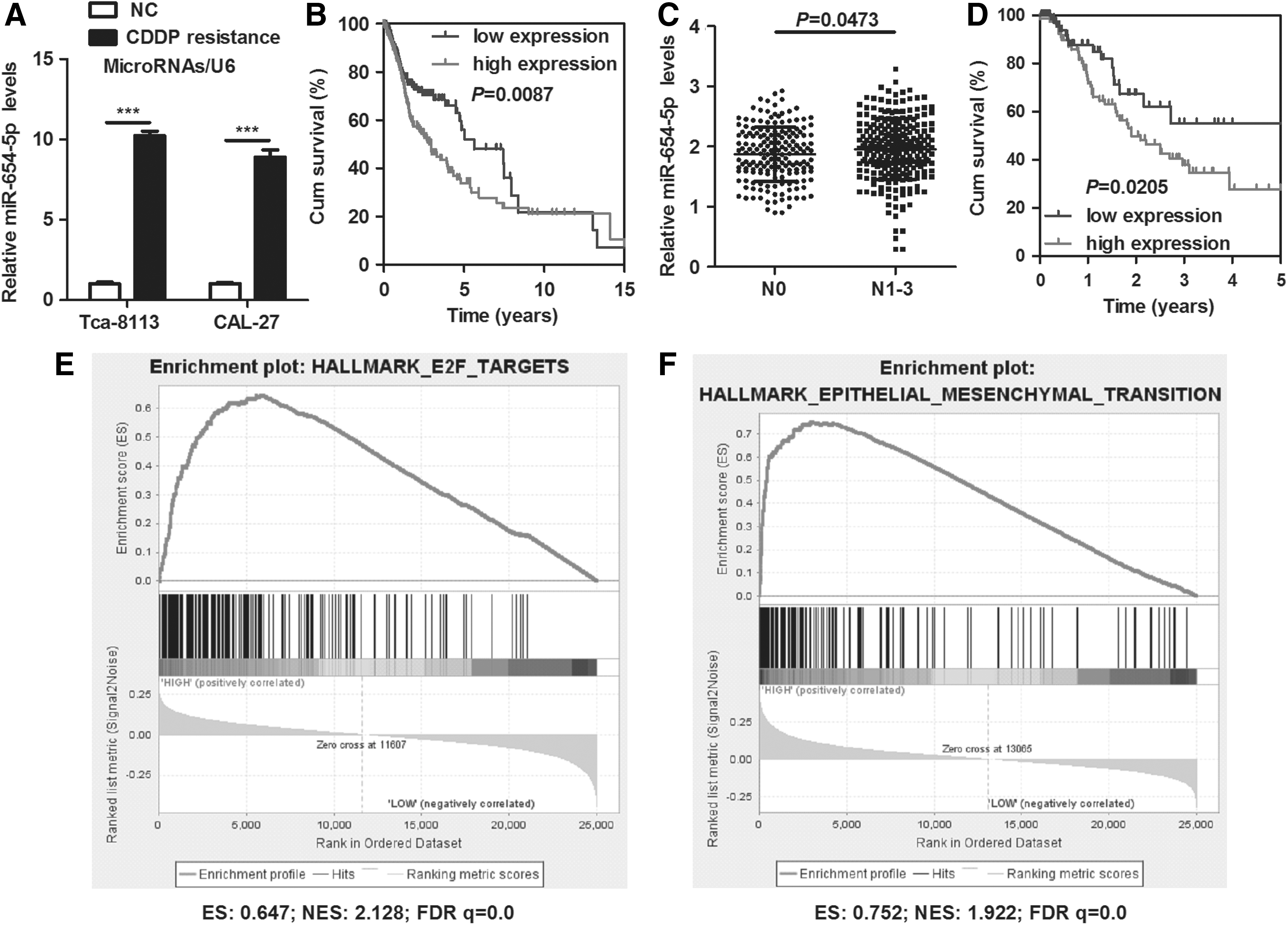
miR-654-5p is associated with poor prognosis in OSCC patients.
miRNA-654-5p regulates EMT to promote proliferation, migration, invasion, and chemoresistance of OSCC cells in vitro and in vivo
To further elucidate the impact of miR-654-5p on OSCC, we established stable miR-654-5p-silenced Tca-8113 and CAL-27 cells via lentiviral infection (Supplementary Fig. S1; Supplementary Data are available online at
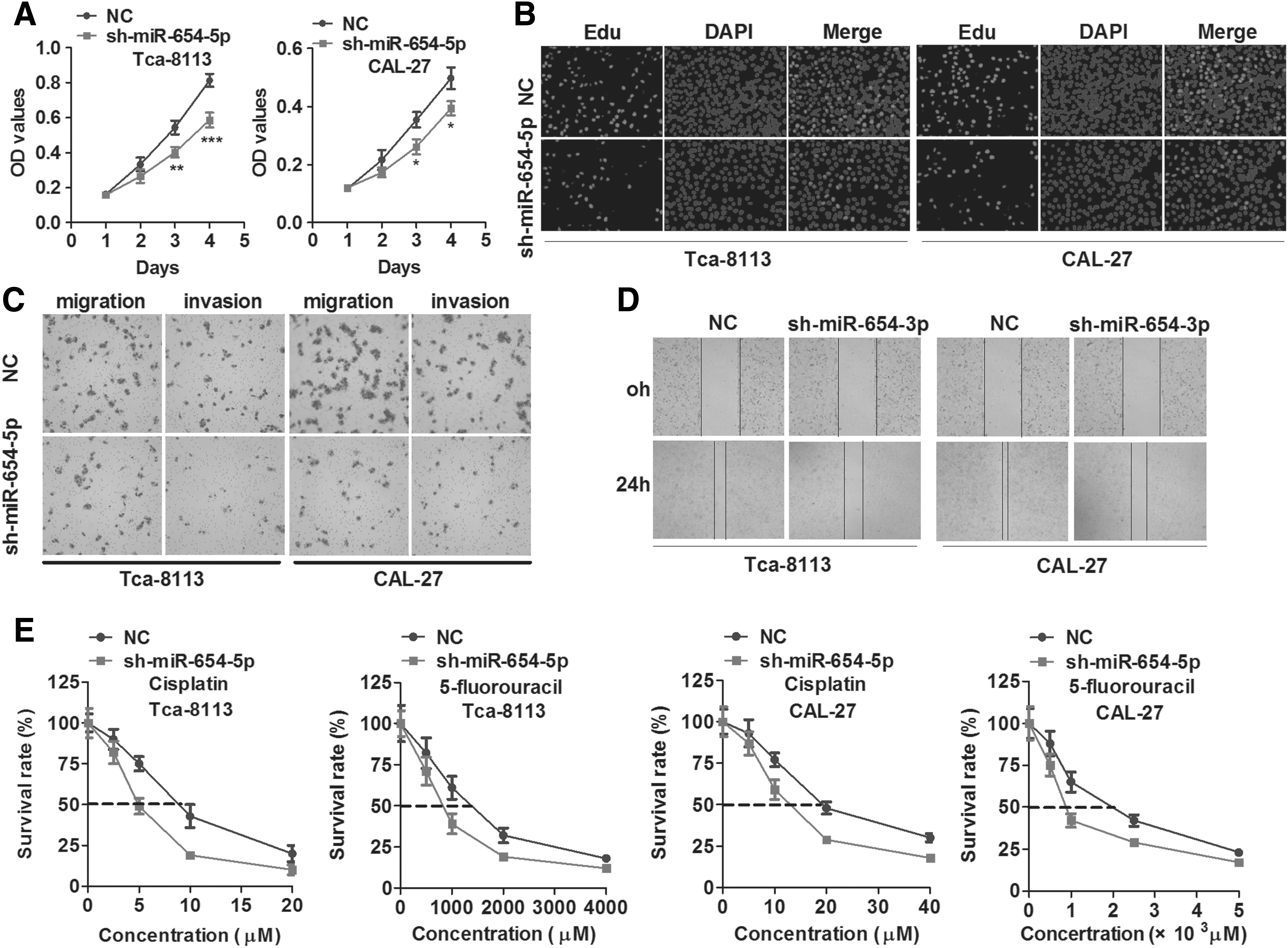
miR-654-5p promotes proliferation, metastasis, and chemoresistance of OSCC cells.
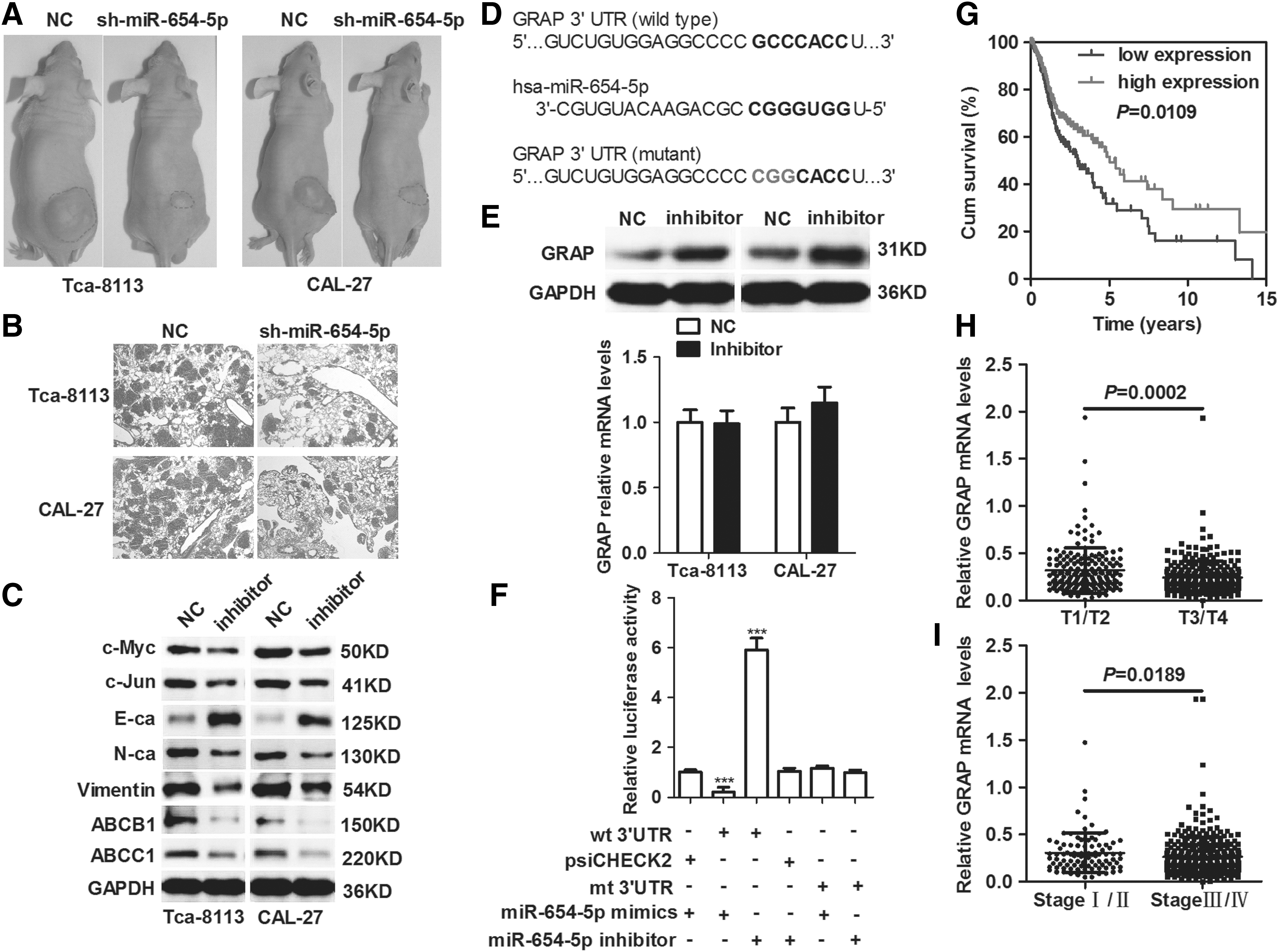
miR-654-5p directly targets GRAP to regulate OSCC progression.
miR-654-5p directly targets Grb2-related adaptor protein
GRAP was predicted to be a direct target of miR-654-5p by five computational tools, namely, miRWalk, DIANAmT, miRanda, PICTAR5, and TargetScan (Fig. 3D). The silencing of miR-654-5p elevated GRAP levels in Tca-8113 and CAL-27 cells (Fig. 3E). However, downregulation of miR-654-5p had no effect on mRNA levels of GRAP in OSCC cells (Fig. 3E). GRAP was further identified as a direct target of miR-654-5p by luciferase reporter assay. Introduction of miR-654-5p mimics impaired GRAP luciferase activity, whereas the transfection of a miR-654-5p inhibitor enhanced the luciferase activity in CAL-27 cells. Moreover, these effects on luciferase activity were abrogated when the cells were cotransfected with a mutated GRAP reporter (Fig. 3F). Survival analysis based on the TCGA HNSC data set revealed that GRAP was correlated with good prognosis in HNSC patients (log-rank test, p = 0.0109, Fig. 3G). Moreover, differential expression analysis suggested that GRAP was downregulated in T1–2 stage versus T3–4 stage HNSC (p = 0.0002) (Fig. 3H); consistently, GRAP was negatively correlated with TNM stage in HNSC patients (p = 0.0189).
miR-654-5p promotes proliferation, migration, invasion, and chemoresistance of OSCC cells through GRAP-mediated Ras/MAPK signaling
Using in vitro gain- and loss-of-function analysis, we observed that inhibition of miR-654-5p decreased cell proliferation, migration, invasion, and chemoresistance; however, subsequent downregulation of GRAP restored the effect in both Tca-8113 and CAL-27 cells (Fig. 4A–C). Consistently, knockdown of miR-654-5p could partly reverse chemoresistance of Tca-8113/CDDP and CAL-27/CDDP cells (Fig. 4D). Subsequently, western blot analysis confirmed that the silencing of miR-654-5p inhibited Ras/MAPK signaling, and the effect could be restored in miR-654-5p knockdown OSCC cells treated with GRAP siRNA (Fig. 4E).
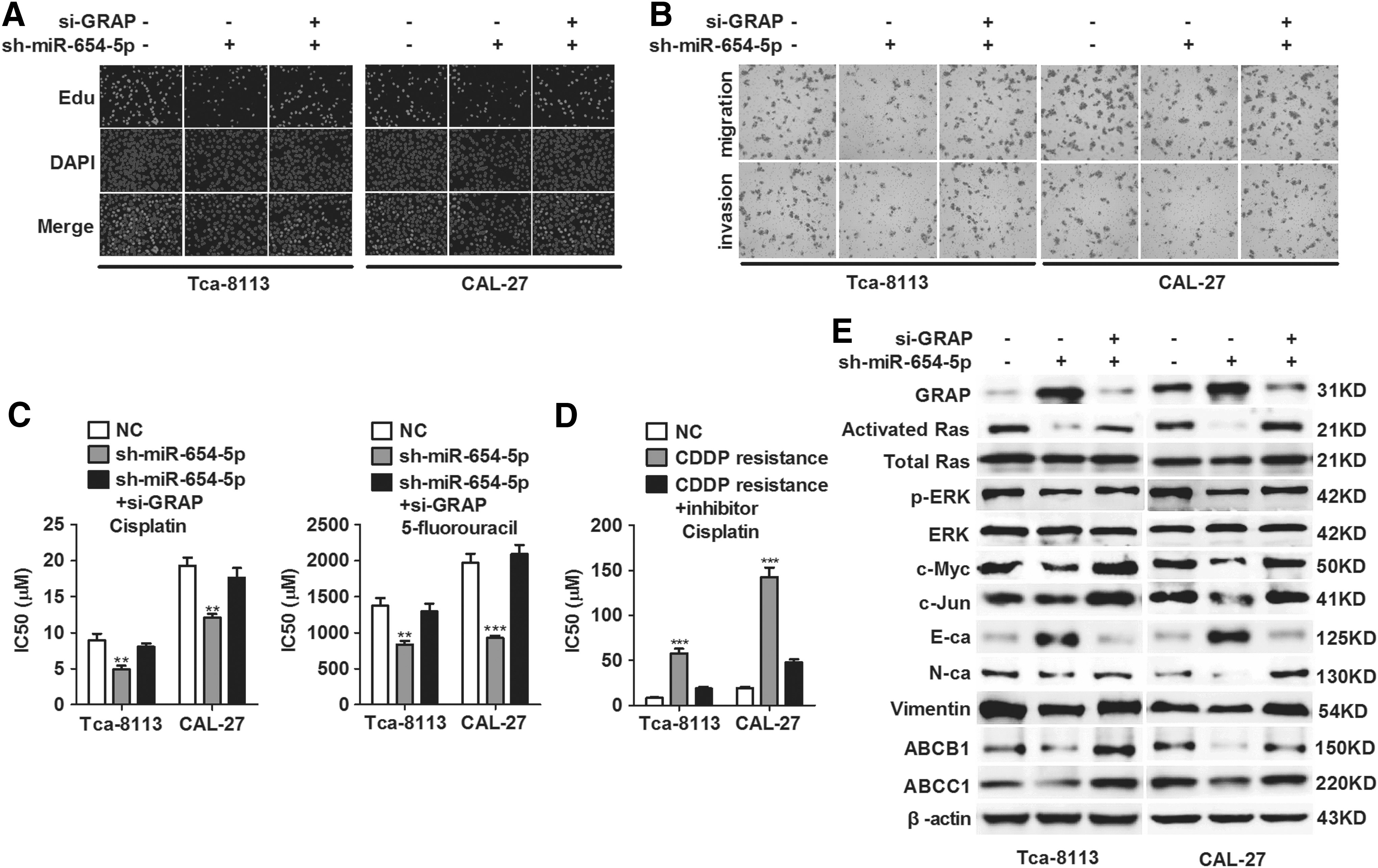
miR-654-5p promotes proliferation, metastasis, and chemoresistance of OSCC cells through GRAP-mediated Ras/MAPK signaling.
Discussion
Previous studies indicated that miR-654-5p and miR-654-3p act as tumor suppressors in breast cancer (Tan et al., 2016), prostate cancer (Ostling et al., 2011), Hodgkin lymphoma (Paydas et al., 2016), and papillary thyroid cancer (Geraldo et al., 2017). However, the roles and molecular mechanisms of miR-654-5p and miR-654-3p in OSCC progression are yet to be reported. In the present work, we demonstrated that miR-654-5p (not miR-654-3p), which was identified as a biomarker, correlated with poor prognosis in patients with OSCC and promoted proliferation, metastasis, and chemoresistance of OSCC. Mechanistically, miR-654-5p stimulated cancer progression by directly targeting GRAP and, subsequently, activating the Ras/MAPK signaling pathway in OSCC.
miRNAs participate in many biological processes, including regulation of proliferation (Bu and Luo, 2017), metastasis (Wang et al., 2017), and chemoresistance (Jin et al., 2016). Resistance to chemotherapy is one of the main reasons that cause treatment failure in OSCC patients, consequently increasing cancer-associated mortality. Therefore, we constructed Tca-8113/CDDP and CAL-27/CDDP cells to identify miRNAs that regulate chemoresistance in OSCC. Combining microarray data with TCGA data, we found that miR-654-5p acts as an onco-miRNA in HNSC, enhancing tumor progression. Data obtained from patient tissues further validated that miR-654-5p was correlated with poor prognosis in OSCC patients. The GSEA, based on TCGA HNSC data, indicated that miR-654-5p modulated the expression of E2F targets and enhanced EMT, which is an important process that leads to cancer metastasis. The E2F family plays a crucial role in the regulation of cell cycle. In addition, molecular mechanisms of chemoresistance include transporter pumps, oncogenes, tumor suppressor gene, mitochondrial alteration, DNA repair, autophagy, exosomes, cancer stemness, and EMT (Zheng, 2017). Therefore, we further determined the impact of miR-654-5p on cell proliferation and metastasis in OSCC. In this study, we demonstrated that miR-654-5p promoted OSCC cell proliferation, metastasis, and chemoresistance in vivo and in vitro. Further investigation into its mechanism showed that miR-654-5p affected the expression of c-Myc and c-Jun to regulate cell proliferation, the expression of E-ca, N-ca, and Vimentin (the makers of EMT) to regulate cell metastasis, and the expression of ABCB1 and ABCC1 to modulate chemoresistance in OSCC cells.
GRAP is a Grb2-like protein, which can couple signals from receptor and cytoplasmic tyrosine kinases to the Ras signaling pathway (Feng et al., 1996; Trub et al., 1997). A previous study suggested that GRAP, unlike Grb2, downregulates signal relay through the Ras/Erk pathway and acts as a negative regulator of T-cell receptor (TCR)-stimulated intracellular signaling (Shen et al., 2002). However, the function of GRAP in cancer has not been elucidated. In our study, we showed that GRAP, which was correlated with good prognosis in HNSC patients, acts as a tumor suppressor in HNSC. In addition, GRAP was negatively correlated with TNM stage in HNSC, indicating its role in the regulation of cancer progression. Intriguingly, GRAP was further verified as the direct target of miR-654-5p. Cell progression and the activity of Ras/MAPK signaling were reversed when knocking down GRAP in miR-654-5p-silenced OSCC cells, suggesting that miR-654-5p directly targets GRAP to facilitate OSCC progression through the Ras/MAPK signaling pathway.
Conclusion
Taken together, our findings suggest that miR-654-5p enhances cell proliferation, migration, invasion, and chemoresistance by directly targeting GRAP and promoting EMT in OSCC cells. The miR-654-5p/GRAP/Ras/Erk signaling pathway in OSCC cells might be a part of the underlying mechanism through which miR-654-5p regulates OSCC progression. miR-654-5p, as a potential biomarker for the clinical diagnosis or prognosis of OSCC, may be an effective anticancer target for the treatment of OSCC.
Footnotes
Acknowledgment
The authors thank Experimental Animal Center of Fujian Medical University for providing us with necessary conditions and facilities to complete this project.
Disclosure Statement
No competing financial interests exist.
References
Supplementary Material
Please find the following supplemental material available below.
For Open Access articles published under a Creative Commons License, all supplemental material carries the same license as the article it is associated with.
For non-Open Access articles published, all supplemental material carries a non-exclusive license, and permission requests for re-use of supplemental material or any part of supplemental material shall be sent directly to the copyright owner as specified in the copyright notice associated with the article.