
Abstract
Premenopausal women have a reduced risk for cardiovascular disease. Estrogen deficiency augments cardiac inflammation and oxidative stress and, thereby, aggravates myocardial fibrosis (MF) and diastolic dysfunction in hypertensive female rats. However, estrogen replacement therapy has no effect on myocardial infarction and cardiac fibrosis in postmenopausal women. Further clinical studies showed that high blood glucose levels in patients with diabetes is an important cause of MF, but the underlying mechanism is unclear. To experimentally address this issue, diabetes mellitus (DM) was induced by injecting streptozotocin and administering a high-fat diet in ovariectomized (OVX) rats. High degrees of fibrosis and apoptosis were detected in the cardiac tissue of these rats, together with increased expression of iNOS. Further treatment with the G protein-coupled estrogen receptor 30 (GPR30) agonist G1 decreased iNOS expression and the apoptosis rate in cardiac tissue significantly and inhibited cardiac fibroblast (CF) proliferation. Similar trends were observed in cultured CFs treated with high concentrations of fat and glucose. In addition, treatment with the iNOS-specific inhibitor W1400 attenuated iNOS and vimentin expression, which is associated with a marked reduction in MF. These results suggest that GPR30 activation inhibits MF in diabetic OVX female rats by suppressing cardiac iNOS activity and consequently NO levels. Thus, GPR30 activation may provide novel cardioprotection strategies for postmenopausal women, especially those with DM.
Introduction
M
Estrogens protect the premenopausal heart from hypertension and ventricular remodeling (Zhao et al., 2014). Estrogen deficiency augments cardiac inflammation and oxidative stress and thereby aggravates MF and diastolic dysfunction in hypertensive female rats (Mori et al., 2011). However, estrogen replacement therapy has no effect on myocardial infarction (MI) and cardiac fibrosis in postmenopausal women (Angeja et al., 2001; Waters et al., 2002). G protein-coupled estrogen receptor 30 (GPR30) is a member of the seven-transmembrane G protein-coupled receptor family that directly binds to estrogen and activates downstream signaling pathways (Prossnitz et al., 2008). Animal studies have shown that GPR30 can reduce complications associated with MI by inhibiting the formation of fibrous scar tissue (Wang et al., 2015). GPR30 provides cardiovascular protection by inhibiting MF (Zhao et al., 2014; Li et al., 2015; Wang et al., 2015). Clinical studies have shown that high blood glucose in patients with diabetes is an important cause of MF (Aguilar et al., 2014). GPR30 plays a critical role in improving glucose-stimulated insulin release by suppressing glucagon and somatostatin secretion (Sharma and Prossnitz, 2016). The GPR30 agonist G1 suppresses MF, whereas the GPR30 inhibitor G15 has the opposite effect (Kang et al., 2012).
Nitric oxide (NO) is synthesized by members of the NO synthase (NOS) family, which includes endothelial NOS, neuronal NOS, and iNOS (Sunico et al., 2008). NO-mediated effects can be beneficial or harmful depending on specific risk factors for a particular disease (Lee et al., 2016). Abnormal iNOS expression is related to the progression of vascular dysfunction and affects the synthesis of the fibroblast matrix (Larsson-Callerfelt et al., 2015). It was previously reported that heart-specific iNOS overexpression in mice stimulated CF proliferation; conversely, inhibition of iNOS decreased MF (Mungrue et al., 2002). Estrogen is known to specifically bind to GPR30 and activate iNOS signaling through epidermal growth factor (Filardo and Thomas, 2005). It is unclear whether iNOS influences MF.
Based on the above findings, we hypothesized that GPR30 may suppress cardiac iNOS activity and that consequent downregulation of NO expression can lead to a reduction in the development of MF. To test this hypothesis, in the present study, we induced diabetes in ovariectomized (OVX) female rats by treatment with streptozotocin (STZ) and a high-fat diet (HFD). Rats were treated with a GPR30 agonist and inhibitor, respectively, and the relationship between iNOS levels in cardiac tissue and degree of MF was examined in the animal model and in primary cultured CFs to determine the role of the estrogen receptor (ER) in MF progression.
Materials and Methods
Reagents
STZ and DAPI were purchased from Sigma-Aldrich (St. Louis, MO). G1 and G15 (GPR30 agonist and inhibitor, respectively) were obtained from Cayman Chemical (Ann Arbor, MI). The NO Detection Kit used for quantification of NO levels in serum was from Elabscience Bioengineering Institute (Wuhan, China). The terminal deoxynucleotidyl transferase dUTP nick-end labeling (TUNEL) Assay Kit was from Roche Diagnostics (Mannheim, Germany). The Bicinchoninic Acid Assay Protein Quantification Kit was purchased from Merck Millipore (Darmstadt, Germany). Primary antibodies against GPR30 (ab39742), ER-α (ab32063), ER-β (ab3576), iNOS (ab15323), and vimentin (ab92547) were purchased from Abcam (Cambridge, MA). Antibodies against β-actin and GAPDH were purchased from Santa Cruz Biotechnology (Santa Cruz, CA). W1400 was from Enzo Life Sciences (Farmingdale, NY).
Animals
Female Sprague-Dawley rats (6 weeks old) were obtained from the animal center of the Fourth Military Medical University (FMMU) and maintained under specific pathogen-free conditions. All animal experiments were approved by the Institutional Animal Care and Use Committee (IACUC) of the Fourth Military Medical University (No. 20170203). Rats were anesthetized with ketamine (90 mg/kg, i.p.) and xylazine (9 mg/kg, i.p.).
All animals were divided into five groups (n = 12). Bilateral ovariectomy was performed on rats under 50 mg/kg (i.p.) pentobarbital sodium anesthesia (OVX rats). Animals were intraperitoneally injected with STZ (40 mg/kg) to induce diabetes mellitus (DM). When the blood glucose level reached 13 mM, the rats were fed a HFD composed of 21.2% protein, 12% fat, 15% sucrose, and 1% cholesterol for 2 months (DM rats) (Srinivasan et al., 2005; Seo et al., 2012; Shi et al., 2015). Control rats were fed normal rodent chow composed of 20% protein and 4.5% fat. Blood glucose concentrations were measured with a glucometer 48 h post-STZ injection. Serum NO levels were determined with an Enzyme-Linked Immunosorbent Assay Kit. G1 stock solution was prepared in dimethyl sulfoxide (DMSO) and diluted with culture medium immediately before the experiment; 0.01% DMSO was used as a control. Diabetic OVX rats (OVX+DM) were treated with G1 (35 μg/kg by intraperitoneal injection) thrice a week for 8 weeks.
CF culture and treatment
CFs from left ventricular myocardial tissues were isolated and collected as previously described (Dubey et al., 1997; Coulombe and Wong, 2004). The purity of cultured CFs was determined as >98% by immunohistochemistry based on expression of the CF marker vimentin. Immunopositive cells from passages 3–5 were used for experiments. Before each experiment, cells were seeded in six-well plates or a laser confocal microscopy dish at a density of 3 × 10⁴ cells/cm2 in DMEM containing 10% fetal bovine serum and cultured for 24 h.
High-glucose/high-fat (HG/HF) DMEM containing 25 mM glucose and saturated free fatty acid palmitate (16:0; 500 μM) was used to mimic the diabetic state (Yu et al., 2015a). Mannitol (20 mM) was used as needed. G1 (10 nM) (Dennis et al., 2009), G15 (1 μM), and W1400 (1 μM) (Okada and Yamawaki, 2015) were added simultaneously to the appropriate groups for 48 h; HG/HF was added 40 h later for a total of 8 h. Cells were fixed with cold methanol for immunocytochemistry or lysed with TRIzol reagent (Qiagen, Valencia, CA) for real-time PCR or trypsinized for western blot analysis.
Cell viability assay
Premature senescence in CFs was induced by doxorubicin (DOX) as previously described (Maejima et al., 2008; Spallarossa et al., 2009). CFs were seeded in a 96-well culture plate. Cell viability was measured after treatment with DOX, HG/HF, and G1 using the 3-(4,5-dimethylthiazol-2-yl)-2,5-diphenyltetrazolium bromide (MTT) assay (Yang et al., 2013). Briefly, after treatment and washing with phosphate-buffered saline, 10 μL of MTT reagent was added to each well at a final concentration of 0.5 mg/mL. After 4 h of incubation, 100 μL of DMSO was added to dissolve the formazan crystals, and the absorbance was measured on a SpectraMax 190 microtiter plate reader (Molecular Device, Sunnyvale, CA) at a wavelength of 490 nm. Cell viability was calculated by dividing the optical density of samples by that of the control group.
Western blot analysis
Primary antibodies against the following proteins were used for western blotting: GPR30, ER/α, ER/β, iNOS, vimentin, β-actin, and GAPDH. Immunoreactivity was detected by enhanced chemiluminescence and quantified by densitometric analysis using the Quantity One image analyzer (Bio-Rad, Hercules, CA).
Analysis of iNOS expression
Real-time PCR was used to determine mRNA levels in CFs and left ventricular myocardial tissues as previously described (Tank et al., 2014). Briefly, total RNA was extracted from left ventricular tissue using TRIzol reagent and reverse-transcribed to cDNA using the ReverTra Ace qPCR RT Kit (Takara Bio, Dalian, China). Real-time PCR was performed using a SYBR Green PCR Kit (Takara Bio) and forward (5′-TCCTCAGGCTTGGGTCTTGTTAG-3′) and reverse (5′-TTCAGGTCACCTTGGTAGG ATTTG-3′) primers for iNOS. The reaction conditions were as follows: 95°C for 30 s followed by 40 cycles of 95°C for 5 s and 60°C for 30 s.
Immunofluorescence and TUNEL assay
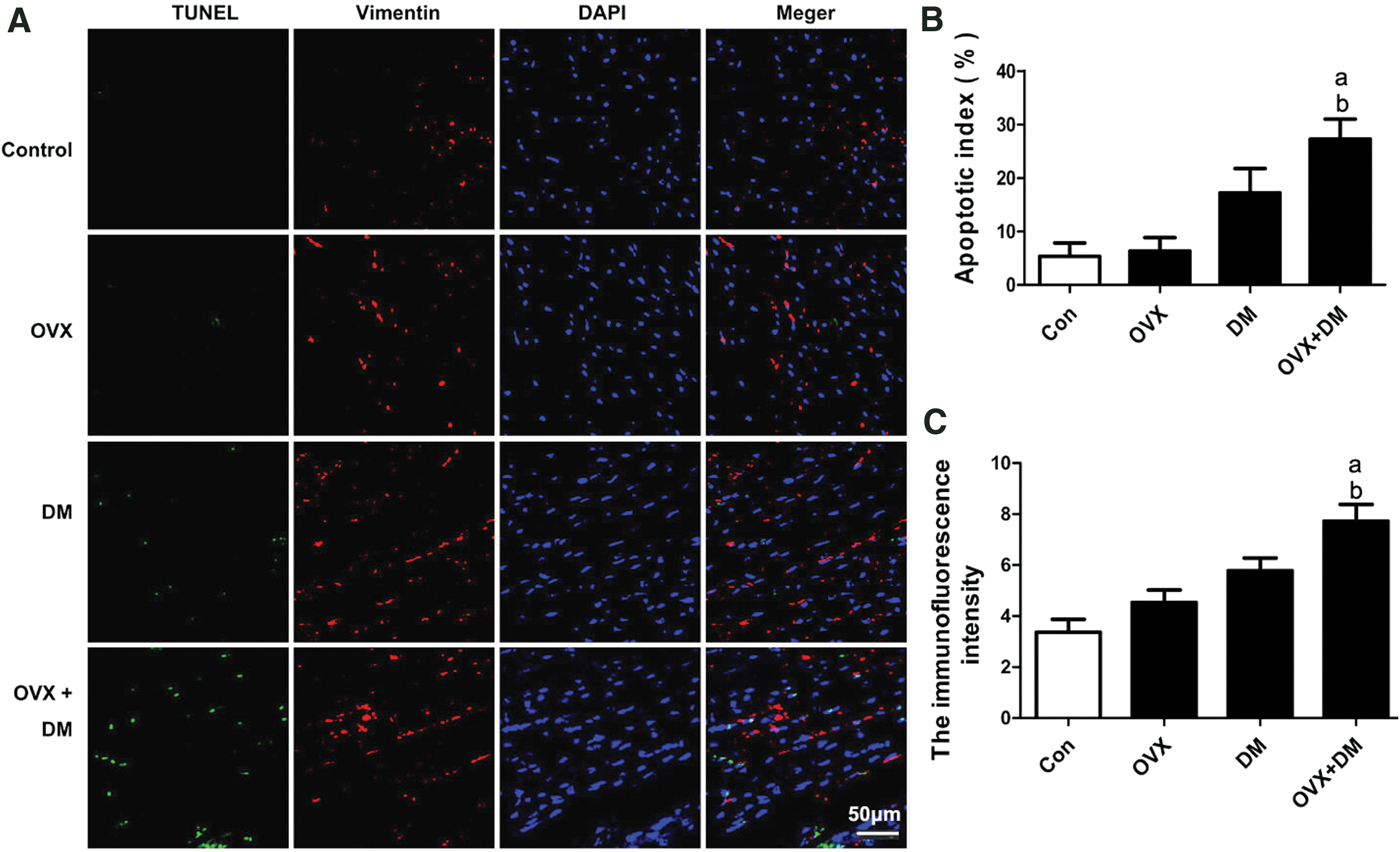
Immunofluorescence (IF) of formalin-fixed and paraffin-embedded left ventricular myocardial tissue was performed as previously described (Yu et al., 2016). Fixed CFs were incubated overnight with anti-vimentin antibody (1:200) at 4°C, followed by Cy3-conjugated goat anti-rabbit IgG (1:100 in blocking solution). TUNEL detection solution was added to the sections, which were incubated at 37°C in the dark for 2 h. Nuclei were stained with DAPI. Sections were visualized using a Fluoview FV100 laser scanning confocal microscope (Olympus, Tokyo, Japan). Cells from 10 randomly selected visual fields were counted to determine the apoptotic index. Images of IF were captured digitally in five random microscope fields from each sample and were analyzed with analysis software (Image Pro-Plus; Image Solutions, Torrance, CA). The index of apoptosis was expressed as the ratio of positively stained apoptotic myocytes to the total number of myocytes counted × 100% (Yu et al., 2015b).
Statistical analyses
Experiments were performed in triplicate. All data are presented as the mean ± SEM. Comparisons between groups were performed using one-way ANOVA with Tukey's multiple-comparison post hoc test, and p < 0.05 was considered significant. All of the statistical analyses were performed with the GraphPad Prism software version 5.0 (GraphPad Software, San Diego, CA).
Results
Effect of DM and OVX on body weight, blood glucose, and triglycerides of rats
After bilateral ovariectomy and/or feeding with a HFD, the body weight, concentration of blood glucose, and blood triglycerides were measured in each group. OVX caused an increase in body weight compared with normal rats (338.7 ± 15.6 g vs. 413.7 ± 13.2 g, p < 0.05); however, when DM was induced by STZ and a HFD, the body weights of the DM (272.7 ± 9.6 g) and OVX+DM (280.3 ± 11.8 g) groups were lower than those of normal rats (p < 0.05) (Fig. 1A). The plasma glucose level was increased in OVX diabetic (23.47 ± 0.74 mM) compared with nondiabetic OVX rats (7.05 ± 0.82 mM) (p < 0.01) (Fig. 1B). Triglyceride levels were similar between DM and DM+OVX groups, with no significant difference (Fig. 1C).

Effects of OVX and DM on body weight, blood glucose, and serum triglyceride.
Effect of DM+OVX on cardiac apoptosis and fibrosis of rats
To determine the pathological significance of OVX and DM, cardiac apoptosis was examined by TUNEL assay, whereas expression of the fibrosis marker vimentin was examined by IF. The number of vimentin-positive cells (Fig. 2A, C) and TUNEL-positive nuclei in myocardial fibroblasts (Fig. 2A, B) was increased significantly in the OVX+DM group compared with other groups (p < 0.05). Bilateral ovariectomy significantly increased the cardiac apoptosis and fibrosis with DM (p < 0.05). We also confirmed the degree of fibrosis in different treatment groups by Trichrome staining. The results are consistent with Figure 2A (Supplementary Fig. S1; Supplementary Data are available online at
(
Effect of DM+OVX on expression level of ERs and iNOS
To determine the potential mechanisms responsible for the increase in apoptosis and fibrosis caused by estrogen deficiency in OVX+DM rats, several signaling molecules were identified. The western blot results for the expression of different proteins are shown in Figure 3. ER/α, ER/β, and GPR30 are ERs (Dennis et al., 2009). Treatment with OVX and OVX+DM significantly decreased GPR30 levels (vs. control rats, p < 0.05). The other ERs, ER/α and ER/β, were downregulated in the OVX and OVX+DM groups (p < 0.05). However, OVX and diabetes induced a significant increase in iNOS (p < 0.05) expression relative to DM rats or normal rats (Fig. 3E). The changes of GPR30 mRNA in OVX and OVX+DM rats displayed similar results as protein level (Supplementary Fig. S2).
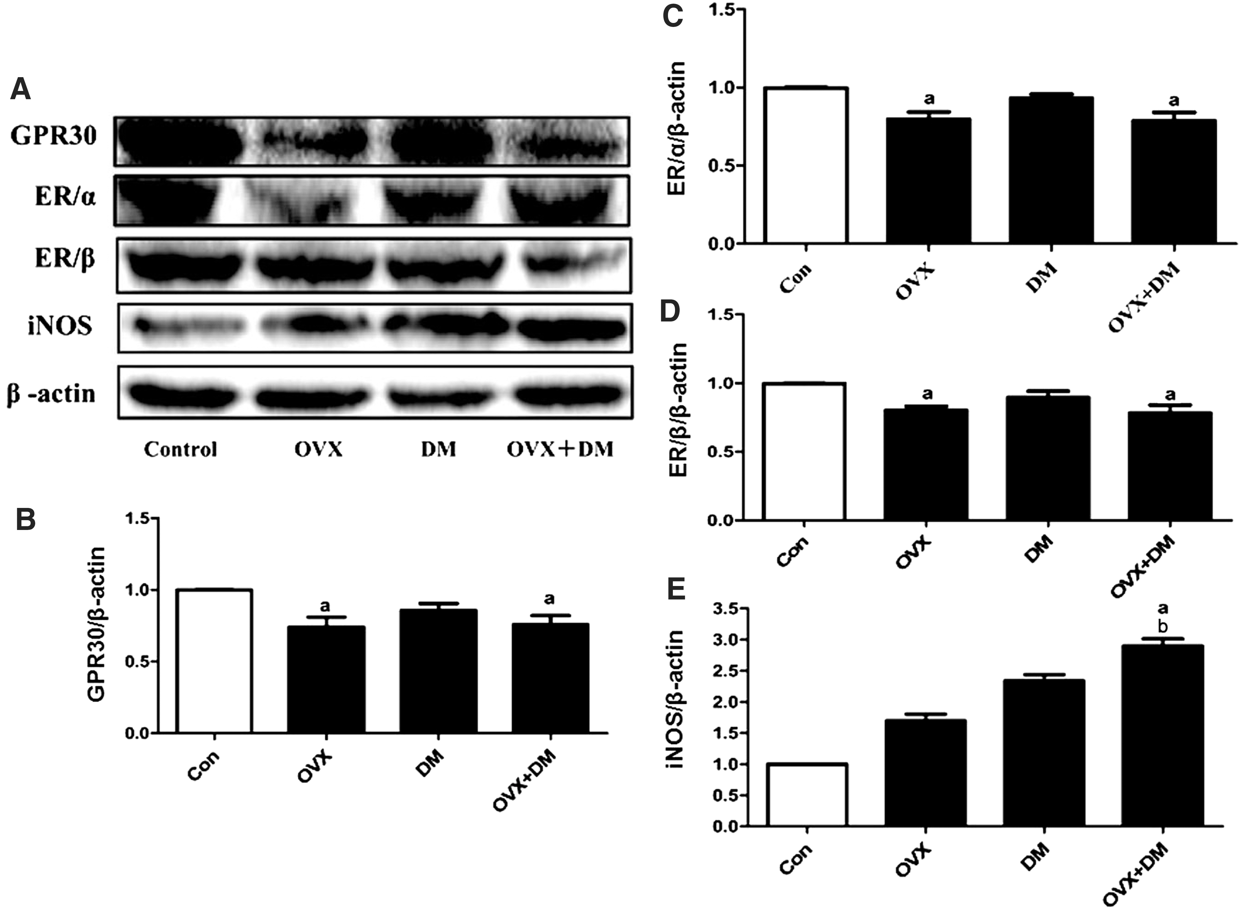
Effect of DM and OVX on expression level of ERs and iNOS. Western blotting was used to analyze the expression levels of GPR30, ER/α, ER/β, and iNOS.
Activation of GPR30 reduces plasma glucose levels and increases body weight in OVX+DM rats
To further determine the role of GPR30 in OVX+DM rats, GPR30 was stimulated by its agonist G1. The body weight of diabetic OVX rats treated with G1 (346.7 ± 17.4 g) increased to a value close to normal (306.7 ± 8.3 g) (Fig. 4A). Treatment with DM induced high plasma glucose and triglyceride levels, which indicated that the DM rat model was established successfully. Similar results were shown in DM rats with OVX treatment. Further addition of the agonist G1 caused no significant effect on plasma glucose or triglyceride levels (Fig. 4B, C).

Effects of G1 on body weight and blood glucose, serum triglyceride, NO levels in OVX + DM rat.
Activation of GPR30 inhibits iNOS expression and reverses the increase of NO levels in cardiac tissue
OVX and diabetes caused an increase in NO levels relative to those in normal rats. However, this effect was inhibited by G1 treatment (OVX+DM+G1 vs. OVX+DM, p < 0.05; Fig. 4D). In the OVX+DM group, GPR30 expression was reduced, but iNOS expression was increased. Further addition of G1 caused an apparent increase in the expression of GPR30 (Fig. 5A, B), and the high levels of iNOS in the OVX+DM group were reduced by the GPR30 agonist G1 (p < 0.05; Fig. 5C, D).

Effect of stimulation of GPR30 on apoptosis, fibrosis, iNOS expression, and NO levels in cardiac tissue.
Activation of GPR30 reduces apoptosis and fibrosis in DM+OVX rats
To assess the impact of stimulation of GPR30 on cardiac apoptosis and fibrosis, vimentin expression and apoptosis in myocardial tissue fibroblasts are shown in Figure 5E. Both apoptosis and fibrosis were alleviated partially by the GPR30 agonist G1. The apoptosis rate in the group treated with G1 decreased to 18.67% ± 2.60% (Fig. 5F), whereas vimentin expression was downregulated by ∼30% (Fig. 5G). Trichrome staining showed the same trend (Supplementary Fig. S1).
Effect of HG/HF on cultured primary CFs
To further determine the specific mechanism involved in GPR30-mediated attenuated apoptosis and fibrosis, primary cultured CFs were used. The primary cultured cardiomyocytes from rats in each group were examined by IF. Cells were positive for vimentin and troponin (Fig. 6A), consistent with their CF identity (Bei et al., 2015). HG/HF treatment decreased CF viability (p < 0.05), and this effect was reversed by addition of G1 (p < 0.05; Fig. 6B). Expression of GPR30 was slightly downregulated in the presence of HG/HF (Fig. 6D), whereas the amount of iNOS was significantly increased (Fig. 6E).

Effect of HG/HF on cultured primary CFs.
Activation of GPR30 inhibits morphological changes associated with fibrosis in CFs by decreasing iNOS and NO levels
CF morphology and vimentin expression are shown in Figure 7. Normal CFs displayed spindle-shaped and elongated morphology with extended pseudopodia and clearly visible nuclei and were surrounded by fibrous filaments. In contrast, the spindle shape was not apparent in the HG/HF group; these cells were broader, with few pseudopodia and fibrous filaments, and showed increased vimentin expression. G1-treated CFs in the HG/HF group were fusiform with visible pseudopodia and lower vimentin expression than those in the HG/HF group. iNOS was upregulated in CFs treated with HG/HF (p < 0.01; Fig. 7B, C), whereas upon addition of G1, both expression of iNOs and levels of NO were downregulated. Although treatment with the GPR30 inhibitor G15 did not alter the morphology of these cells, iNOs expression was further increased. When the iNOS-specific inhibitor W1400 was added to CFs treated with HG/HF, iNOS was downregulated along with vimentin. Simultaneous treatment with W1400 and G1 synergistically reduced the levels of both proteins (Fig. 7E–G).

Effect of stimulation of GPR30 on morphological changes associated with fibrosis in CFs.
Discussion
Upregulation of iNOS in myocarditis (i.e., ischemic and dilated cardiomyocytes) is an important part of the immune response, as it increases the production of peroxynitrite, which is closely related to inflammatory cell infiltration, MF, hypertrophy, and dilatation (Mungrue et al., 2002). Although iNOS overexpression in mice rarely leads to heart failure, in cardiomyocytes it is sufficient to cause cardiomyopathy (Mungrue et al., 2002). It has been reported that CFs have an iNOS/NO system (Gustafsson and Brunton, 2000). Myocardial NO production and abnormal NOS activity caused by loss of estrogen is a major mechanism underlying the development of MF in postmenopausal women. Our study demonstrated that NO levels are significantly altered in the myocardial tissue of female diabetic rats after ovariectomy.
In this study, we found that body weight increased after ovariectomy and that only diabetic rats showed weight loss. Further OVX treatment has no apparent effect on body weight. All of these results are consistent with earlier reports (Li et al., 2012). Triglyceride and blood glucose levels were similar among treated rats, confirming that the animal model was successfully established. Treatment with G1 not only abrogated the weight loss in the OVX+DM group but also reversed the increase in blood sugar level in these rats. Some of the effects of G1 may have resulted from improvement of the animals' metabolic status rather than directly from changes in cardiac physiology. When OVX+DM rats were administered G1, there was a reduction in NO levels in the myocardial tissue, which was largely due to high basal expression of iNOS.
Although OVX+DM rats displayed a higher level of iNOS/NO along with apparent apoptosis and higher levels of fibroblasts compared to normal rats, addition of the GPR30 agonist G1 decreased iNOS expression significantly, attenuated apoptosis, and improved fibroblast content in myocardial tissue. This indicated that iNOS plays an important role in MF induced by OVX+DM treatment. To determine the mechanism involved in GPR30-mediated attenuated apoptosis and fibrosis, primary cultured CFs from rats were treated with HG/HF simulated DM induction. IF and morphological analyses revealed that vimentin expression and the degree of fibrosis increased in CFs along with iNOS upregulation, reducing cell viability.
The GPR30 levels were unaltered between normally cultured CFs and those with HG/HF treatment, but high iNOS levels were induced by HG/HF. Further addition of G1 mitigated fibrosis such that the CF morphology returned to normal (Fig. 7A) and CF viability was improved (Fig. 6B). Treatment with the GPR30 inhibitor G15 in combination with HG/HF induced higher iNOS expression than HG/HF alone (Fig. 7C, D) and aggravated fibrosis (Fig. 7A). Addition of the iNOS-specific inhibitor W1400 to CFs treated with HG/HF reduced vimentin expression; when W1400 was administered in combination with G1, iNOS and vimentin were downregulated, which was associated with a marked reduction in MF (Fig. 7E–G). These results indicate that GPR30 inhibits MF in OVX diabetic rats by regulating iNOS expression.
In the literature, the importance of iNOS signaling in myocardial diseases is controversial. For example, use of left coronary artery ligation as MI model in mice with or without inactivation of the iNOS gene resulted in no difference in cardiac TGF-beta1 expression, myofibroblast population, collagen synthesis/degradation, and collagen volume between wild-type and iNOS-knockout mice (Lu et al., 2008). Therefore, analyzing only iNOS expression to explain the cardioprotective effects of GPR30 agonists seems to be insufficient. Therefore, we performed in vitro experiments with specific iNOS inhibitors (W1400) to determine whether the OVX/DM effects (treated with HG/HF) are reversed by iNOS inhibition. Addition of W1400 in the HG/HF group significantly downregulated iNOS and vimentin, indicating the inhibition of W1400 for the OVX+DM effects. A recent study evaluating the effect of the GPR30-selective agonist G1 in doxorubicin-induced cardiotoxicity in male rats, the effects on several other important mediators, including TNF-α, IL-1β, LDH, ROS levels, p-c-jun, BAX, CTGF, iNOS, and COX2, were analyzed (De Francesco et al., 2017). Therefore, we plan to determine the changes in those indicators in further experiments and comprehensively evaluate their role.
In summary, OVX combined with DM upregulated iNOS in rat myocardial cells, resulting in MF. GPR30 suppressed this effect by inhibiting iNOS expression. Thus, low estrogen levels, NO production in myocardial cells, and abnormal NOS activity are important mechanisms underlying the development of MF in postmenopausal women. These findings provide a basis for the prevention and treatment of MF in this group, although a more detailed investigation of the precise signaling mechanisms involved is needed.
Footnotes
Acknowledgments
This work was supported by the Natural Science Foundation of China (No. 81570330, 81470500, and 81570232) and the Natural Science Foundation of Shaanxi (No. 2016SR053).
Disclosure Statement
No competing financial interests exist.
References
Supplementary Material
Please find the following supplemental material available below.
For Open Access articles published under a Creative Commons License, all supplemental material carries the same license as the article it is associated with.
For non-Open Access articles published, all supplemental material carries a non-exclusive license, and permission requests for re-use of supplemental material or any part of supplemental material shall be sent directly to the copyright owner as specified in the copyright notice associated with the article.