
Abstract
We investigated the effects of cigarette smoke (CS) and resveratrol intake on the modulation of bone repair-related genes through epigenetic mechanisms at the global and gene-specific levels, after 30 days of calvarial defects were created, in rats. The samples were assigned to three groups as follows: no CS, CS, and CS/resveratrol. After evaluation of global (5 hmC) changes and epigenetic and transcription regulation at gene-specific levels, CS group showed increased 5 hmC and Tets transcripts with demethylation at Rankl and Trap promoters (p ≤ 0.01), linked to their increased gene expression (p ≤ 0.001). These modifications were reverted in the CS/resveratrol group. Opposite patterns were observed among CS and CS/resveratrol for epigenetic enzyme transcripts with higher levels of Dnmts in the CS/resveratrol (p ≤ 0.01). No CS and CS/resveratrol demonstrated similar gene expression levels for all Tets and bone-related genes. Resveratrol reverts epigenetic and transcription changes caused by CS at both global and gene-specific levels in bone-related and epigenetic machinery genes, emphasizing the resveratrol as biological modulator for CS in rats.
Introduction
C
Biological mediators acting as balancers minimizing the CS's adverse effects on bone is a relevant topic of study. Resveratrol, a polyphenol used in complementary medicine, presents several biological effects, including outcomes from bone tissue studies. Even in the presence of CS or AhR ligands, resveratrol reverts the inhibition of osteogenesis (Andreou et al., 2004; Baur and Sinclair, 2006; Allard et al., 2009; Shakibaei et al., 2011; Singh et al., 2000; Franck et al., 2018) modulating some key genes directly implicated on bone healing. However, the mechanisms and molecules upstream to bone-related genes, triggered by resveratrol, are not completely understood.
Resveratrol was identified as a direct activator of the silent information regulator (SIRT)1, by Howitz et al. (2013). The SIRT family consists of protein deacetylases, which are capable of catalyzing the deacetylation of lysine residues on histones (histone deacetylases, HDACs) (Tanner et al., 2000; McKinsey et al., 2002; Stünkel and Campbell, 2011) causing effects on epigenetic regulation and gene transcription. A broad range of roles has been pointed out for SIRT1, and at bone level, recent publications have highlighted its role in regulating the balance between the osteoclastic and osteoblastic activity, promoting osteogenic differentiation, contributing to bone formation (Casarin et al., 2014; Edwards et al., 2013; Qu et al., 2016; Zhang et al., 2016; Franck et al., 2018). As SIRT1 is a HDAC, the resveratrol might, indirectly, trigger changes in other epigenetic machinery enzymes, which in turn, might act in concert in the regulation of bone related genes.
Epigenetics refers to a change in gene that is passed on through cell division, but it is not involved in the DNA sequence change (Tang et al., 2009), acting through chemical modifications of the DNA and histone proteins, instead, that is, DNA methylation/hydroxymethylation and histone modifications. DNA methyltransferases (DNMTs) are enzymes responsible for catalyzing the DNA methylation; the ten eleven translocation (TET) enzymes are implicated in hydroxymethylation (5 hmC), participating in the DNA demethylation (Tahiliani et al., 2009). The HDACs promote chromatin condensation through deacetylation and, consequently, negative transcription regulation occurs (Goldberg et al., 2007). However, chromatin modifications such as DNA methylation/hydroxymethylation and histone modifications are dynamic epigenetic mechanisms that act in concert to promote gene expression regulation and, therefore, closely related to tissue remodeling and regeneration.
Bone tissue regeneration is modulated at multiple levels, and the epigenetic regulation may add another level to this complex cascade of events. However, the epigenetic mechanisms and molecules triggered by CS and resveratrol on bone tissue regeneration are not completely understood. Since epigenetic alterations are cell specific and highly sensitive to environmental stimulations, bone tissue recently formed in calvarial defects is particularly interesting to better understand the CS and resveratrol mechanisms for future clinical applications. The present investigation was designed to investigate the effects of CS inhalation and resveratrol intake on the modulation of bone repair-related genes through epigenetic mechanisms at the global and gene-specific levels.
Materials and Methods
Animals
Ten-week-old male Wistar rats were exposed to a 12-h light/12-h dark cycle of equal periods and had access to ad libitum water and food (Labina; Purina1, Paulínia, Brazil) in the animal facility of the Paulista University. All efforts were made to minimize suffering. The experimental procedure was approved by the Paulista University Animal Care and Use Committee (no. 226/14). This study was conducted according to the ethical rules of the Brazilian College of Animal Experimentation (COBEA) and according to the Guide for the Care and Use of Laboratory Animals of the National Institutes of Health.
Groups and treatment
No CS intakers (n = 15): The animals were not exposed to CS and received placebo solution.
CS intakers (n = 15): The animals were exposed to CS and received placebo solution.
CS/resveratrol intakers (n = 15): The animals were exposed to CS and received 10 mg/kg resveratrol (Sigma-Aldrich Ltda, São Paulo, SP, Brazil).
After 2 days of adaptation, in which the rats were exposed to CS for periods of 5 min on the first day and 7 min on the second day, the CS and CS/resveratrol intakers were exposed, in an acrylic chamber, to the inhalation of 10 cigarettes, during 8 min, thrice a day for 37 days (7 days before/30 days after surgical procedure), according to Franck et al. (2018). The no CS animals were submitted to the same daily conditions as the CS intakers, except that they were never exposed to CS (Fig. 1). The placebo solution consisted of the same proportions of Tween 80 and water as used in the preparation of resveratrol. Resveratrol and placebo solution were administrated daily using gavage for 30 days after surgery (Casarin et al., 2014).
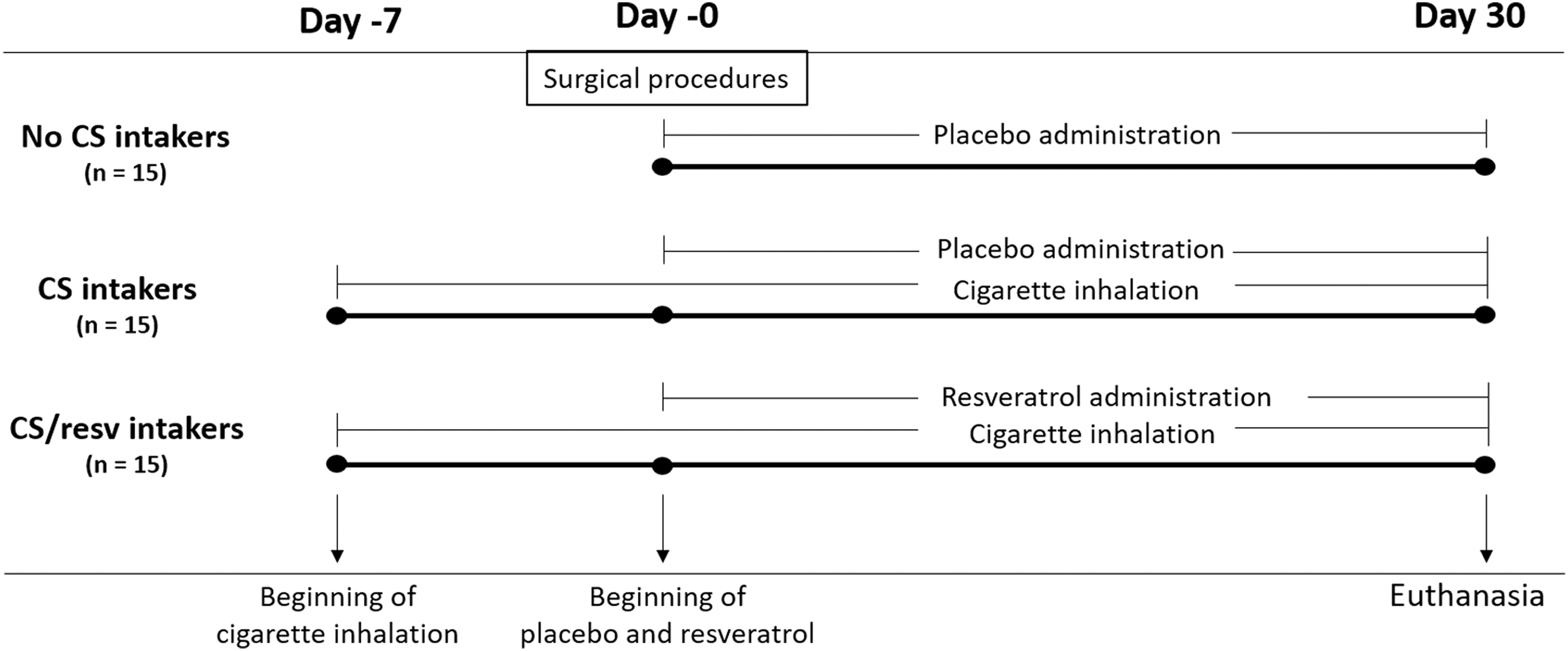
Diagram of the experimental design. CS, cigarette smoke.
Surgical procedures and postoperative period
Calvarial defects were performed under general anesthesia (ketamine and xylazine hydrochloride), and two critical-sized defects of 5 mm in diameter were made on the parietal bone with a trephine drill (Implacil de Bortoli, São Paulo, Brazil) (Franck et al., 2018). Thirty days after surgery, the animals were euthanized by CO2 inhalation. The newly formed bone from calvaria was removed after reopening the surgical site, and the samples were stored in RNAlater (Ambion, Inc., Austin, TX) at −70°C.
Gene expression analysis
Total RNA was isolated by TRIzol method (Gibco BRL; Life Technologies, Rockville, MD) and stored at −70°C. The RNA concentration was determined using a spectrophotometer (NanoDrop 1000; NanoDrop Technologies LLC, Wilmington, NC). Total RNA was DNase-treated (Turbo DNA-free; Ambion, Inc.), and 1 μg was used for complementary cDNA synthesis, using the First-Strand cDNA Synthesis Kit (Roche Diagnostic Co., Indianapolis, IN), in accordance with the manufacturer's recommendations.
The Sirt1, Dnmts, Tets, Ahr, Rankl, and Trap mRNA expression analyses were conducted in a real-time PCR apparatus (LightCycler 96 Real Time PCR System; Roche Diagnostics GmbH, Mannheim, Germany) with a SYBR Green Kit (FastStart Essential DNA Green Master; Roche Diagnostic Co., Indianapolis, IN). Results were expressed as relative amounts of the target gene using glyceraldehyde-3-phosphate dehydrogenase as the inner reference gene.
Primers were designed (Primer3, Simgene) (Untergasser et al., 2012) and confirmed by the in silico PCR (Genome Bioinformatics, University of California, CA). For further analysis of secondary structures and annealing temperatures, a primer and probe designer software were used (Beacon Designer; Premier Biosoft International, Palo Alto, CA). The details of the primers are presented in Table 1.
Global hydroxymethylation analysis
After isolation of the total RNA from the aqueous phase, the DNA was isolated from the interphase and phenol-chloroform layer (Gibco BRL; Life Technologies, Rockville, MD), following the manufacturer's recommendations. The DNA concentration was estimated using the same spectrophotometer as for mRNA samples. The DNA samples were stored at −20°C.
For global hydroxymethylation levels, the Quest 5-hmC DNA Elisa Kit (Zymo Research Corp., Irvine, CA) was used, and 100 ng DNA of each sample was plated, according to manufacturer's recommendation. The control DNA set containing a specified percentage of 5 hmC, supplied by the manufacturer, was used to determine the hydroxymethylation quantification, and the 450 nm absorbance was read in VersaMax microplate reader (Molecular Devices, Sunnyvale, CA), at 450 nm.
Quantitative PCR assay for gene-specific DNA methylation and hydroxymethylation
Genomic DNA was initially treated with T4-β-glucosyltransferase (T4-BGT) (New England Biolabs, Beverly, MA), which adds glucose moiety to 5-hmC to distinguish among DNA methylation and hydroxymethylation. For each sample, three tubes containing 400 ng gDNA each were processed (1 × NE buffer 4, 40 mM UDP glucose, 1 U T4-BGT) in a final volume of 20 μL and incubated at 37°C for 1 h, followed by 10 min at 65°C. Subsequently, samples were digested with methylation-independent MspI or methylation-sensitive HpaII restriction enzymes (New England Biolabs) or H2O (control), to a final volume of 25 μL at 37°C for 1 h. Tubes containing the HpaII restriction enzyme were submitted to additional incubation for 10 min at 65°C, for enzyme inactivation. Subsequently, 40 ng of gDNA was subjected to 40 amplification cycles using 0.5 μM of gene-specific primers and gene-specific qPCR conditions in a final reaction volume of 10 μL.
Primers for Rankl (F-5′AGGAACATGAAGCGGGAGG3′ and R-5′GAGGAAGGGAGAGAGCGATC3′) and Trap (F-5′CTGCACGAGTTGTGAAGAGG3′ and R-5′CCTGGGACTCTGAATCTTGG3′) genes were chosen at regulatory regions and confirmed according to described for gene expression analysis. qPCRs were performed as described for mRNA expression. Each sample was run in three technical replicates. The comparative Ct method was used in analysis, and samples were normalized by setting the control reaction (only treated with T4-BGT, without enzyme digestion) as a calibrator. A CpG island, with a genomic size of 270 bp, was found at chr15:60512549-60512818 position, in the Rankl gene, containing 29 CpGs with a CpG observed-to-expected ratio at 1.14. The primers were designed to amplify a 207-bp region containing two CCGG sites for MspI or HpaII digestion, specifically at chr15:60512696 and chr15:60512715 positions, located at the promoter region vicinity and first exon. For the Trap gene, a CpG island, with a genomic size of 268 bp, was found at chr8:23148529-23148796 position, containing 22 CpGs with a CpG observed-to-expected ratio at 0.75. The primers were designed to amplify a 162-bp region containing one CCGG site for MspI or HpaII digestion, specifically at chr8: 23148555 position located at the promoter region vicinity. Technical data of analyzed regions in the Rankl and Trap genes are demonstrated in the Figure 2.
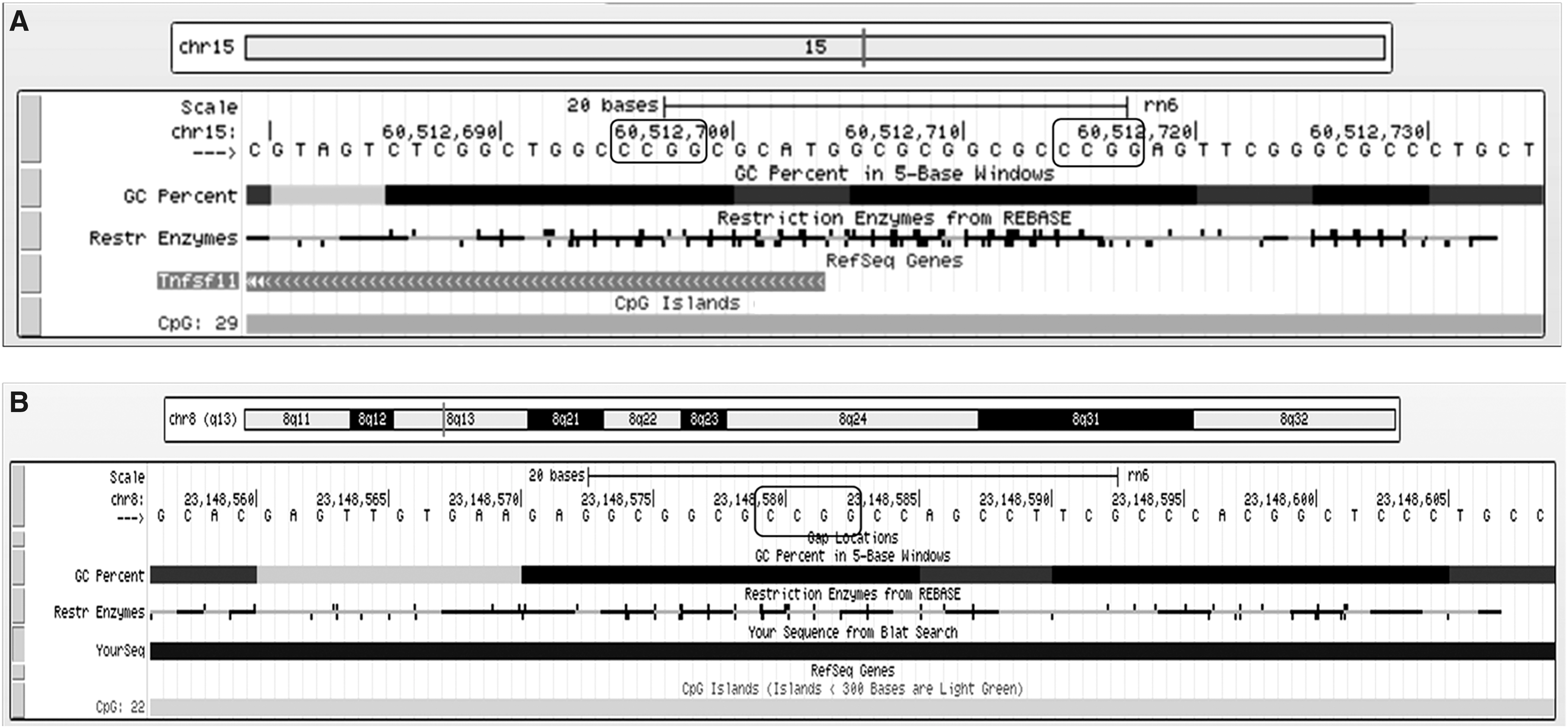
Technical data of the sequence used for Rankl
Statistical analysis
Data were initially examined for normality by Shapiro–Wilk test and expressed as mean ± standard deviation. One-way analysis of variance (ANOVA α ≤ 0.05) followed by pairwise multiple-comparison test (Tukey) (GraphPad Prism 5—GraphPad Software, Inc., San Diego, CA) was used to identify the difference among groups.
Results
Effect of CS and resveratrol on gene expression
Results demonstrated a large (three times) increase in the Sirt1 expression in those animals treated with resveratrol (CS/resveratrol intakers × CS intakers p ≤ 0.01; CS/resveratrol intakers × no CS intakers p ≤ 0.001). Ahr gene expression was significantly increased in the CS and CS/resveratrol, being almost thrice more when comparing to the no CS group (p ≤ 0.001) (Fig. 3).
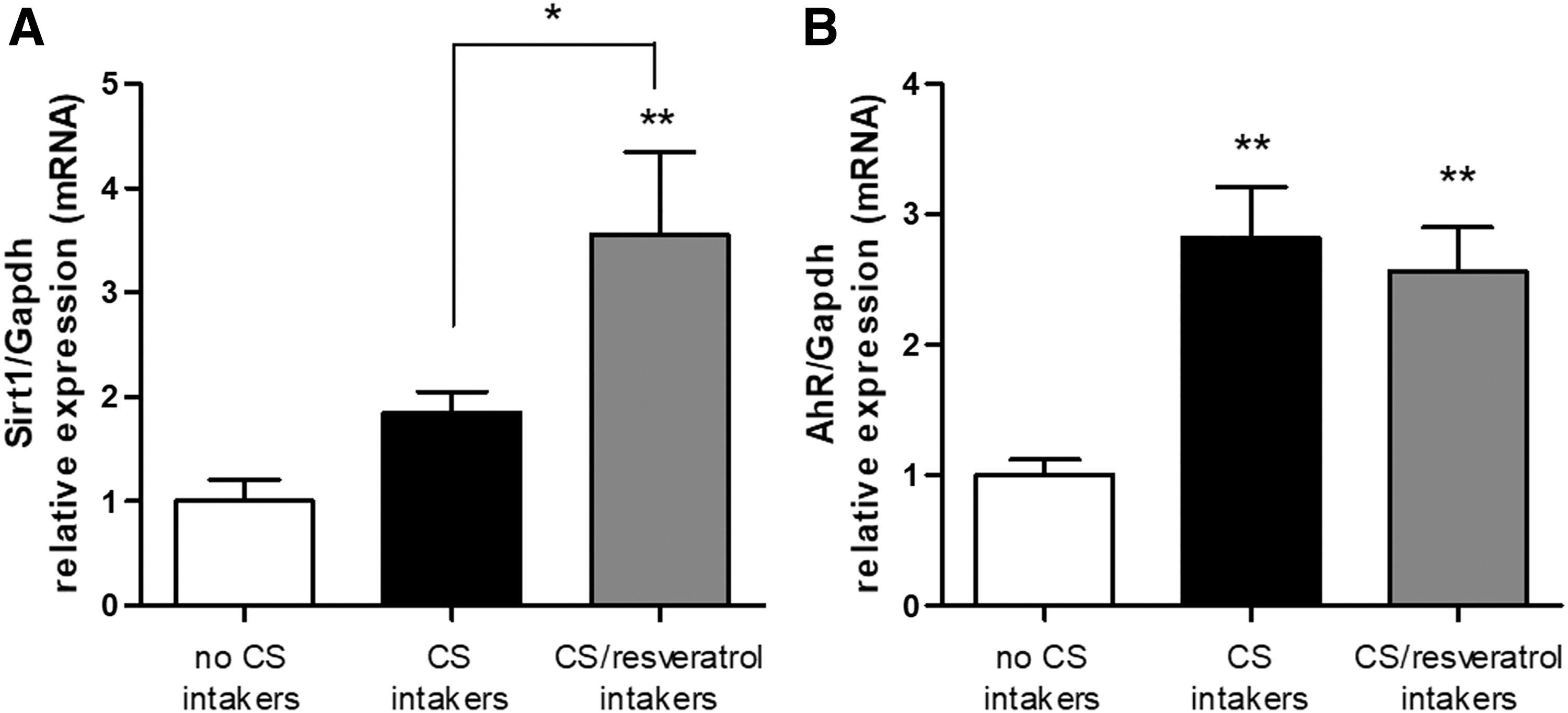
Changes in transcript levels of Sirt1 and Ahr after cigarette smoking inhalation and resveratrol intake. Sirt1
The expression of Dnmt1 was not significantly modulated, but the expressions of de novo DNA methyltransferases Dnmt3a (CS/resveratrol × CS p ≤ 0.05; CS/resveratrol × no CS p ≤ 0.001) and Dnmt3b (p ≤ 0.001) were increased after resveratrol intake (Fig. 4A–C). Dnmt3a demonstrated an 85% increase in resveratrol-treated animals, reaching the Dnmt1 gene expression levels, the most highly expressed DNA methyltransferase in these samples (Fig. 4D). The Dnmt3b was the least expressed DNA methyltransferase, and although it showed a 200% increase in the CS/resveratrol, the Dnmt3b did not reach the Dnmt1 and Dnmt3A transcript levels (Fig. 4D).
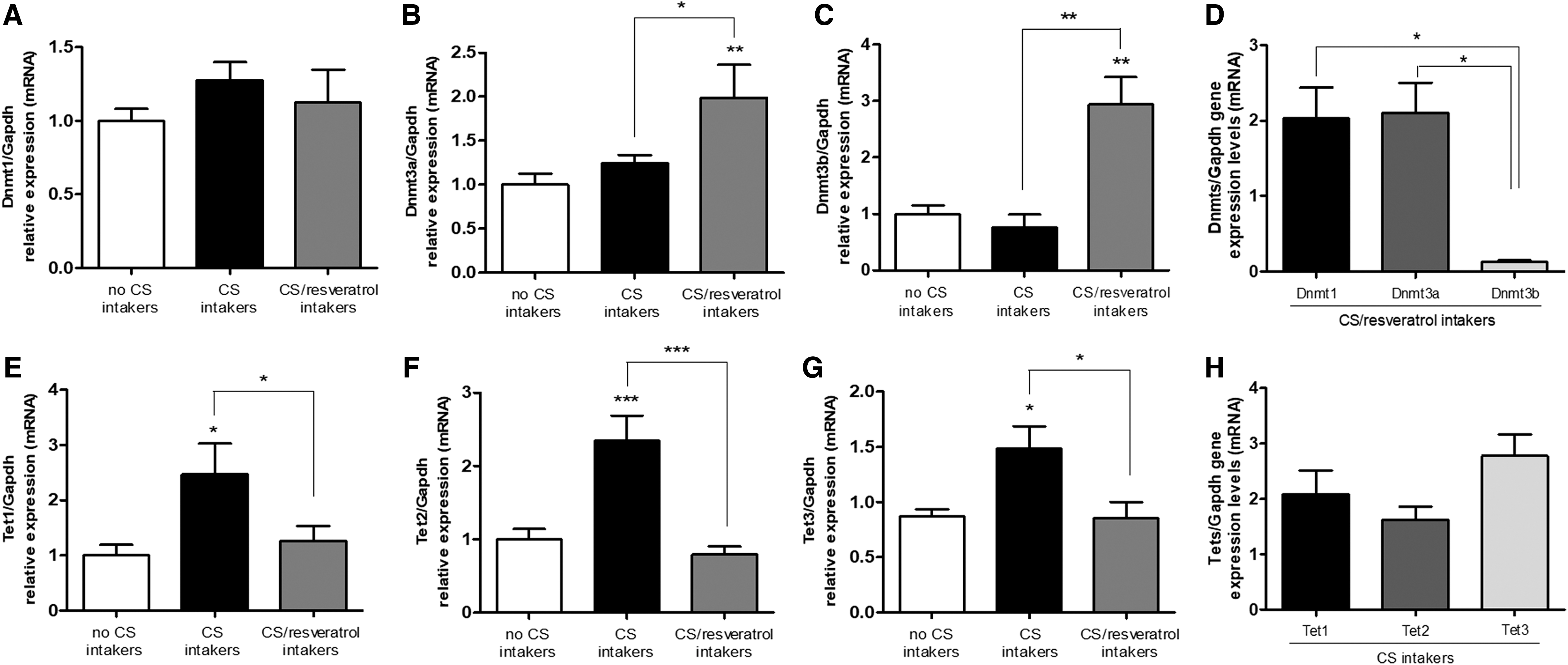
Changes in transcript levels of epigenetic machinery genes after cigarette smoking inhalation and resveratrol intake. Dnmt1
All Tets have responded, increasing their gene expression levels in the CS intakers, being almost twice, comparing to CS/resveratrol and no CS intakers. These increased levels were not observed in the CS/resveratrol animals; on the contrary, they presented similar levels to rats from the no CS intakers group (Fig. 4E–G). These differences for CS results in the Tets gene expression were statistically significant for Tet1 (p ≤ 0.05), Tet2 (p ≤ 0.0001), and Tet3 (p ≤ 0.05), the most highly expressed Tet in the samples (Fig. 4H).
Results demonstrated a significant increase (two times) in the transcript levels in the CS intakers group for Rankl and Trap (p ≤ 0.001), compared to no CS and CS/resveratrol intakers. The mRNA levels of Rankl and Trap were decreased in the animals exposed to CS and treated with resveratrol, compared to CS rats (p ≤ 0.001) and similar to no CS (Fig. 5B, C).

Global effects on DNA hydroxymethylation after cigarette smoking inhalation and resveratrol intake and changes in transcript levels and DNA modifications at gene-specific regulatory elements in osteogenic genes. Results were analyzed in newly formed bone tissue from calvaria, 30 days after defects were created.
Effect of CS and resveratrol on global DNA hydroxymethylation levels
Since TETS enzymes are involved in the DNA hydroxymethylation, we further investigate whether the increase in the Tets mRNA levels in the CS was correlated with an increase in the DNA hydroxymethylation global levels. The global DNA hydroxymethylation was assessed and demonstrated a statistically significant increase of ∼57%, in CS exposed rats (p ≤ 0.05), in line with the increased Tets transcript levels. When rats were exposed to CS and treated with resveratrol, the DNA hydroxymethylation global levels were reduced comparing to CS and similar to no CS intakers (p ≥ 0.05) (Fig. 5A).
Gene-specific changes in DNA methylation and hydroxymethylation after CS and resveratrol intake
The analyzed regions are within the promoters (±1.5 kB from the transcription start site), showing increased density of CpG sites. The CS-triggered decreases in methylation (12%) and increases in hydroxymethylation (144%) in the Rankl were statistically significant (p ≤ 0.001) and probably related to changes in gene expression (Fig. 5D). Furthermore, the CS inhalation also had effects in the Trap gene, and the absolute values showed a decrease of 10% (p ≤ 0.05) in the DNA methylation and a milder increase of 5% in the DNA hydroxymethylation levels, comparing to no CS intakers (Fig. 5E). An opposite pattern was observed among methylation and transcript levels and the higher the methylation levels, the lower the transcript levels and vice versa. There was no change in DNA methylation and hydroxymethylation levels at these genes in the CS/resveratrol intakers, when comparing to no CS intakers, demonstrating similar results between the groups. Again, the results from CS intakers' samples are in the opposite direction of the CS/resveratrol samples (Fig. 5).
Discussion
Epigenetic regulation is related to modifications in gene expression determined by environmental exposure, and these dynamic changes may occur in response to factors such as diets, aging, and toxins (Edwards and Myers, 2007). Therefore, CS and resveratrol may modulate gene expression through epigenetic mechanisms. Function and activity of bone cells might be affected by perturbation of epigenetic programs, contributing to pathogenesis of bone diseases (Park-Min, 2017); thus, having a better knowledge of epigenetic regulation would be relevant for premature diagnosis and therapeutic approaches in the future (Park-Min, 2017). Previous studies have pointed out changes in bone-related genes, in the presence of resveratrol and CS; however, the epigenetic regulation is still not fully recognized. In this study, we focused on epigenetic mechanisms that could be related to transcript changes in bone-related genes. Our results point to distinct patterns in epigenetic and transcript levels among CS rats treated or not with resveratrol. Importantly, resveratrol reverted the epigenetic and transcription changes caused by CS inhalation at both global and bone-related genes. It should be underlined although that, in general, the observed results of rats exposed to CS and treated with resveratrol resembled those from the nonexposed rats.
Previous results from our group showed that resveratrol optimized the repair of calvarial bone defects, increasing the gene expression of bone remodeling markers, in rats exposed to CS. The remaining bone defects of CS/resveratrol and no CS rats were similar and significantly reduced, compared to CS rats (Franck et al., 2018). The results of the present investigation are in line with these previous observations, and we demonstrated that epigenetic markers and mechanisms might act favoring the previous clinical outcomes observed, whereas Rankl and Trap showed changes at DNA methylation and hydroxymethylation linked to increased transcript levels in CS. These molecular results might contribute to differences observed in bone defect repair.
A significant increase in the Sirt1 and Ahr transcript levels was observed in the presence of resveratrol and CS, respectively, confirming the effectiveness of the administration in activating receptors. Shakibaei et al. (2011) suggested that the SIRT-1 activation, mediated by resveratrol, plays a relevant role in inhibiting RANKL-activated p300 acetyltransferase during osteoclastogenesis. In this study we showed epigenetic changes at Rankl and Trap promoters linked to a decrease in their transcripts, suggesting an additional mechanism for resveratrol/Sirt1 to that previous one described (Shakibaei et al., 2011) for controlling Rankl and Trap and, ultimately, regulating the bone balance. These results are in line with previous studies, when positive results of resveratrol/SIRT1 in bone metabolism were demonstrated, in the presence of CS (Shakibaei et al., 2011; Casarin et al., 2014; Edwards et al., 2013; Qu et al., 2016; Zhang et al., 2016; Franck et al., 2018). In fact, the characteristic of reversing the effects of CS by resveratrol was observed very often in the present investigation.
The role of DNA methylation and DNMTs in bone cells remains elusive; however, DNMTs could bind HDAC and mediate formation of repression complex surrounding promoter regions (Tang et al., 2009). Our results demonstrated that animals treated with resveratrol showed increased Dnmt3a, Dnmt3b, and Sirt1 transcript levels, which might be linked to the decrease in the Rankl and Trap gene expression, comparing to CS. Furthermore, the role of DNA hydroxymethylation and TETs in bone cells is not completely understood. Recently, it was shown that after stimulation with RANKL, monocytes become demethylated for TRAP gene, with an increase in the recruitment of TET2 at the promoter (de la Rica et al., 2013). In this study, we observed that CS inhalation stimulated a demethylation at both Rankl and Trap promoters with an increase in their transcripts. In addition, we observed an increase in Tets gene expression and global 5 hmC levels. Although we did not perform TET protein assays, the link between increased global 5 hmC levels and Tets transcripts and demethylation at Rankl and Trap promoters might exist, since DNA methylation changes during osteoclast differentiation are associated with TET2-mediated demethylation (de la Rica et al., 2013). Importantly, the increase in both global 5 hmC and Tets gene expression in the CS animals could lead to a relaxed state of chromatin with decreased DNA methylation, which may be required for modulating the transcripts of downstream genes, such as Rankl and Trap.
Genome-wide studies have pointed to CS being associated with changes in DNA methylation/hydroxymethylation patterns in health and several types of cancer (Coulter et al., 2013; Tsaprouni et al., 2014; Huang et al., 2015; Ambatipudi et al., 2016; Zhu et al., 2016). To date, it is known that a physical association between TET proteins and areas of relaxed chromatin do exist, affecting gene transcription. TET proteins are susceptible to environmental perturbations; hydroquinone, a carcinogen found in CS, causes an increase in TET1 activity and global 5 hmC in human embryonic kidney cell cultures (Gao et al., 2016), suggesting a link among carcinogen exposures, changes in the 5 hmC status, and TET1 activity. We did not analyze TETs activity; however, high levels of global 5 hmC and Tets transcripts in CS animals were addressed, in accordance with the hypothesis of CS modulating the global 5 hmC and Tets expression, corroborating the previous results from embryonic kidney cell cultures (Gao et al., 2016), whereas the increase in the global 5 hmC was also observed in this study, in bone tissue.
Conclusions
The results point to resveratrol reverting epigenetic and transcription changes caused by CS inhalation at both global and gene-specific levels in bone-related and epigenetic machinery genes. For the first time, the epigenetic regulation of Rankl and Trap genes was demonstrated in rats under CS and resveratrol intake, and it was linked to changes in their gene expression. These results emphasize the resveratrol as a biological modulator for CS in rats, and it might be considered a therapeutic agent to protect bone tissue from CS deleterious effects.
Footnotes
Acknowledgments
This work was supported by the São Paulo Research Foundation—FAPESP (Denise Carleto Andia—Grant number 2013/09650-8 and Fernanda Vieira Ribeiro—Grant number 2014/09480-8).
Disclosure Statement
No competing financial interests exist.