




Abstract
DNA condensed agents can improve the transfection efficiency of the cationic liposome delivery system. However, various condensed agents have distinct transfection efficiency and cellular cytotoxicity. The object of this study was to screen the optimal agents with the high transfection efficiency and low cytotoxicity from four polymer compressive materials, polyethylenimine (PEI), chitosan, poly-
Introduction
The research of gene therapy in various diseases is getting increasing attention with the development of the theories and technologies of gene therapy. However, developing a safe and efficient vector delivery system is one of the greatest difficulties in gene therapy (Chira et al., 2015). Generally, gene delivery systems are divided into viral vectors and nonviral vectors. Nonviral vectors including cationic liposomes and cationic polymers have attracted more attention than viral ones due to their good biodegradability, low immune response, and low toxicity. However, there are still some problems limiting their application, such as the relatively low transfection efficiency and the instability in serum.
Studies showed that nucleic acid compression could improve the transfection efficiency and the stability of the nucleic acid–cationic liposome complex (Friedrich et al., 2016). Many materials are available for nucleic acid compression. The basic mechanism of the compression is utilizing the interaction between the positive charge of the compressive materials and the negative charge of DNA with the electrostatic force. Polyethylenimine (PEI) is an organic macromolecule with high cationic charge density. Chitosan is the only one of natural polysaccharides with a large quantity of basic amino polysaccharides, which is linked by β (1.4)-glycosidic bonds between
It has been reported that through precompressing DNA, the cationic polymers can combine to liposomes to form liposomes–polymers–DNA complexes, which could effectively improve the transfection efficiency (Pires et al., 1999). Polymers such as protamine can compress DNA to be uniform and compact forms and improve the transfection efficiency of liposomes. However, the systematic comparison of these polymers in combination with liposomes as gene delivery vehicles has not been explored. Therefore, to further enhance the transfection efficiency and decrease the cytotoxicity of nonviral gene vectors, we utilized PEI, chitosan, spermidine, and PLL as nucleic acid compacts and combined with cationic liposomes to form ternary complexes in this study. The particle size, gel blocking experiments, in vitro transfection, cytotoxicity and other experiments were utilized to evaluate the effect of these four polymers on transfection efficiency and cell cytotoxicity. Furthermore, the effects of endocytosis inhibitors on the efficiency of nucleic acid delivery were examined, and the mechanism was elucidated from the perspective of endocytosis.
Materials and Methods
Materials
DOTAP (2,3-dioleoyloxy-propyl-trimethylammonium) were purchased from Corden Pharma Switzerland LLC. Cholesterol, PEI (25 kDa), chitosan, spermidine, and PLL were obtained from Sigma. pCDH-miR145-GFP plasmid (pDNA) was constructed by our own lab.
Cell culture
Human embryonic kidney transformed 293 (293T) was purchased from Shanghai Institutes for Biological Sciences and incubated in DMEM with 10% fetal bovine serum (FBS) and 1% antibiotic solution (penicillin–streptomycin, 10,000 U/mL) at 37°C in a humidified atmosphere containing 5% CO2.
Preparation of the cationic liposomes
The cationic liposomes were prepared by the thin-film dispersion method. A lipid mixture of DOTAP and cholesterol (molar ratio of 3:1) was dissolved in chloroform. The solvent was evaporated under vacuum by rotary evaporation for 30 min at 45°C to form the thin lipid film. The obtained lipid film was hydrated with 5% glucose. Then, we held the extruder to extrude liposomes 10 times, each through two stacks of progressively decreasing polycarbonate membranes with the pore sizes of 400, 200, and 100 nm (ME-25S). The liposomes were filtered to sterilize through 0.22 μm filter membranes and stored at 4°C.
Preparation of liposome–polymer–DNA complexes
The polymer–DNA nanoparticles were prepared by adding the polymer to an equal volume of plasmid DNA at different weight ratios. After incubation for 10 min at room temperature, the polymer–DNA complexes were mixed with an equal volume of liposome and then incubated for 20 min at room temperature to prepare the liposome–polymer–DNA nanoparticles (abbreviated as Lipo–polymer–DNA).
Measurement of particle size and zeta potential
The particle size and zeta potential of the liposomes, polymers, and liposome–polymer–DNA complexes were measured by using a laser particle size analyzer, Zetasizer Nano ZS90 (Malvern Instruments, Malvern, United Kingdom) after being dispersed in deionized water.
The gel retardation assay
The gel retardation assay was used to evaluate the DNA binding ability. The complexes including 1 μg DNA at various mass ratios of liposome–polymer were loaded onto individual wells of 1% agarose gel. Electrophorese was carried out in TBE buffer at 120 V for 30 min, and stained with ethidium bromide.
In vitro transfection experiment
The cells were seeded in a 12-well plate at a density of 1.5 × 10⁵ cells per well and incubated for 24 h. Before transfection experiments, the incubated cells were washed twice with PBS solution and the medium was replaced with serum-free DMEM. Then, the complexes were added into cells. After 6 h incubation, the medium was replaced with fresh medium. Cells were further incubated for 48 h before the transfection efficiency analysis. The transfection efficiency was analyzed by using the fluorescence microscope (Zeiss) and flow cytometer (BD Biosciences).
Cytotoxicity assay
293T cells were seeded at a density of 1 × 10⁴ cells per well in 96-well plates and incubated for 24 h. The culture medium was removed, and the cells were treated with liposome–polymer–DNA complexes in serum-free DMEM. The medium was replaced with 100 μL DMEM containing 10% FBS after 6 h incubation. After 48 h, the cells were incubated with 10 μL MTT reagent for 4 h, the formazan was dissolved by DMSO and the optical density at 570 and 630 nm was measured by using a microplate reader.
Intake inhibition experiments
Cells were treated with the endocytosis inhibitors containing 10 μg/mL chlorpromazine, 10 μg/mL cytochalasin D, 25 μg/mL nystatin, and 80 mM sodium chlorate in serum-free medium for 1 h at 37°C. Subsequently, Lipo–PEI–DNA or Lipo–spermidine–DNA was added and the transfection efficiency analysis was carried out. Simultaneously, we investigated the effect of temperature on the uptake of nanoparticles. After adding the Lipo–PEI–DNA and Lipo–spermidine–DNA, they were incubated at 37°C for 6 h. The transfection efficiency was analyzed. Transfection inhibition rate was described as (1 − experimental group/control group) × 100%.
Statistical analysis
All experiment results were represented as means ± standard deviations and calculated with the GraphPad prism 5.0 software.
Results
Different weight ratios of liposome, polymer, and DNA can condense the compound to different particle size
The particle size is one of the most important parameters for evaluating nanoparticles (Chen et al., 2016). Furthermore, the particle size can crucially influence the transfection efficiency of cationic liposomes. The particle sizes of the liposome–polymer–DNA complexes containing different amounts of polymers were determined, among which the mass ratio of immobilized liposome/DNA was 3:1 (Tao et al., 2016). The liposome–DNA complex was used as the control. The results showed that the liposome–polymer–DNA complex had a smaller particle size than those of the liposome–DNA or polymer–DNA complex (Fig. 1A–D). It means that precompression remarkably reduced the particle size of the complex. Although the change in zeta potential was not obvious, the zeta potential was the highest when the ratio of Lipo-PEI-DNA, Lipo-chitosan-DNA, Lipo-PLL-DNA, and Lipo–spermidine–DNA were 3:0.1:1, 3:0.8:1, 3:0.2:1, and 3:1:1, respectively (Fig. 1E). The optimal particle size was selected to employ into subsequent transfection efficiency and cytotoxicity experiments.
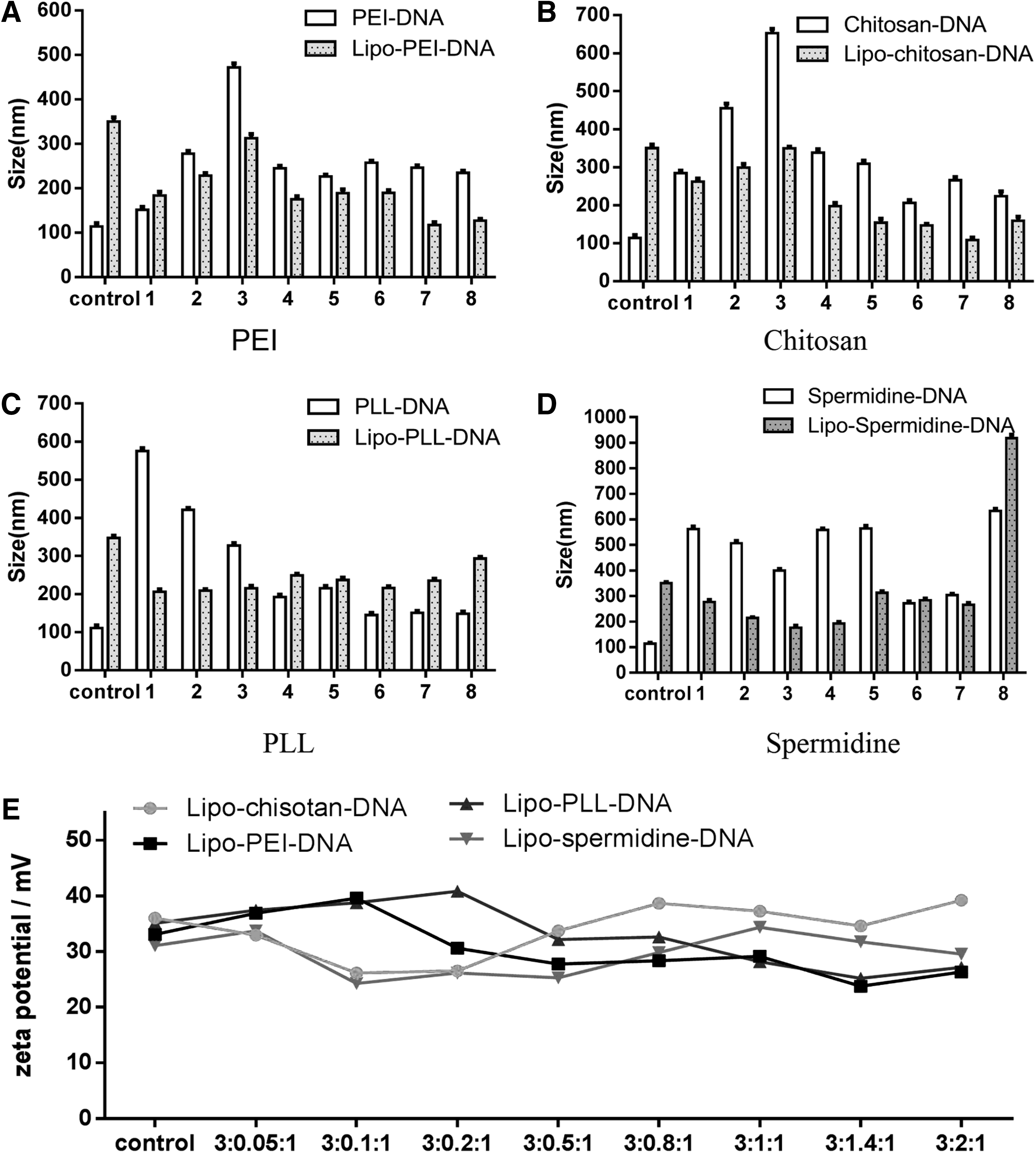
The particle sizes and zeta potential of polymer condensed materials–DNA or liposome–polymer condensed materials–DNA. The particle sizes
Spermidine had the weakest binding capacity to DNA among four polymer compressive agents
The gel retardation assays are widely used to determine the binding capability of cationic liposomes to DNA. As shown in Figure 2, the migration distance of DNA in the complex were altered when the nucleic acid complex were formed at different mass ratios of polymer compressive materials and DNA complex. The reason is that DNA partially or completely combined with the cationic compound results in slowing down of the electrophoresis speed and embodies in the short migration distance of DNA. For PEI and chitosan (Ahmed and Aljaeid, 2016), free DNA was almost impossible to be seen on agarose at polymer/DNA ≥0.5. It indicated that DNA was completely combined with or packaged by PEI and chitosan when polymer/DNA ≥0.5. For PLL, free DNA was rarely seen on the agarose at PLL/DNA ≥1.4, indicating that PLL was completely bound to the DNA at this ratio. However, the migration distance of Lipo–PLL–DNA complex was shorter than that of the naked plasmid on agarose at PLL/DNA <1.4, indicating that PLL was incompletely bound to DNA. Spermidine, incompletely encapsulated the nucleic acids, even though the spermidine–DNA mass ratio was >3, but the migration distance of the nucleic acids in the complex was reduced. This phenomenon probably was caused by the small molecule cationic characteristic of spermidine, which made it to not completely encapsulate DNA.

The gel retardation assays to determine the DNA binding capability of various polymers. The polymers were PEI
The transfection efficiency of Lipo–PEI and Lipo–spermidine copolymers were higher than those of Lipo–chitosan and Lipo–PLL
The transfection efficiency (Chettab et al., 2015) is the best indicator to evaluate the quality of nucleic acid delivery vectors. The transfection efficiency was determined by measuring the expression level of GFP protein, which depended on the weight ratio of liposome, compressed materials, and DNA of the complex in 293T cells (Figs. 3 and 4). The expression levels of GFP protein in liposome–polymer–DNA complex groups showed higher efficient transfection than those of the liposome–DNA complex groups when compression materials and DNA were formed at a certain ratio. For PEI (Zhang et al., 2013) as a compression material, the transfection efficiency was significantly increased from 30.0% to 45.5% (p < 0.01) when the mass ratio of liposome–PEI–DNA was 3:0.1:1. Chitosan as a compression material, the transfection efficiency was significantly increased from 30.0% to 42.4% (p < 0.05) when the mass ratio of liposome–chitosan–DNA was 3:0.8:1. For PLL as the nucleic acid compression material, the transfection efficiency was markedly enhanced from 30.0% to 36.8% (p < 0.05) when the weight ratio of liposome-PLL–DNA was 3:0.2:1. For spermidine as a compression material, the transfection efficiency was significantly improved from 30% to 47.4% when the mass ratio of liposome–spermidine–DNA was 3:1:1 (p < 0.01). Taken together, Lipo–PEI, Lipo–chitosan, Lipo–PLL, and Lipo–spermidine had higher transfection efficiency than those in the control groups (45.5%, 42.4%, 36.8%, and 47.4%, respectively) and spermidine was the most efficient transfection agent among these four compression agents, PEI came second.

The representative images of GFP fluorescence in the transfection experiments. The green fluorescence of GFP protein in plasmid was detected by a fluorescence microscope. The condensed materials of PEI
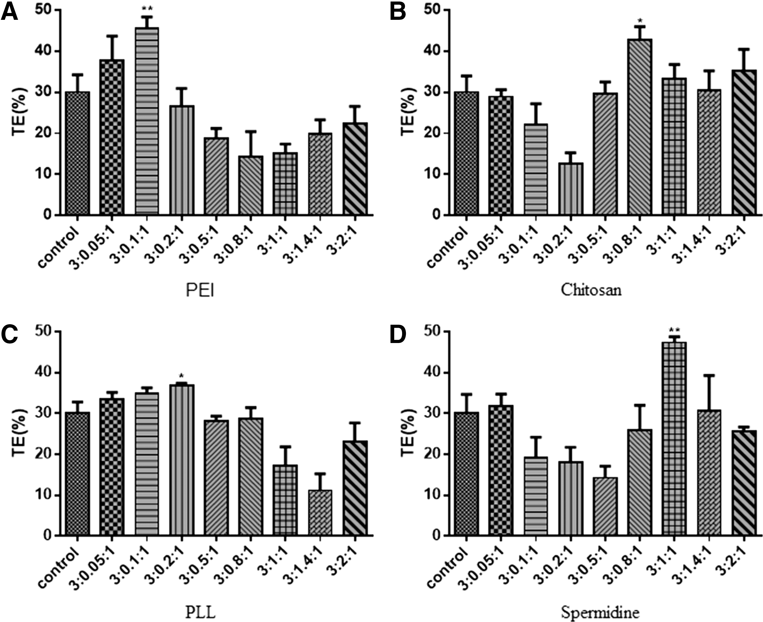
Transfection efficiency of liposome–polymer condensed agents–DNA nanoparticles in 293T cells. The condensed materials of PEI
Liposome–polymer–DNA complex had low cytotoxicity to 293T cell line
Improving safety is a major challenge to the preparation of nonviral vectors. The gene transfection efficiency of cationic copolymers is often limited by their cytotoxicity. In this experiment, the toxicity of the copolymers was evaluated using 293T cells by MTT assay. As shown in Figure 5, liposome–polymer–DNA complex showed a lower toxicity than that in the control groups, while ∼60% cell viability was maintained at the 3:1:1 ratio of liposome–PLL–DNA in 293T cells. Moreover, each component of liposome–polymer–DNA complex was very low toxic to 293T cells. The results showed that PEI, chitosan, PLL, and spermidine did not have significant effect on the cytotoxicity of the carrier, and all the compressed materials had good compatibility with 293T cells.
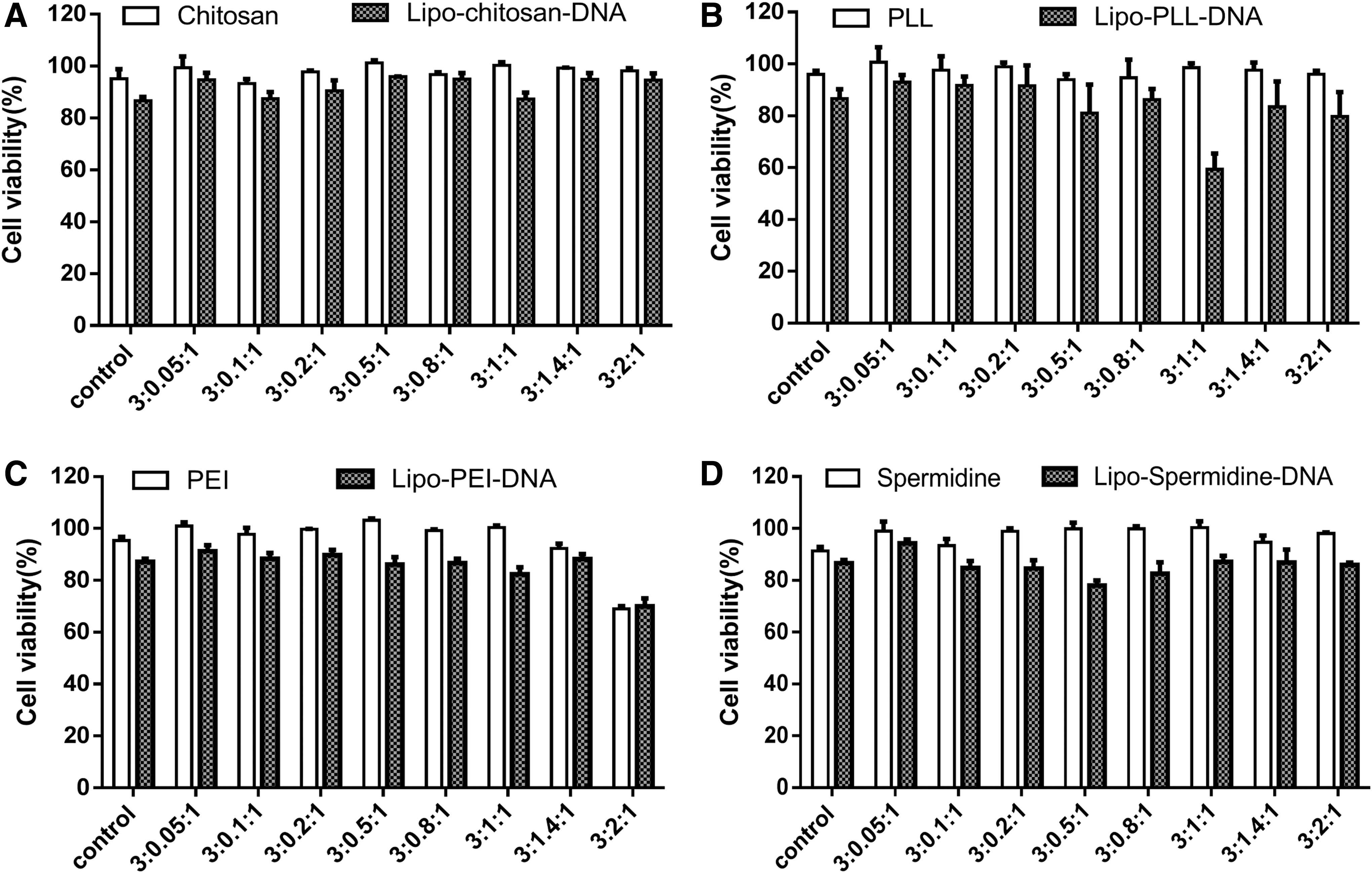
The cell viability of 293T cells transfected with different liposome–polymer condensed agents–DNA.
Lipo–spermidine–DNA and Lipo–PEI–DNA were mainly dependent on caveolin-mediated endocytosis pathway
As mentioned above, we verified that spermidine and PEI were superior compression materials than the other two. Therefore, to further clarify the delivery effect of spermidine and PEI, we explored the endocytic pathways of them in 293T cells., The transfection efficiency of Lipo–PEI and Lipo–spermidine copolymers under the action of endocytic channel inhibitors in 293T cells were determined and the fluoresce stainings were also shown (Fig. 6). The transfection efficiency of Lipo–PEI copolymer decreased by 39.2% and the decrement of Lipo–spermidine was 18.5% after treating with chlorpromazine in 293T cells (Fig. 7). After treating with cytochalasin D, the transfection efficiency of Lipo–spermidine and Lipo–PEI copolymers increased by 27.4% and 11.48%, respectively, and the cell growth rate of transfected by Lipo–spermidine–DNA was larger (Fig. 7). While treating by nystatin, the transfection efficiency of Lipo–spermidine and Lipo–PEI copolymers were decreased by 46.05% and 51.88%, respectively. However, after treating with sodium chlorate, the transfection efficiency of Lipo–spermidine and Lipo–PEI decreased by 32.68% and 46.33% in 293T cells, respectively. Together, the results indicated that Lipo–spermidine–DNA and Lipo–PEI–DNA entered into 293T cells and were mainly dependent on caveolin-mediated endocytosis pathway.

The representative morphology of 293T cells transfected by liposome–polymer condensed agents–DNA with each channel inhibitor treatment. Chlorpromazine, nystatin, cytochalasin D, and sodium chlorate were used as clathrin inhibitor, caveolin inhibitor, actin depolymerization agent, and ATPase inhibitor, respectively. Lipo–spermidine–DNA, liposome combined with spermidine and DNA; Lipo–PEI–DNA, liposome combined with PEI and DNA; Upper in each group, under the bright microscope field of vision; Lower in each group, under the fluorescence microscope field of vision; Control (the control group), Lipo–spermidine–DNA or Lipo–PEI–DNA without various channel inhibitor treatments.

The transfection efficiency in 293T cells by Lipo–spermidine–DNA and Lipo PEI–DNA with various channel inhibitor treatments. After transfection with Lipo–spermidine–DNA
Discussion
Using nonviral vectors as vehicles transferring genes is a safe and effective method in gene therapy. Cationic liposomes and micelles are important components of nonviral delivery vectors. Although cationic liposomes have the advantages of low immune response and good biodegradability, their uses are limited due to less transfection efficiency, high cytotoxicity, and instability in serum. DOTAP–cholesterol liposome has proven to be one of the most potent gene delivery vectors, however, it still faces two major challenges of difficult escape from endosomes and difficult entry into the nucleus. Therefore, this study explored four compressive materials, PEI, PLL, chitosan, and spermidine combined with DOTAP–cholesterol liposome to improve the transfection efficiency and cellular cytotoxicity. Our results verified that liposome–spermidine was the optimal gene delivery vehicle among these four compressive agents at 3:1:1 of the weight ratio of liposome–spermidine–DNA, where endocytosis pathway was probably caveolin dependent.
Cationic polymers are also currently used as gene delivery vehicles. Polymer–DNA complexes were self-assembled by using the cationic groups of the cationic polymers in combination with the negative charge of the phosphate groups on the DNA. However, the low gene delivery efficiency and the high toxicity limit their use. PEI, chitosan (Richardson et al., 1999; Mumper and Rolland, 2014), PLL (Forsman et al., 2017), and spermidine as four classical cationic polymers have been widely used to design the carrier delivery vehicles.
Spermidine (Zhang et al., 2017) is a polyamine compound. Goytia et al. (2015) reported that spermidine could induce DNA aggregation to achieve further compression. Spermidine could increase the DNA replication and transcription to improve its transfection efficiency in cells (Raspaud et al., 2005). However, the stability of the complex was affected by the distance between amino groups of the linear spermidine and DNA or RNA. Therefore, the combination of spermidine and liposome improve the transfection efficiency for its property of compression DNA. In this research, we found that spermidine modified by DOTAP–cholesterol liposomes could conjunct with the phosphate groups on the DNA though it was difficult to completely encase the DNA (Fig. 2D) and the transfection efficiency was the best, which increased by 47.4% compared to the control group and other three compressive agents. Although spermidine has the weaker binding capability to DNA, combining with liposomes helps to improve its ultimate transfection efficiency.
PEI can promote the escape of DNA endosomes because of its proton sponge effect to avoid DNA degradation by lysosomes and thereby DNA is protected to achieve a more effective delivery of genes. However, it is more toxic to cells if used as a carrier alone. Jiang et al. (2010) reported that cholesterol-modified PEI (PEI–cholesterol) can effectively reduce their own cytotoxicity and enhance transfection efficiency in A549 and MCF-7 cells. The use of PEI-mediated delivery of miR-145 achieved a good anticolon cancer therapeutic efficacy in mouse models (Ahmed and Narain, 2013). PEIs were classified as linear PEI and branched PEI depending on their structure. Branched PEI reflected higher transfection ability with higher toxicity than the linear one. However, the different molecular weight of branched PEI has distinct cytotoxicity and 25 kDa branched PEI was the most effective and widely used PEIs for its relatively low toxic and high transfection capability. Therefore, the combination of 25 kDa branched PEI and liposomes were used as a gene delivery vector in our study.
Particle size is a key parameter of nanoparticle associated with the delivery effect of nonviral vectors. The transfection efficiency of the large particle size of liposome–polymers complex was higher than that of the small particle size of complex (Masotti et al., 2009). However, our results found that the size of the liposome–polymers complex did not always positively correlate with its transfection efficiency. The particle size of the composite reached the maximum when the ratio of liposome–PEI–DNA was 3:0.2:1, while the transfection efficiency was not optimal. The transfection efficiency of the complex was the best when the ratio of liposome–PEI–DNA was 3:0.1:1. So did chitosan and other compressed materials. Results of this study might reveal that mechanisms of gene vector enter into cells were different when different types of complexes were used; and it was also possible that complexes at this ratio were more likely to escape lysosomes.
Zeta potential is an important physicochemical parameter of nanoparticles. The ratio with the highest transfection efficiency of the four materials was similar with the ratio with that of highest zeta potential, which indicated that higher the surface potential of the particles higher the tendency of transfection efficiency. This may be due to the fact that the surface of the complex is positively charged, where positive potential is higher and the ability to bind to the negative charge on the surface of the cell membrane is stronger, so it is more advantageous for transfection (Fig. 1).
Chlorpromazine, nystatin, cytochalasin D, and sodium chlorate often act as clathrin inhibitor, caveolin inhibitor, actin depolymerization agent, and ATPase inhibitor, respectively. With nystatin treatments, the transfection efficacy of Lipo–PEI–DNA and Lipo–spermidine–DNA complexes in 293T cells were significantly inhibited than those of the control groups, while cytochalasin D, chlorpromazine, and sodium chlorate were not. Therefore, the endocytosis pathway of Lipo–spermidine and Lipo–PEI copolymers was probably caveolin dependent. The in-depth study of these pathways may be beneficial to fundamentally improve the design of nonviral vectors.
Footnotes
Disclosure Statement
The authors declare no conflict of interest.
Funding Information
The work was supported by Nantong People's Livelihood Science and Technology Plan (No. MS12018032). It was partly financially supported by the National Key Research and Development Program of China (No. 2016YFC0800906) and the Technology Research Program of the Ministry of Public Security (No. 2016JSYJA32).