
Abstract
The long noncoding RNA cancer susceptibility candidate 2 (CASC2) has been shown to play a crucial role in cancer cell chemoresistance. However, its function and underlying molecular mechanism in hepatocellular carcinoma (HCC) chemoresistance remain unknown. In this study, we used cisplatin (DDP)-resistant HCC cells to investigate CASC2 function and its underlying mechanism. The results demonstrated that CASC2 expression was significantly reduced in HCC tissues and cells, especially in DDP-resistant HCC tissues and cells. Lower CASC2 expression was strongly correlated with shorter survival times in patients with HCC. Functionally, CASC2 overexpression sensitized DDP-resistant Huh7/DDP and SMMC-7721/DDP cells to DDP. Mechanically, CASC2 improved the sensitivity of HCC cells to DDP through sponging miR-222. Taken together, these findings suggested that overexpression of CASC2 overcame DDP resistance in HCC by regulating miR-222 expression, thereby providing a potential therapeutic strategy for overcoming HCC cell chemoresistance.
Introduction
Hepatocellular carcinoma (HCC), the most common liver malignancy, has a high mortality rate and poses a threat to human health (El-Serag and Mason, 1999; Shen et al., 2016; Zhang et al., 2017). Despite remarkable therapeutic advances, the prognosis of patients with HCC remains poor due to the acquisition of multidrug resistance (Lebwohl and Canetta, 1998). Cisplatin (DDP) is one of the most common and effective chemotherapeutic drugs used for the treatment of various cancers (Giaccone, 2000; Galanski, 2006). However, DDP-based chemotherapy is often limited by intrinsic and acquired resistance, which ultimately leads to chemotherapy failure (Koberle et al., 2010; Johnsson et al., 2014). Therefore, there is an urgent need to understand the pathogenesis of HCC chemoresistance and develop novel effective therapeutic strategies for improving the outcome of patients with HCC.
Long noncoding RNAs (lncRNAs) are nonprotein-coding transcripts longer than 200 nucleotides (Martens-Uzunova et al., 2014; Sun et al., 2015). Recently, lncRNAs have been shown to be aberrantly expressed in multiple cancers, and their roles in tumorigenesis, progression, and drug resistance have been elucidated (Palmieri et al., 2017; Lin and Yang, 2018). Cancer susceptibility candidate 2 (CASC2), located on chromosome 10q26, has been reported to function as a tumor suppressor in human malignancies (Wang et al., 2017a). Previous studies have confirmed that CASC2 is dramatically underexpressed in several malignancies, such as HCC (Baldinu et al., 2007), endometrial cancer (Gao et al., 2019), and prostate cancer (Ambros, 2001). However, the function of CASC2 in HCC remains uncertain.
MicroRNAs (miRNAs), a new class of small noncoding RNAs (18–25 nucleotides), exert their function by directly binding the 3′untranslated region of mRNAs, resulting in inhibition of mRNA translation and degradation (Carrington and Ambros, 2003; Bartel, 2004; Guo et al., 2015). It has been shown that lncRNAs interact with miRNAs and modulate the expression of their target genes (Wang et al., 2014; Feng et al., 2017). For instance, a previous study showed that CASC2 might function as a competing endogenous RNA (ceRNA) to upregulate phosphatase and tensin homolog expression through sponging miR-18a, thus enhancing the sensitivity of cervical cancer cells to DDP (Feng et al., 2017). Jiang et al. (2018) revealed that CASC2 functioned as a miR-193a-5p sponge to sensitize glioma cells to temozolomide cytotoxicity by inhibiting autophagy and promoting cell death. CASC2 overexpression has been shown to enhance the DDP sensitivity of gastric cancer cells through sponging miR-19a, thereby serving as a new therapeutic target for chemotherapy-resistant gastric cancer (Li et al., 2018). The molecular mechanism by which CASC2 regulates DDP resistance in HCC, however, remains unclear.
In this study, we aimed to investigate the functional role of CASC2 and its underlying molecular mechanism in the DDP resistance of HCC cells. Our results showed that CASC2 expression was downregulated in HCC tissues and cells, suggesting that the CASC2/miR-222 axis might be a novel target for overcoming DDP resistance in HCC.
Materials and Methods
Collecting samples
We collected 40 paired tumor and adjacent normal tissue samples from HCC patients after approval by the Ethics Committee of the Central Hospital of Petrochina. Samples were obtained from patients who underwent surgical resection at the Department of Hepatobiliary Surgery, Central Hospital of Petrochina. All patients signed the consent form. The tissue samples were immediately stored in liquid nitrogen until further use (Supplementary Table S1).
Cell culture and transfection
Human normal liver cell line (HL-7702) and human liver cancer cell lines (Huh7 and SMMC-7721) were obtained from American Tissue Culture Collection (ATCC, Manassas, VA). All cell lines were cultured in Roswell Park Memorial Institute-1640 medium (HyClone, Logan, UT) supplemented with 10% fetal bovine serum (Gibco) and incubated at 37°C with 5% v/v CO2. The DDP-resistant cell lines, Huh7/DDP and SMMC-7721/DDP, were established by exposing their parent cells to increasing concentrations of DDP for 12 months.
Empty pcDNA3.1 vector (Vector) and CASC2-overexpressing pcDNA3.1-CASC2 vector (CASC2) were synthesized by GeneCopoeia (Guangzhou, China). miR-con, miR-222, anti-miR-con, and anti-miR-222 were purchased from GenePharma (Shanghai, China). The cells were cotransfected with Vector or pcDNA3.1-CASC2 and miR-con, miR-222, anti-miR-con, or anti-miR-222 using Lipofectamine 2000 (Invitrogen, Shanghai, China). Cells were collected for further assays 48 h after transfection.
Isolation of RNA and quantitative real-time polymerase chain reaction
Total RNA was extracted from tissues or cells using TRIzol reagent (Invitrogen) according to the manufacturer's instructions. Total RNA was reverse transcribed into cDNA using PrimeScript RT Reagent Kit (TaKaRa, Tokyo, Japan). Quantitative real-time polymerase chain reaction (qRT-PCR) was performed to determine the expression of CASC2 and miR-222. Glyceraldehyde 3-phosphate dehydrogenase (GAPDH) and U6 were used as the internal reference genes for CASC2 and miR-222, respectively. The relative expression of CASC2 and miR-222 was calculated by the 2−ΔΔCt method.
DDP sensitivity assay
To determine the viability of cells after DDP treatment, 3-(4, 5-dimethyl-2-thiazolyl)-2, 5-diphenyltetrazolium bromide (MTT) assay was performed. The transfected cells were seeded in 96-well plates. After 24 h of incubation at 37°C, Huh7/DDP and SMMC-7721/DDP cells were treated with different concentrations of DDP (0.5, 1, 2, 4, 8, 16, 32, and 64 μg/mL) for 48 h. MTT solution (10 μL; 5 mg/mL) was then added into each well, and the cells were incubated for 4 h. Dimethyl sulfoxide (DMSO) (200 μL) was added into each well to dissolve the formazan crystals. The optical density was measured at 490 nm. The viability of the control cells was defined as 100%. The sensitivity of HCC cells to DDP was assessed using the half maximal inhibitory concentration (IC50).
Flow cytometry
Flow cytometry was used to assess cell apoptosis. The transfected Huh7/DDP and SMMC-7721/DDP cells were incubated with 2 μg/mL of DDP for 48 h, collected, and washed with 1 × phosphate buffer saline. The cells were then resuspended in 1 × binding buffer and stained with 5 μL Annexin V-FITC and 5 μL propidium iodide for 10 min at room temperature in the dark. Following this, cell apoptosis was measured by fluorescence-activated cell sorting.
Luciferase reporter assay
Two luciferase reporters containing the wild-type CASC2 (CASC2-WT) or mutant-type CASC2 (CASC2-MUT) were generated to analyze the relationship between CASC2 and miR-222. Huh7/DDP and SMMC-7721/DDP cells were seeded in 24-well plates and incubated for 24 h. Cells were then cotransfected with either of the two luciferase plasmids and miR-222 mimics or miR-con (100 ng/well) using Lipofectamine 2000 (Invitrogen). At 48 h post-transfection, luciferase activity was measured using the Dual Luciferase Reporter Assay System (Promega, Madison, WI) as per the manufacturer's protocol.
Statistical analysis
The values represent the mean ± standard deviation of at least three independent experiments. Statistical analysis of all data was carried out using SPSS 20.0 software (IBM, NY). A p value of <0.05 was considered statistically significant.
Results
lncRNA CASC2 was downregulated in DDP-resistant HCC tissues and cells
To investigate the functional role of CASC2 in HCC, the expression level of CASC2 in HCC tumor tissues (n = 40) and adjacent normal tissues (n = 40) was evaluated by qRT-PCR. Results showed that CASC2 expression was significantly decreased in HCC tumor tissues compared with that in adjacent normal tissues (Fig. 1A). Next, the expression of CASC2 in the tumor tissues of DDP-sensitive and DDP-resistant HCC patients was further analyzed by qRT-PCR. The results showed that CASC2 expression was remarkably downregulated in DDP-resistant patients compared with that in DDP-sensitive patients (Fig. 1B). In addition, to assess CASC2 expression in HCC cell lines, qRT-PCR was performed in the liver cancer parental cell lines (Huh7 and SMMC-7721), their corresponding DDP-resistant cell lines (Huh7/DDP and SMMC-7721/DDP), and human normal liver cell line (HL-7702). Compared with that in HL-7702 cells, CASC2 expression was significantly decreased in Huh7 and SMMC-7721 cells (Fig. 1C, D). More importantly, the expression of CASC2 in Huh7/DDP and SMMC-7721/DDP cells was lower than that in their parental cells (Fig. 1C, D). Kaplan–Meier survival analysis showed that the survival rate of HCC patients with high CASC2 expression levels was higher than those with low CASC2 expression levels (p = 0.0219) (Fig. 1E). Taken together, these results suggested that CASC2 may be involved in the DDP resistance of HCC cells.

Expression of CASC2 was downregulated in HCC tissues and cells.
CASC2 overexpression enhanced the sensitivity of DDP-resistant HCC cells to DDP
To evaluate whether CASC2 overexpression reverses the DDP resistance of Huh7/DDP and SMMC-7721/DDP cells, the IC50 value of DDP in Huh7/DDP and SMMC-7721/DDP cells and their parental cells was determined by the MTT assay. As demonstrated in Figure 2A and B, the IC50 values of DDP were obviously higher in SMMC-7721 and Huh7 cells than those in SMMC-7721/DDP and Huh7/DDP cells. To further investigate the functional roles of CASC2 in DDP-resistant HCC cells, we established two stable CASC2-overexpressing resistant cell lines by transfecting pcDNA3.1-CASC2 into SMMC-7721/DDP and Huh7/DDP cells. The results of qRT-PCR analysis showed that the expression levels of CASC2 were markedly increased in pcDNA3.1-CASC2-transfected SMMC-7721/DDP and Huh7/DDP cells compared with their parent cells (Fig. 2C, D). As expected, the results revealed that CASC2 overexpression remarkably improved the sensitivity of SMMC-7721/DDP and Huh7/DDP cells to DDP (Fig. 2E, F). Next, we evaluated the effects of CASC2 overexpression on DDP-induced apoptosis in SMMC-7721/DDP and Huh7/DDP cells using flow cytometry. As shown in Figure 2G and H, SMMC-7721/DDP and Huh7/DDP cells transfected with pcDNA3.1-CASC2 showed a significant increase in DDP-induced apoptosis compared to cells transfected with pcDNA3.1. These findings indicated that overexpression of CASC2 could overcome DDP resistance in DDP-resistant HCC cells.
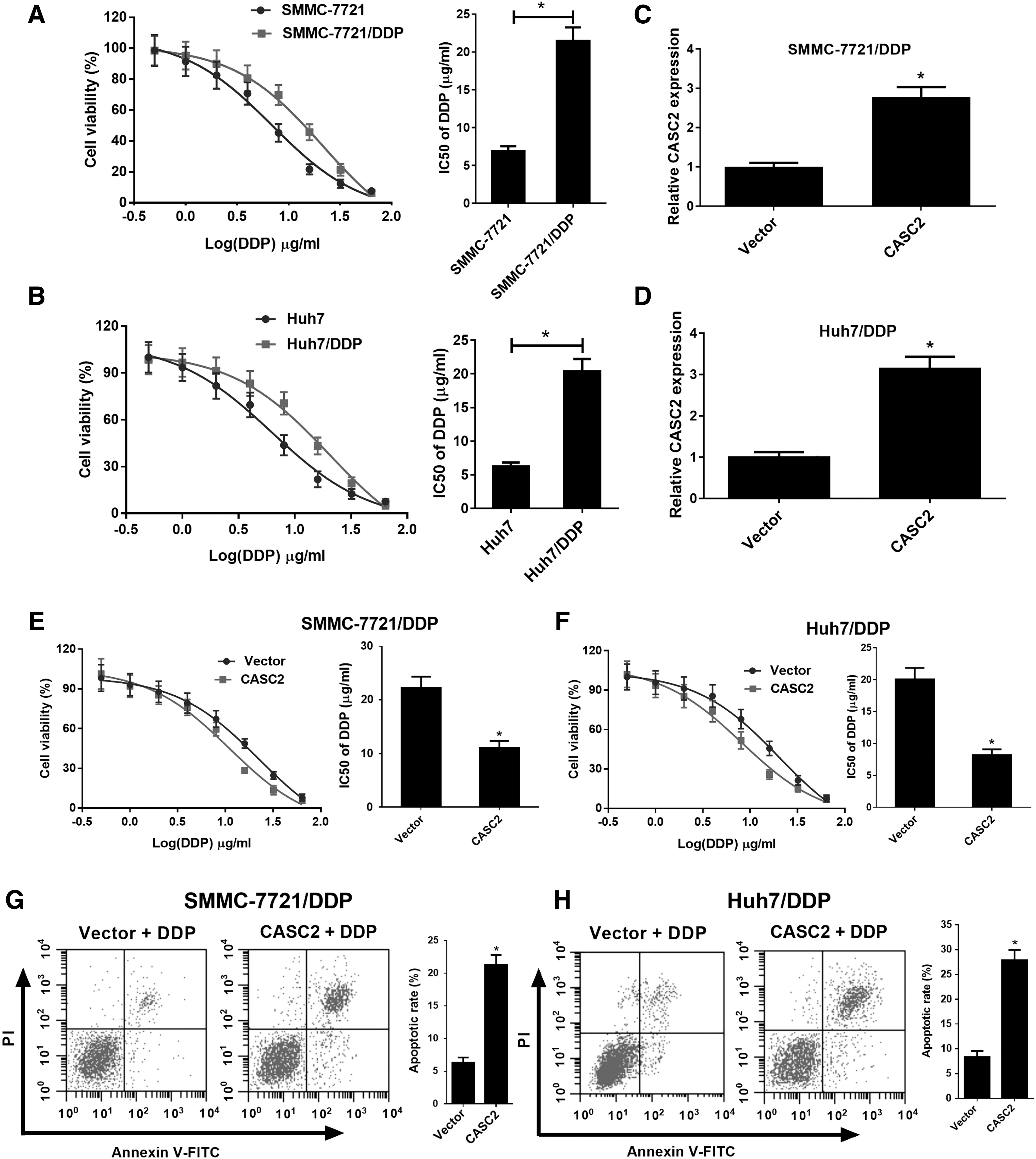
CASC2 overexpression enhanced the DDP sensitivity of SMMC-7721/DDP and Huh7/DDP cells.
CASC2 sponges miR-222 in HCC cells
Next, we investigated the possible mechanism by which CASC2 mediated DDP resistance in HCC cells. The online software miRcode was used to identify the miRNA targets of CASC2 (
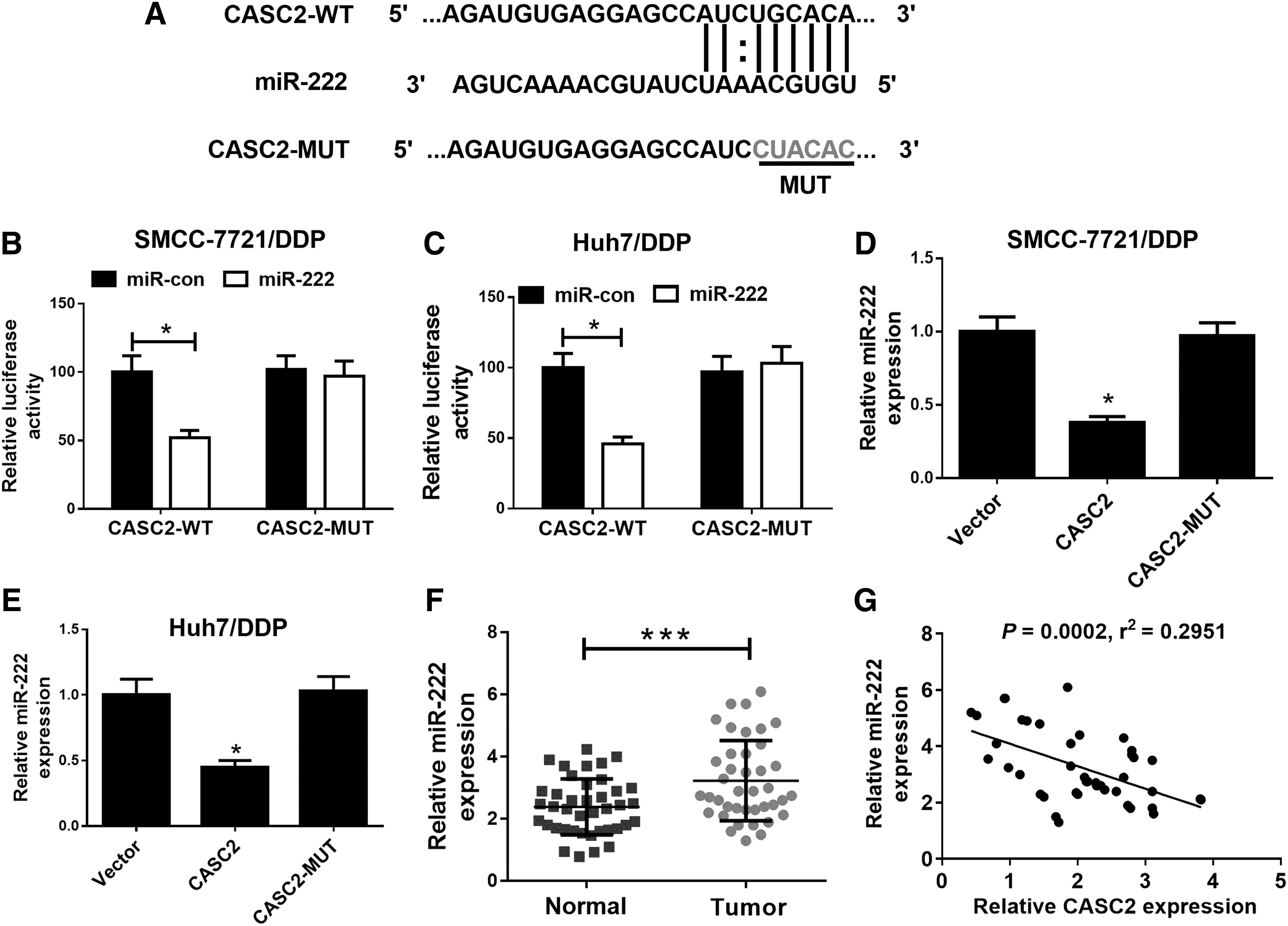
CASC2 acted as a miR-222 sponge in HCC cells.
CASC2 overexpression overcame DDP resistance in DDP-resistant HCC cells through suppressing miR-222 expression. SMMC-7721/DDP and Huh7/DDP cells were cotransfected with Vector or pcDNA3.1-CASC2 and miR-con, miR-222, anti-miR-con, or anti-miR-222, and miR-222 expression was determined by qRT-PCR
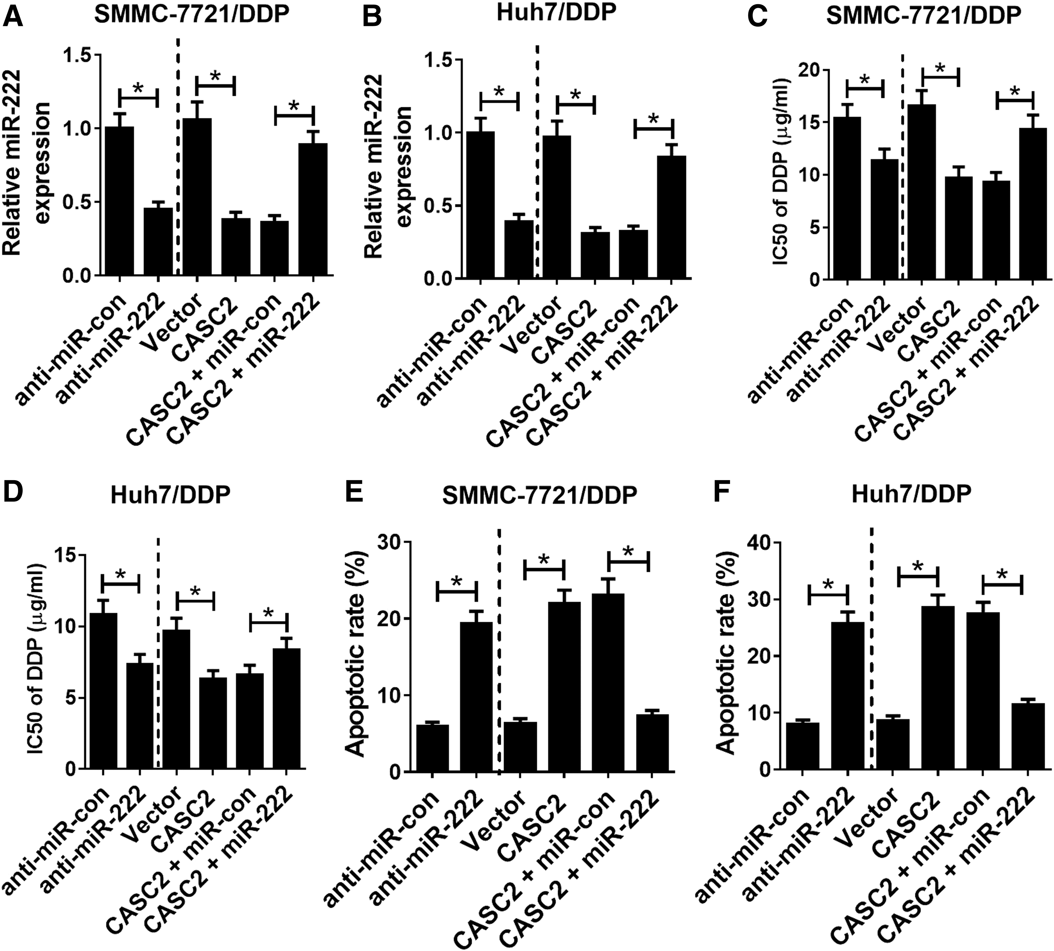
Overexpression of CASC2 sensitized DDP-resistant HCC cells to DDP through suppressing miR-222 expression
The above results indicated that miR-222 can directly bind to CASC2. Hence, we further explored the role of CASC2 in enhancing the sensitivity of HCC cells to DDP through regulating miR-222 expression. Results of qRT-PCR showed that transfection with anti-miR-222 inhibited miR-222 expression in SMMC-7721/DDP and Huh7/DDP cells. Furthermore, miR-222 overexpression abolished the inhibitory effect of CASC2 on miR-222 expression in SMMC-7721/DDP and Huh7/DDP cells (Fig. 4A, B). The results of MTT assay demonstrated that inhibition of miR-222 expression improved the sensitivity of SMMC-7721/DDP and Huh7/DDP cells to DDP (Fig. 4C, D). We further analyzed the effect of CASC2 and miR-222 on DDP-induced apoptosis. Flow cytometry results showed that anti-miR-222 transfection significantly increased DPP-induced cell apoptosis in SMMC-7721/DDP and Huh7/DDP cells exposed to 2 μg/mL DDP, while upregulation of miR-222 suppressed CASC2-induced SMMC-7721/DDP and Huh7/DDP cell apoptosis (Fig. 4E, F). These findings confirmed our hypothesis that overexpression of CASC2 improves DDP sensitivity in DDP-resistant HCC cells through suppressing miR-222 expression.
Discussion
The role of dysregulated lncRNAs in the chemoresistance of various cancers has received recent attention (Wang et al., 2012, 2019; Zhao et al., 2016; Jiang et al., 2018; Jin et al., 2018). Studies have suggested that CASC2 plays a key role in DDP resistance. In the present study, we found that CASC2 expression was significantly decreased in DDP-resistant HCC tissues and cells. HCC patients with low CASC2 expression had shorter survival rates, which prompted us to further study the potential role of CASC2 in the chemoresistance of HCC. DDP treatment resulted in decreased miR-222 expression in human HCC cells. Moreover, we observed that CASC2 overexpression enhanced the DDP sensitivity of SMMC-7721/DDP and Huh7/DDP cells by promoting cell apoptosis, suggesting that upregulation of CASC2 can enhance the sensitivity of HCC cells to DDP. Hence, CASC2 may serve as a promising therapeutic target for overcoming DDP resistance in HCC.
Elucidating the molecular mechanisms underlying DDP resistance may help contribute to the development of effective therapies to overcome DDP resistance. Our results revealed that CASC2 expression was downregulated in HCC tissues and cells. CASC2 has been reported to act as a tumor suppressor, and its expression has been shown to be downregulated in many cancers (Wang et al., 2015; Liao et al., 2017; Yu et al., 2019). Numerous studies have reported that abnormal expression of CASC2 is implicated in chemoresistance in various cancers. For instance, lncRNA maternally expressed gene 3 was shown to act as a ceRNA to sponge miR-21-5p, thus repressing miR-21-5p expression and enhancing the sensitivity of nonsmall cell lung cancer cells to DDP (Wang et al., 2017b). Overexpression of CASC2 enhanced the sensitivity of cervical cancer cells to DDP by acting as a miR-21 sponge (Feng et al., 2017). Moreover, downregulated miR-21 was shown to increase the sensitivity of melanoma cell line A375 to DDP (Zhang et al., 2018). Furthermore, CASC2 has been shown to function as a ceRNA by sponging miR-18a in colorectal cancer to suppress tumor cell proliferation (Huang et al., 2016).
In our study, we demonstrated that overexpression of CASC2 contributed to the sensitivity of SMMC-7721/DDP and Huh7/DDP cells to DDP. The precise mechanism by which downregulation of CASC2 led to DDP resistance in HCC remains unclear. Hence, we explored the functional role of the CASC2/miR-222 axis in the sensitivity of HCC cells to DDP. The ceRNA hypothesis suggests that lncRNAs function as miRNA sponges to repress the function of miRNA (Wang et al., 2017b; Jiang et al., 2018). The online software miRcode was utilized to predict the potential miRNA target of CASC2. Growing evidences suggest that miR-222 functions as a key oncogene. For instance, a previous study reported that miR-222 overexpression may be a key contributor to tumorigenesis (Wong et al., 2010). Moreover, miR-21, miR-31, miR-122, miR-221, and miR-222 expression has been shown to be significantly upregulated in liver cancer (Galardi et al., 2007; Karakatsanis et al., 2013). A recent study demonstrated that acquired DDP resistance was associated with decreased expression of CASC2 and could be partially reversed by overexpression of miRNA in a gastric cancer cell line (Li et al., 2018). Furthermore, miR-222 overexpression was shown to dramatically antagonize DDP-induced autophagy in bladder cancer cells, thereby attenuating DDP-induced cell death (Zeng et al., 2016). In the present study, our data demonstrated that CASC2 acted as a miR-222 sponge in SMMC-7721/DDP and Huh7/DDP cells. Overexpression of miR-222 reversed the inductive effect of CASC2 upregulation on the DPP sensitivity of SMMC-7721/DDP and Huh7/DDP cells. Moreover, an inverse correlation between miR-222 and CASC2 expression was observed in HCC tissues and cells. Taken together, these data demonstrated that CASC2 inhibition sensitized DDP-resistant HCC cells to DDP through sponging miR-222.
Conclusion
In conclusion, we identified the key role of CASC2 in regulating DDP resistance in HCC and its underlying mechanism. Our results confirmed that overexpression of CASC2 enhanced the DDP sensitivity of HCC cells through sponging miR-222, thereby providing a promising therapeutic strategy to overcome DDP resistance in HCC.
Ethical Approval
This study was approved by the Ethics Committee of the Central Hospital of Petrochina (20140128).
Footnotes
Disclosure Statement
No competing financial interests exist.
Funding Information
No funding was received.
Supplementary Material
Supplementary Table S1
References
Supplementary Material
Please find the following supplemental material available below.
For Open Access articles published under a Creative Commons License, all supplemental material carries the same license as the article it is associated with.
For non-Open Access articles published, all supplemental material carries a non-exclusive license, and permission requests for re-use of supplemental material or any part of supplemental material shall be sent directly to the copyright owner as specified in the copyright notice associated with the article.