
Abstract
Vascular smooth muscle cells (VSMCs) of ascending aorta and TBX18 + sinus node both originated from the second heart field. The study explored whether ascending aortic smooth muscle cells in vitro could be reprogrammed into pacemaker-like cells with human TBX18. In the study, VSMCs were infected with TBX18, and then cocultured with neonatal rat ventricular cardiomyocytes (NRVMs) in vitro. By overexpressing TBX18, the transfected VSMCs expressed high levels of hyperpolarization-activated cyclic nucleotide-gated channel 4 (HCN4), insulin gene enhancer binding protein 1, and human dwarf homeobox gene SHOX2, cardiac troponin I, and low level of connexin 43. In addition, funny current (If) was recorded by patch clamp appeared the time and voltage dependence in TBX18 group, which the amplitude of If density was from −5.164 ± 0.662 pA/pF to −0.765 ± 0.358 pA/pF (n = 14). Furthermore, TBX18-transfected VSMCs coupled with NRVMs showed typical action potential of pacemaker-like cells and the beating rate was faster (178.00 ± 7.55 bpm, p < 0.05) compared with other groups. In conclusion, our study indicated that transcription factor TBX18 could reprogram VSMCs into pacemaker-like cells in vitro.
Introduction
Bradycardiac arrhythmias in cardiovascular diseases, including sick sinus syndrome, sinus arrest, and atrioventricular block, could cause syncope and circulatory collapse. Current therapies for bradyarrhythmia rely on electronic pacemaker implantation. However, there are a lot of drawbacks about electronic devices, such as limited battery lifetime, lack of autonomic responsiveness, susceptible infection, and so on (Munshi and Olson, 2014; Boink et al., 2015). To replace electronic pacemakers, a biological pacemaker could be considered a better treatment theoretically. A biological pacemaker could provide a new physiological pacemaker by gene therapy and cell therapy, respond to human physiological changes, and be suitable for human growth environment. These points provide strong rationale for creating a biological pacemaker.
Sinoatrial node (SAN) comprised <1000 pacemaker cells and other mesenchymal cells, which can produce native impulses that cause cardiac contraction. Embryonic development of SAN is regulated by a variety of transcription factors and signaling systems. Transcription factor TBX18 is a member of the TBX subfamily of the T-box family, which is involved in the formation and development of sinus node head. Furthermore, TBX18 plays a key role in regulating the sinus node-specific markers, such as hyperpolarization-activated cyclic nucleotide-gated channel 4 (HCN4), insulin gene enhancer binding protein 1 (ISL1), human dwarf homeobox Gene SHOX2, connexin 43 (Cx43), and so on (Blaschke et al., 2007; Wiese et al., 2009). Recently, reprogramming neonatal rat ventricular cardiomyocytes (NRVMs) with TBX18 has been carried out in vitro and in vivo, and the results demonstrated the effectiveness and feasibility for creating a biological pacemaker (Kapoor et al., 2013). Studies (Sun et al., 2007; Sawada et al., 2017) have showed that VSMCs of ascending aorta and sinus node originate from a common cardiac progenitor. In this study, we explored whether VSMCs of the ascending aorta could be reprogrammed into pacemaker-like cells by human transcription factor TBX18 in vitro.
Materials and Methods
Culture of ascending aortic smooth muscle cells and NRVMs
All experimental procedures and animals conformed to the Institutional Guidelines for the Care and Use of Laboratory Animals at Wuhan University. This study was approved by the Experimental Animal Committee of Wuhan University (Hubei, China; no. WDRM20180802). After intraperitoneal injection of anesthetized rats (2% pentobarbital sodium), the male SD rat (Wuhan University Animal Testing Center), about 150–200 g, was exposed to the chest and abdomen. After the heart and aorta were fully exposed, the ascending tissue was cut along both ends and placed in fresh DMEM-F12 medium. After cleaning the intima and adventitia of aorta, the blood vessel was cut into pieces of 1 × 1 mm sizes and attached to a clean 25 cm2 culture dish. After 40 min, 20% serum medium was slowly added to the culture dish and incubated in a 37°C incubator. After 5 days, spindle cells were observed around the tissue pieces under an inverted microscope, which are the original vascular smooth muscle cells (VSMCs). Once the cells reached 80–90% fusion, adherent cells were passaged. VSMCs were identified by immunofluorescence using mouse anti-SMα-actin (α-SMA) (cat. no. BM002; Boster, Wuhan, China) and anti-smooth muscle myosin heavy chain 11 (MHC) antibodies (Cat. no. ab683; Abcam, Cambridge, MA). The passaged VSMCs were cultured into a six-well plate in a 5% CO2 incubator at 37°C for 48 h. After being fixed for 30 min with 4% polyaddition, PBS solution containing 0.1% Triton X-100 was added and placed in a room at 4°C for 10 min. And the plate was incubated with PBS containing 4% BSA for 30 min at 37°C. Finally, primary antibody was added in the plate overnight, CY-3 labeled secondary antibody was added for 30 min the next day, and stained with 4′,6-diamidino-2-phenylindole (DAPI) 1 μg/mL for 5 min. Also, under the fluorescence microscope, the ratio of positive stained cells was calculated.
NRVMs were cultured according to a previous study (Choi et al., 2010). Briefly, NRVMs were isolated from 1- to 3-day SD rats (Wuhan University Animal Testing Center) and were digested with 0.125% trypsin (Beyotime Institute of Biotechnology, Shanghai, China) at 37°C for 10 min. The tissue was repeatedly digested with a mixture that included 0.125% trypsin and 0.08% collagenase II (Sigma). When this digestive process ended, the single NRVMs were harvested by anchorage dependence and cultured in six-well plates with fresh DMEM/F-12 containing 15% FBS and 1% penicillin/streptomycin.
Construction of human TBX18 adenoviral vector
The adenoviral backbone vector pHBAd-MCMV-GFP (Shanghai Hanheng Biotechnology Co., Ltd., Shanghai, China) was digested with BamH I and Not I, and the ORF sequence of TBX18 was amplified by PCR. The recombinant fragment was ligated to the vector to obtain recombinant adenovirus plasmid pHBAd-MCMV-GFP-TBX18. The pHBAd-MCMV-GFP-TBX18 was transformed into a DH5a competent strain, and the transformed TBX18 plate was picked. The plasmid was extracted and identified by restriction enzyme digestion, and amplified with 293 cells until a large amount of purified TBX18 adenovirus vector was obtained.
Infection with hTBX18 for VSMCs and NRVMs
When the passaged VSMCs in 24-well plates were fused to 50–70%, the adenovirus carrying the TBX18 gene was added at different multiplicity of infection (MOI) values (MOI = 0, 20, 30, 40, 60, and 80) in DMEM/F-12. Then, the plate was cultured in a CO2 incubator at 37°C for 4–6 h and clean 10% serum medium was added after washing thrice with 4°C PBS. The VSMCs were observed by fluorescence microscopy (Olympus, Japan) at 24, 48, and 72 h after infection, and the transfection rate of VSMCs was detected by flow cytometric analysis. Finally, the optimal MOI was selected. And the same way that ascertained the optimal MOI of NRVMs was conducted.
VSMCs were randomly divided into TBX18 group, GFP group, and Null group, NRVMs into TBX18 group and GFP group. According to the optimal MOI, VSMCs and NRVMs were transfected with TBX18 adenovirus and GFP adenovirus, respectively. When the cells were incubated in 37°C, 5% CO2 incubator for 4–6 h, the suspension was replaced with the fresh medium containing 10% fetal serum. Then, the cells were continued to culture in 37°C, 5% CO2 incubator for 4–5 days. Finally, VSMCs and NRVMs transfected with Ad-GFP and Ad-TBX18 were analyzed using the Flow cytometer. The detection was conducted at least thrice.
Real-time quantitative PCR
Quantitative PCR was performed to detect the mRNA expression of the cardiac-specific genes. When the cells were transfected for 4–5 days, total RNA was extracted from Null group, GFP group, and TBX18 group. The first-strand cDNA was Synthesized by PrimeScript™RT reagent Kit with gDNA Eraser (TaKaRa company) and the primers by Invitrogen Biotechnology (Shanghai, China). RT-qPCR was finished using the StepOne™ Real-Time PCR system (Life Technologies, Carlsbad, CA). Also, the results of RT-qPCR were compared with the way of the 2−ΔΔCT and analyzed with glyceraldehyde 3-phosphate dehydrogenase (GAPDH) used as a reference gene. All primers and reaction conditions are shown in Table 1. In addition, at the 3th, 4th, 5th, 7th, 10th, and 14th day, the sinus node-specific genes (TBX18, HCN4, and SHOX2) were detected by PCR in GFP group and TBX18 group, respectively, which tested the time of continuous expression for relative genes by adenovirus transfection. Finally, the results were repeated at least three times.
Polymerase Chain Reaction Primers Used in This Study
Western blot
After 4–5 days of TBX18 transfection, the total protein was extracted with RIPA lysis buffer (Cat. no. AS1004; Aspen Biological, Wuhan, China). The protein concentration was detected using a BCA Protein Concentration Assay Kit (cat. no. AS1086, Aspen Biological). The protein loading of each sample was 40 μg onto a gel for sodium dodecyl sulfate-polyacrylamide gel electrophoresis (SDS-PAGE; Cat. no. AS1012, Aspen Biological). When separated, the proteins were transferred to a nitrocellulose membrane, and then incubated with the primary antibody against TBX18 (Cat. no. ab115262; Abcam), HCN4 (Cat. no. ab32675; Abcam), SHOX2 (Cat. no. ab55740; Abcam), Cx43 (Cat. no. ab11370; Abcam), cardiac troponin I (cTNI, Cat. no. ab47003; Abcam), and ISL1 (Cat. no. abs132916; Absin Bioscience, Inc., Shanghai, China) overnight at 4°C. Also, HRP secondary antibody (Cat. no. AS1107 and AS1093; Aspen Biological) was added at room temperature for 30 min. Finally, chemiluminescence detection (ECL; Beyotime Institute of Biotechnology) was performed and repeated thrice, and then the data with GAPDH as an internal reference were analyzed.
Immunofluorescence
The cells transfected with TBX18 and GFP adenovirus, respectively, for 4–5 days were washed with 4°C PBS and fixed with 4% paraformaldehyde (Cat. no. AS1018; Aspen Biological) for 20 min at room temperature. Then, the cells were incubated with the primary antibody anti-HCN4 (Cat. no. ab32675; Abcam) overnight at 4°C and with CY3, the labeled goat anti-rat secondary antibody (Cat. no. BA1034; Boster, Wuhan, China), for 60 min at room temperature. Nuclei were stained with DAPI for 5 min. The fluorescent images were observed with a fluorescence microscope (Leica Microsystems GmbH, Solms, Germany). Images were selected randomly of three visual fields in three samples to observe the positive rates of HCN4 expression.
Co-culture of NRVMs and transfected VSMCs
When transfected with TBX18 and GFP for 4 days, VSMCs above three groups were added into the six-well plates with NRVMs at a ratio of 1:8, respectively, for co-culture conditions. Finally, there were four groups, TBX18-transfected VSMCs and NRVMs (TBX18 group), GFP-transfected VSMCs and NRVMs (GFP group), none-transfected VSMCs and NRVMs (Null group), and only NRVMs group. And gap junction protein was detected by immunofluorescence when cell clusters were formed between transfected VSMCs and NRVMs. The beating rate was observed after 1, 3, and 5 days, respectively. Finally, the co-culture experiment was performed thrice to validate the results.
Electrophysiological recordings
After being co-cultured with NRVMs for 4 days, the electrophysiological characterization of transfected VSMCs with green fluorescence was detected by patch-clamp technique with an Axon patch-clamp amplifier 700B (Molecular Devices LLC, Sunnyvale, CA). A digital 700AD/DA converter and 6.0.4 patch clamp (both Axon Instruments, Union City, CA) were used for recording and analyzing the data. The funny current (If) and action potential (AP) were recorded with following extracellular fluid (mM): NaCl 135, KCl 5.4, CaCl2 1.8, MgCl2 1.0, glucose 10, Bacl2 2.0, and HEPES 5.5 (pH 7.35) with NaOH, and pipette solution contained (mM) the following: K+-ATP 110, KCl 20, CaCl2 5.0, MgCl2 5.0, HEPES 10, and EGTA 10 (pH 7.3) with KOH. The impedance of fluid-filled electrode was 7–10 MΩ. Using the whole-cell clamp-recorded If current, the Clampex program was applied to the sample. The sampling frequency was 10 kHz, and the filtering rate was 5 kHz. The voltage of recording mode of If current started from −30 mV and decreased to −130 mV with each sweep 10 mV, and holding potential was set at −40 mV. Then, If current and the change of If current were observed when CsCl (4 mM) was added. Also, APs were recorded by current clamp.
Statistical analysis
Data are expressed as mean ± standard error of the mean (SEM). Statistical comparisons among multiple groups were estimated by one-way analysis of variance with SPSS 21.0 software (IBM Corp., Armonk, NY). p < 0.05 was considered statistically significant.
Results
Culture of VSMCs and NRVMs
Primary VSMCs were observed around the tissue block on about the 5th day. These cells that had a triangular or long spindle shape were mountain-like growth under a microscope when they were cultured for 8 days (Fig. 1A). When they reached 80%-90%, the cells could be passaged. The cells adhered for 24 h and grew rapidly. And the 3–5th generation cells were used for experiments. According to the results of α-SMA and MHC immunofluorescence, VSMCs showed red fluorescence and nuclear staining blue fluorescence, respectively (Fig. 1B, C). After the third generation, VSMCs accounted for above 90% of the total number of cells. After being cultured for 48 h, NRVMs began to beat and were triangular under a light microscope (Fig. 1D).

Morphology of VSMCs and NRVMs, and transfection efficiency by different MOI values.
The optimal MOI and transfection efficiency
VSMCs transfected for 24 h were observed under a fluorescence microscope and detected by flow cytometry. Detected by the flow cytometric analysis, the transfection efficiency was 73.33 ± 6.543 at MOI = 20 and was >80% at MOI ≥30 (Fig. 1E). According to the fluorescence intensity and the death and floating of transfected cells, MOI = 30 was selected as the optimal MOI for VSMCs (Fig. 1F, G). An MOI = 10 was determined for NRVMs according to the above method (Fig. 1H).
Flow cytometric analysis of positive cells of HCN4
The percentage of HCN4-positive cells was detected by flow cytometry, in which VSMCs and NRVMs were transfected with Ad-TBX18 and Ad-GFP, respectively (Fig. 2A). The positive cells for HCN4 of Ad-TBX18-VSMCs were 23.233% ± 1.739% higher compared with Ad-GFP -VSMCs (4.300 ± 1.058; p < 0.05; Fig. 2B). The positive cells for HCN4 of Ad-GFP-NRVMs were 5.267% ± 0.702%, which were lower compared with Ad-TBX18-NRVMs (25.800% ± 3.466%; p < 0.05; Fig. 2B). In addition, there was no significant difference between Ad-TBX18-VSMCs and Ad-TBX18-NRVMs (p > 0.05; Fig. 2B).
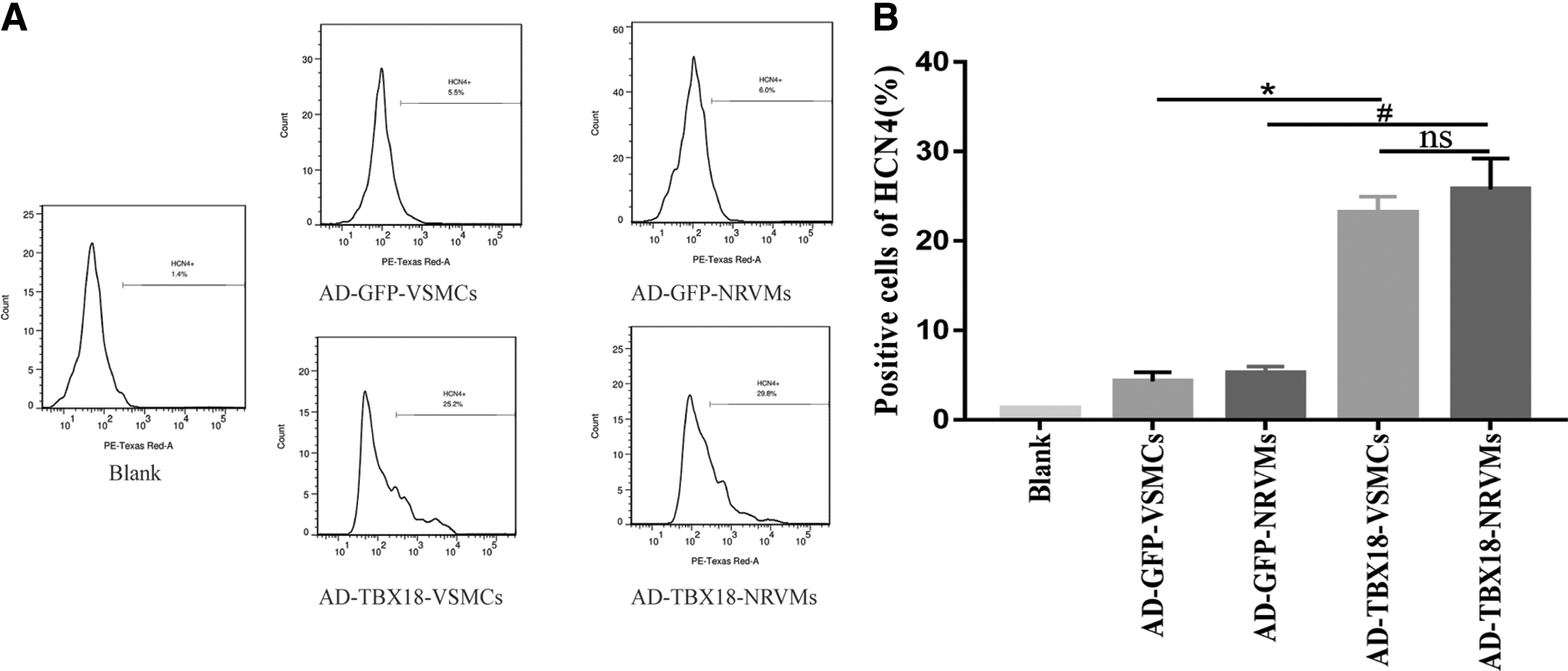
Comparison of positive cells of HCN4 by flow cytometry for VSMCs and NRVMs transfected with Ad-TBX18 and Ad-GFP, respectively.
Expression of correlative mRNA
The TBX18 mRNA was verified by RT-qPCR (Fig. 3A), and the results of RT-qPCR indicated that the mRNA expression levels of HCN4, ISL1, SHOX2, and cTNI were significantly higher in the TBX18 group than in GFP group and Null group (p < 0.05, Fig. 3B). In turn, the expression level of Cx43 mRNA was distinctly lower in the TBX18 group compared with the GFP group (p < 0.05; Fig. 3B). And the statistical analysis revealed that no significant differences were found between the GFP and Null groups. Furthermore, after being transfected for 3, 4, 5, 7, 10, and 14 days, the mRNA expression levels, such as TBX18, HCN4, and SHOX2, showed a peak on the 3th–5th day, which decreased sharply down from the 7th day (Fig. 3C, D).
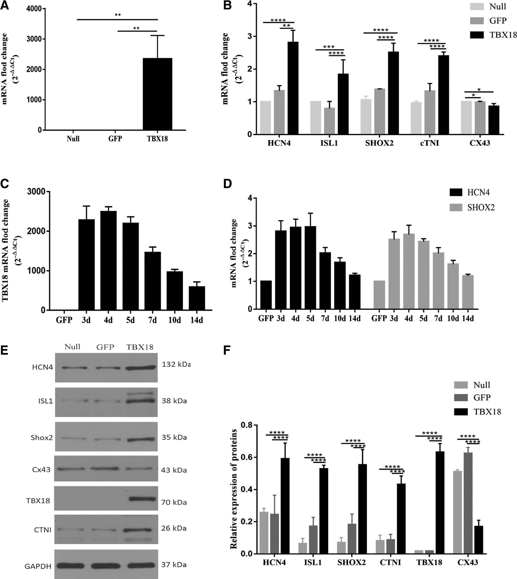
The mRNA and protein expression levels of associated transcription factors by RT-qPCR and WB. (n = 3)
Expression of relevant proteins
According to the results of Western Blot, the protein expression levels of HCN4, ISL1, SHOX2, and cTNI were upregulated clearly in the TBX18 group compared with the GFP group (p < 0.05, Fig. 3E, F; Supplementary Figure S1); in contrast with the GFP group, the expression of Cx43 protein was reduced in the TBX18 group (p < 0.05, Fig. 3E, F; Supplementary Figure S1). And there were no significant differences statistically between the GFP and Null groups.
Immunocytochemical analysis of HCN4 protein
When transduced for 4–5 days, VSMCs have possessed the characteristic of pacemaker-like cells. As is shown in Figure 4, the expression of HCN4 (red) was obvious in the TBX18 group cells, whereas it was hardly seen in the GFP group cells.
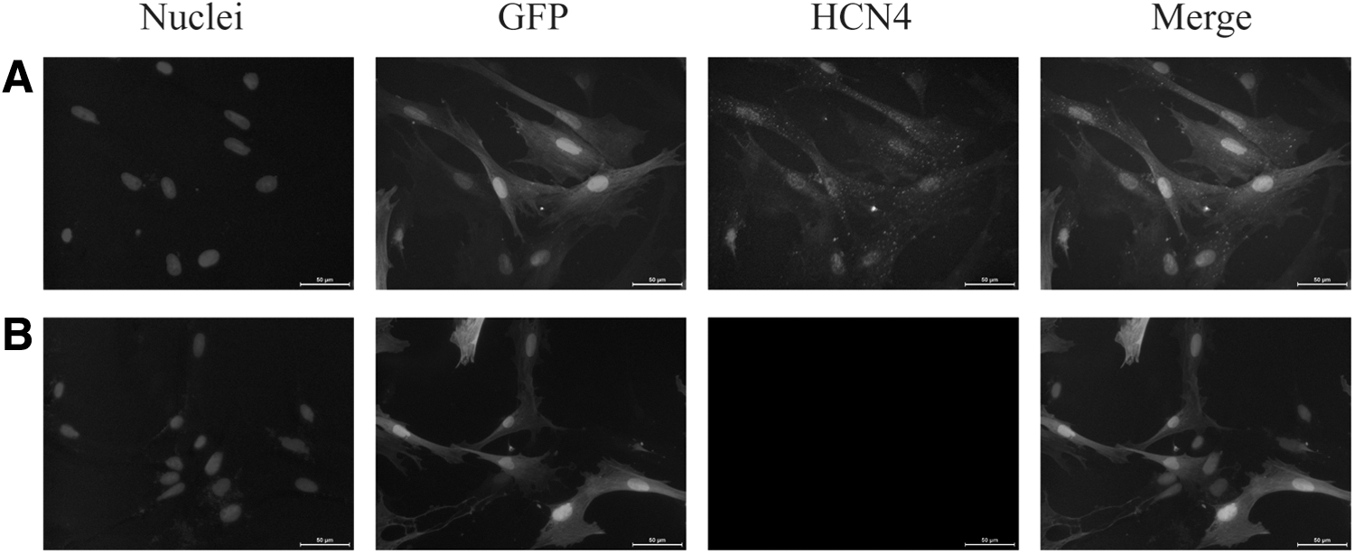
HCN4 protein immunofluorescence of transduced VSMCs. (magnification, × 200)
Morphology and beating rate of co-cultured cells
For 2 days in co-culture conditions, the Ad-TBX18-transfected VSMCs proliferated decreasingly and the majority of VSMCs connected with NRVMs that formed cell clusters (black arrows, Fig. 5A). Every cluster produced synchronous beating after 3 days and the morphology of VSMCs coupled with NRVMs was spindly and quadrangular by fluorescence microscopy (red arrows, Fig. 5B). Immunofluorescence showed that Cx43 protein (red) was clearly detected at membranes of Ad-TBX18-transfected VSMCs (green) and NRVMs, which formed gap junctions between them (white arrows, Fig. 5C). TBX18-transfected VSMCs in co-culture systems had a higher beating rate compared with the GFP group, Null group, and NRVMs. The beating rate of the cells reached 178.00 ± 7.55 bpm (n = 3) in the TBX18 group, 154.00 ± 2.00 bpm (n = 3) in the GFP group, 154.33 ± 1.53 bpm (n = 3) in the Null group, and 147.67 ± 2.52 bpm (n = 3) in the alone NRVMs group after 3 days. And there were statistical differences between the TBX18 group and the other three groups (Fig. 5D).
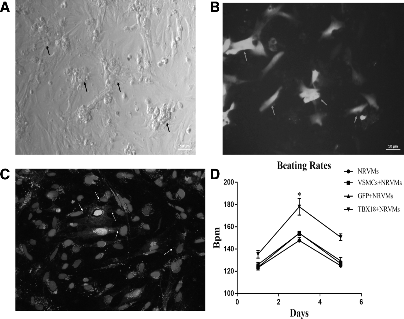
The cell clusters and beating rate of cells in co-culture conditions.
Electrophysiological characterization
If current was examined in a voltage-clamp mode. Spindle cells with whole cell and green fluorescence were used for electrophysiological analysis (Fig. 6A, B). The results of whole-cell patch clamp showed that If current was not recorded in the null group and the GFP group. However, If current recorded in TBX18 group was activated and appeared the time and voltage dependence (Fig. 6C, D). After adding 4 mM CsCl to the extracellular fluid, If current was blocked significantly (Fig. 6C). However, when the CsCl was cleaned with normal fluid, If current recovered rapidly. And the amplitude of current density ranged from −5.164 ± 0.662 pA/pF to −0.765 ± 0.358 pA/pF (n = 14) (Fig. 6D). When VSMCs were cocultured with NRVMs for 5 days, the APs of transfected VSMCs and NRVMs were recorded by c-clamp. It is evident that slow 4 phase depolarization rates and short AP durations were like pacemaker cells APs in VSMCs infected with TBX18 (Fig. 6E). However, the APs of GFP-VSMCs and NRVMs showed typical ventricular-like APs (Fig. 6E).
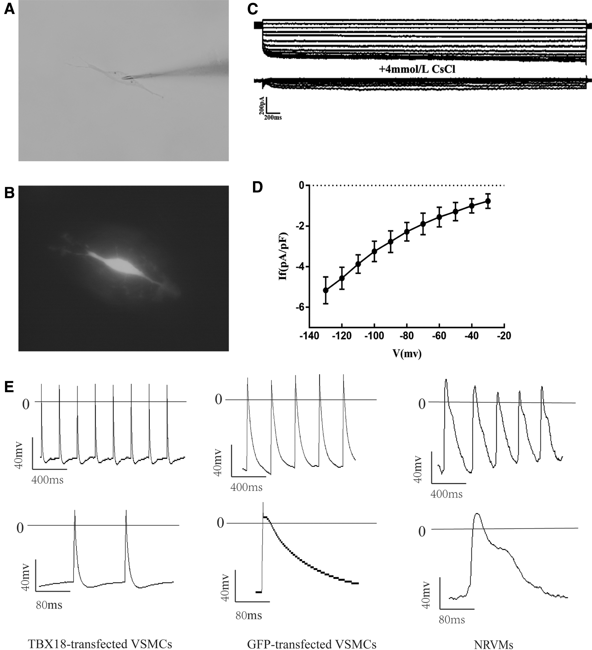
Electrophysiological recordings by patch clamp in co-culture systems for 5 days.
Discussion
This study showed that VSMCs of ascending aorta by overexpressing TBX18 could be reprogrammed into pacemaker-like cells in vitro. We present the following evidences: (1) The gene and protein expression levels of ISL1, SHOX2, HCN4, and cTNI were significantly upregulated and Cx43 expression was downregulated in TBX18-transfected VSMCs. (2) Electrophysiological studies showed that If current was presented and appeared the time and voltage dependence in TBX18-reprogrammed VSMCs in co-culture systems, which was sensitive to Cs+. The APs for VSMCs infected with TBX18 showed slow 4 phase depolarization, similar with that of SAN-like cells. And beating rate of the cells cocultured with NRVMs was much faster in the TBX18 group than other groups.
During cardiac embryonic development, the second heart field not only differentiates into the arterial pole (right ventricle and outflow tract) but also differentiates into the sinus node, endothelial cell lineages, and smooth muscle cells (Waldo et al., 2005; Kattman et al., 2006; Moretti et al., 2006; Bu et al., 2009; Andersen et al., 2018). Recently, Sawada et al. (Harmon and Nakano, 2013; Sawada et al., 2017) demonstrated that smooth muscle cells of the aortic root and ascending aorta were extended by the second heart field and cardiac neural crest. Furthermore, the second heart fields could differentiate into the pacemaker cells of SAN and atrioventricular node (Sun et al., 2007). Therefore, pacemaker cells and ascending aortic smooth muscle cells were both derived from the second heart field cells. Therefore, this study explored whether VSMCs of the ascending aorta could be reprogrammed into pacemaker-like cells by human transcription factor TBX18 in vitro.
Embryonic development of SAN is influenced by multiple transcription factors (Christoffels et al., 2010; Barbuti and Robinson, 2015). During specific differentiation of pacemaker cells, transcription factor TBX18 is the first gene to be expressed, and TBX18 was only expressed in the head region of sinus node in mature cardiomyocytes (Wiese et al., 2009). Cardiac troponins included troponin I (cTNI), troponin C (cTNC), and troponin T (cTNT) proteins, which were considered myocardial makers (Kociol et al., 2010). In this study, we found that cTNI was expressed in VSMCs of the ascending aorta after transfection with TBX18, indicating that the transfected cells have had the characteristics of cardiomyocytes. Transcription factor ISL1, SHOX2, and HCN4 were regarded as sinus node-specific markers, which were both regulated by TBX18. Previous studies (Blaschke et al., 2007; Barbuti and Robinson, 2015; Ye et al., 2015) demonstrated that SHOX2 could inhibit the expression of chamber gene NKX2.5 and activate transcription factor ISL1, which participate in the formation of SAN. And that ISL1 could also regulate sinus node development and interact with HCN4 (Liang et al., 2015; Vedantham et al., 2015). Previous studies (Liu et al., 2007; Brioschi et al., 2009; Wahl-Schott et al., 2014) demonstrated that HCN4 mainly distributed in SAN. In these results, we found that the mRNA and protein expression levels of ISL1, SHOX2, and HCN4 were significantly increased in the TBX18 group, indicating that the transfected cells had the phenotype of SAN-like cells. Furthermore, If current encoded by HCN4 channel was the basis of phase 4 automatic depolarization of pacemaker cells (Marionneau et al., 2005; Mommersteeg et al., 2007; DiFrancesco, 2010). And we indicated that the specific If current was detected in transfected cells by TBX18, further demonstrating that the transfected cells had the characteristics of SAN-like cells. Moreover, gap junction proteins mediated direct cardiomyocyte coupling (electrical and metabolic) that regulated heart homeostasis and function. And three main isoforms of connexin distributed in the heart: Cx40, Cx43, and Cx45. Cx43 is the most abundant connexin in working cardiomyocytes, which is fewer in SAN cells (Jansen et al., 2010). Our result showed that connexin 43 mRNA and protein were distinctly decreased in the TBX18 group, showing that the gap junction appeared to change between the reprogrammed VSMCs toward the SAN-like cells. In addition, the beating rate of TBX18-overexpressing VMSCs was faster compared with other groups in co-culture systems, and most importantly, the transfected cells by TBX18 exhibited sinoatrial-like APs, which do not appear in the remaining groups.
In conclusion, this study suggested that VSMCs of ascending aorta with TBX18 could be reprogrammed into pacemaker-like cells in vitro. However, there are still some limits in this experiment: (1) adenovirus functions a relatively short time and has high innate immunogenicity (Thaci et al., 2011; Merentie et al., 2016), so we need to find a carrier that works longer and safer in future studies; (2) The duration of these transduced pacemaker-like cells is not verified and tested in vivo. Therefore, more studies should be conducted to further verify the long-term efficacy in vitro and in vivo experiments of these transfected cells.
Footnotes
Acknowledgment
The authors are appreciative to Wuhan University School of Basic Medical Science for their assistance in conducting the experiments.
Disclosure Statement
The authors declare no conflict of interests.
Funding Information
This research was funded by the Fundamental Research Funds for the Central Universities of China (grant no. 2042015kf0229) and the Technical Innovation Project of Hu Bei Province of China (grant no. 2016ACA153).
Supplementary Material
Supplementary Figure S1.
References
Supplementary Material
Please find the following supplemental material available below.
For Open Access articles published under a Creative Commons License, all supplemental material carries the same license as the article it is associated with.
For non-Open Access articles published, all supplemental material carries a non-exclusive license, and permission requests for re-use of supplemental material or any part of supplemental material shall be sent directly to the copyright owner as specified in the copyright notice associated with the article.