




Abstract
This study aimed to investigate the expression and clinical significance of chemokine-like factor-like MARVEL transmembrane domain-containing family member 2 (CMTM2) in hepatocellular carcinoma (HCC) tissues. Bioinformatics analysis showed that CMTM2 was downregulated in HCC tissues and correlated with vascular invasion of HCC patients. The immunohistochemistry (IHC) method found that CMTM2 expression was negative in 29/75 (38.67%) cases of HCC tissues and 13/75 (17.33%) cases of paracancerous tissues, also showed a significantly lower expression of CMTM2 in HCC tissues than the paired paracancerous tissues (p < 0.05). Moreover, CMTM2 expression was significantly correlated with tumor grade of HCC patients (p < 0.05), but had no relationship with other clinicopathological features. In addition, multivariate logistic regression analysis indicated that tumor grade was an independent risk factor for CMTM2 expression. The survival time of HCC patients between high and low expression of CMTM2 had no difference by bioinformatics analysis, but the IHC result showed that the negative expression of CMTM2 was related to a poor prognosis of HCC patients. Further COX regression analysis showed that CMTM2 expression was an independent protective factor for the prognosis of HCC patients. We identify that CMTM2 is downregulated in HCC tissues and correlated with the prognosis of HCC patients, suggesting a potential tumor suppressor role of CMTM2 in HCC progression.
Introduction
Hepatocellular carcinoma (HCC) is one of the most common malignant tumors worldwide. There are about 700,000 new cases and 600,000 HCC patients die each year (Siegel et al., 2017). Among the male patients of malignant tumors, younger than 60 years of age in China, the incidence and mortality rate of HCC was the highest one (Zhu et al., 2015). Various hepatitis virus infection, liver alcoholic injury, liver metabolism, and autoimmune diseases are key factors resulting in HCC carcinogenesis (Farazi and DePinho, 2006). As the most important cause of HCC, hepatitis B virus (HBV) infects about 93 million people in China. Many HBV-infected patients develop into HCC through the process of liver fibrosis and liver cirrhosis (Xie, 2017). If HCC can be diagnosed in the early stage, HCC patients can achieve relatively good results from comprehensive treatment, including tumor surgical resection, liver transplantation, transcatheter arterial chemoembolization, and radiotherapy. However, due to lack of effective early diagnostic markers, the hidden onset of HCC causes many patients detected and treated at middle and late stage, leading to the high recurrence and poor prognosis. At present, the overall diagnosis and treatment of HCC in China is still not optimistic, it needs improved efficiency of HCC diagnosis and treatment urgently.
As a newly found gene family, chemokine-like factor-like MARVEL transmembrane domain-containing family members (CMTM) implicate in the pathogenesis of immune, reproductive systems and various tumors (Li et al., 2010; Mezzadra et al., 2017; Bei et al., 2018). CMTM1 was found to affect cell proliferation and lead to tumorigenesis of nonsmall cell lung cancer (Si et al., 2017). Apart from regulating the cell cycle and proliferation of tumor cells (Plate et al., 2010), CMTM4 was reported to participate in immune escape through the synergistic protection of PD-L1 (Mezzadra et al., 2017). Low expression of CMTM5 was found in many tumors and involved in the signaling pathways related to tumorigenesis and tumor development (Zhang et al., 2014; Xiao et al., 2015). CMTM6 was another CMTM member involved in immune escape through PD-L1 protection and identified as a potential therapeutic target of tumors (Burr et al., 2017). Recently, CMTM7 was found downregulated in liver cancer tissues and had a negative relationship with TNM staging and metastasis (Huang et al., 2019). In our previous studies, we have clarified that CMTM4 and CMTM6 play an important role in HCC progression (Bei et al., 2017; Zhu et al., 2019). These findings provide a new way to study the development and gene therapy for tumors.
Unlike the other CMTM members, the role of CMTM2 (alias name CKLFSF2) in tumors has not been well explored. CMTM2 (alias name CKLFSF2) is highly expressed in human testicular tissues and thought to be a pivotal protein for male reproduction (Liu et al., 2007). In a recent study, CMTM2-deficient mice were shown to have small testis, lose the organized epithelium architecture of seminiferous tubules, produce virtually no sperm, and be sterile due to a late, complete arrest of spermiogenesis (Zhang et al., 2019). However, the serum level of testosterone in CMTM2 transgenic mice was significantly higher than that of the wild-type mice (Liu et al., 2012). Moreover, compared with the wild-type mice, the sperm concentration and activity rate of CMTM2 transgenic mice were still at a high level after cyclophosphamide exposure (Liu et al., 2013). In addition, CMTM2 was found to influence HIV-1 transcription to a certain extent through AP-1 and CREB pathway (Song et al., 2010), and affect the transcriptional activity of androgen receptor in PC-3 and HeLa cells (Li et al., 2006). In an expression screen of chemokine and chemokine receptor profiles in metastatic salivary adenoid cystic carcinoma (SACC), CMTM2 was more highly expressed in tissues of patients without tumor recurrence or perineural invasion compared to those with tumor recurrence, suggesting an important role of CMTM2 in the development of tumor metastasis in SACC (Mays et al., 2016).
In this study, bioinformatics and immunohistochemistry (IHC) methods were used to detect the expression of CMTM2 in HCC and paired paracancerous tissues. The relationship between CMTM2 expression and clinicopathological features of HCC patients was also analyzed to explore the clinical significance of CMTM2 in HCC diagnosis and prognosis.
Materials and Methods
Bioinformatics analysis
CMTM2 expression data in HCC tissues were obtained from the National Center for Biotechnology Information Gene Expression Omnibus (NCBI GEO) and The Cancer Genome Atlas (TCGA) database via cBioPortal platform. We used the UCSCXenaTools package in R to download TCGA clinical data and paired liver cancer clinical data with CMTM2 expression data by the samples' ID. After natural logarithmic conversion of CMTM2 expression data to accord with normal distribution, the clinic data between groups were compared by independent sample t-test or ANOVA test. High and low CMTM2 expression groups were set by the median of CMTM2 expression value. We used survival package in R for survival analysis. Kaplan–Meier analysis was used for overall survival (OS) time and relapse-free survival time from TCGA's liver cancer patients.
Tissue specimens
HCC and paired paracancerous tissues were collected from 75 patients diagnosed with HCC by pathological and clinical methods in the Affiliated Hospital of Guilin Medical University between 2015 and 2016. These patients received liver surgery without radiotherapy or chemotherapy before. The clinicopathological features of these patients are shown in Table 1 and Supplementary Table S1. All tissue specimens and clinicopathological data were obtained with the informed consent of the patients and approved by the Ethics Committee of the hospital. This study was based on the Declaration of Helsinki in 1975 and approved by Ethical Committees of Guilin Medical University (GLMC2014003).
Association Between the Expression of CMTM2 and Clinicopathological Features of Hepatocellular Carcinoma Patients
Bold values indicate significance.
AFP, alpha-fetoprotein; HBV, hepatitis B virus.
Immunohistochemistry
The IHC was performed after tissue specimens were integrated into a tissue chip by Fanpu company (Guilin, China). After IHC standard steps for baking, dewaxing, antigen repair, peroxidase blocking, and goat serum incubation, the tissue chip was incubated with rabbit polyclonal antibody against CMTM2 (NBP1-59439, Novus, CO) overnight at 4°C. Then, anti-rabbit second antibody was added and incubated at 37°C for 30 min. Finally, diaminobenzidine solution was given to develop color and hematoxylin was added to restain.
The IHC results were determined under microscope by two professional pathologists. The average staining intensity and percentage of positive stained cells in five visual fields were recorded and counted. The scores for percentage of positive stained cells were 0 for none, 1 for ≤10%, 2 for 11−50%, 3 for 51–75%, and 4 for >75%, respectively. The scores for staining strength were 0 for no staining, 1 for light brown, 2 for medium brown, and 3 for dark brown. Finally, the scores for staining strength and percentage of positive stained cells were multiplied to obtain the total expression score. The expression score ≤4 was defined as negative expression and >4 was defined as positive expression.
Statistical analysis
All the data were analyzed on SPSS 20.0. The expression of CMTM2 between HCC and paired paracancerous tissues was compared by chi-square test. The relationship between the expression level of CMTM2 and the clinicopathological features of HCC patients was analyzed by chi-square test and multivariate logistic regression analysis. Kaplan–Meier method was used for survival analysis. p < 0.05 was considered as statistically significant.
Results
Expression of CMTM2 in HCC tissues
We first analyzed the expression of CMTM2 in HCC tissues from GEO and TCGA databases. As shown in Figure 1A and B, the expression of CMTM2 in HCC tissues was significantly lower than that of normal liver tissues in GSE6764 (p < 0.05), but there was no difference of CMTM2 expression between HCC tissues and normal liver tissues in TCGA database. It has been reported that there is copy number variation of CMTM2 in tumors (Choi et al., 2018). Therefore, we also compared the copy number of CMTM2 in HCC tissues and normal liver tissues in these online databases. However, we found no difference between copy number of CMTM2 in HCC tissues and normal liver tissues (Fig. 1C–E).
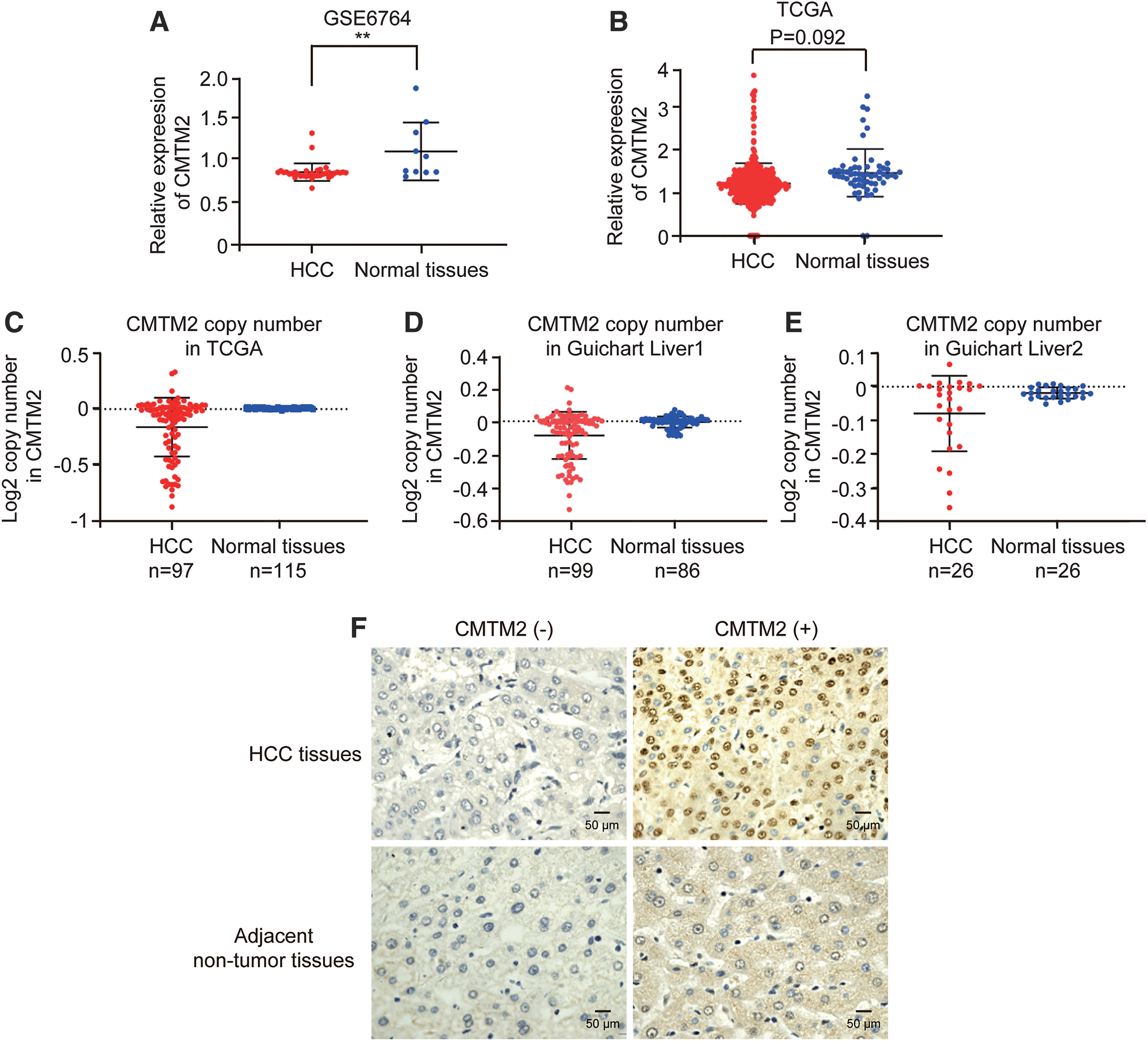
Expression of CMTM2 in HCC tissues.
To further detect the expression of CMTM2 in HCC tissues, we performed IHC assay to verify the results of online databases. As shown in Figure 1F, we can see that CMTM2 mainly located in cell membrane and cytoplasm. Moreover, the negative expression of CMTM2 was found in 29 cases of 75 HCC tissues (38.67%) and 13 cases of 75 paracancerous tissues (17.33%), respectively. Statistical analysis showed that the negative expression of CMTM2 in HCC tissues was more than that of paracancerous tissues (p < 0.05, Table 2). These results suggest that CMTM2 is downregulated in HCC tissues.
CMTM2 Expression in Paired Hepatocellular Carcinoma Tissues and Adjacent Nontumor Tissues
p < 0.05, p value is based on the McNemar χ 2 test.
CMTM2, chemokine-like factor-like MARVEL transmembrane domain-containing family member 2; HCC, hepatocellular carcinoma.
The relationship between CMTM2 expression and the clinicopathological features of HCC patients
We also analyzed the relationship between the expression of CMTM2 and the clinical characteristics of HCC patients from online databases and IHC results. It was found that the expression of CMTM2 was related to the tumor grade and vascular invasion of HCC patients, especially, there was a significant correlation between CMTM2 expression and vascular invasion from TCGA database (p < 0.01, Fig. 2). The 75 HCC patients were divided into CMTM2-positive and -negative expression groups according to the IHC results. As shown in Table 1, the expression of CMTM2 was significantly correlated with tumor grade (p < 0.05), while had no relationship with other clinicopathological features of HCC patients, such as gender, age, smoking, alcohol intake, HCC family history, HBV infection, liver cirrhosis, serum alpha-fetoprotein, tumor diameter, tumor number, TNM stage, and metastasis. Multivariate logistic regression analysis further showed that the expression of CMTM2 was related to the pathological grade of HCC, and its odds ratio (OR) value was 0.374 (p < 0.05, Table 3). These results indicate that the expression of CMTM2 is correlated with vascular invasion and tumor grade of HCC patients.
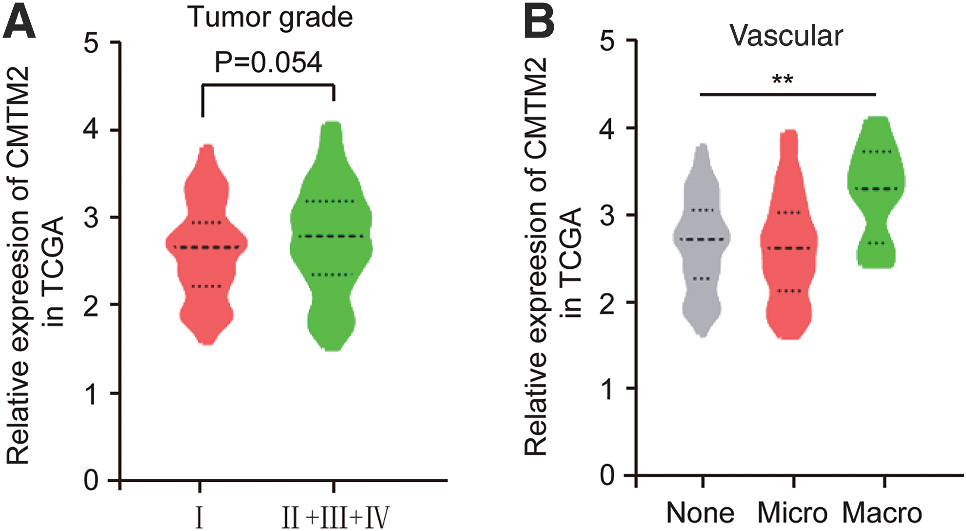
The relationship between CMTM2 expression and the clinicopathological features of HCC patients analyzed from TCGA database.
Multivariable Logistic Regression Analysis of CMTM2 Expression in Hepatocellular Carcinoma Patients
Bold values indicate significance.
Adjusted for age and gender.
CI, confidence interval; OR, odds ratio.
The relationship between CMTM2 expression and the prognosis of HCC patients
The relationship between the expression of CMTM2 and the prognosis of HCC patients was analyzed online first by Kaplan–Meier method. As shown in Figure 3A and B, from TCGA database, there was no significant difference of OS time and disease-free survival time after tumor surgery between CMTM2 high- and low-expression groups. However, the OS time of CMTM2 negative expression group was significantly shorter than that of CMTM2-positive expression groups from the IHC result (Fig. 3C, p < 0.05). It was also found that CMTM2 was an independent protective factor for HCC through COX regression analysis after adjusting for gender and age (p < 0.05, OR = 0.392, Table 4).
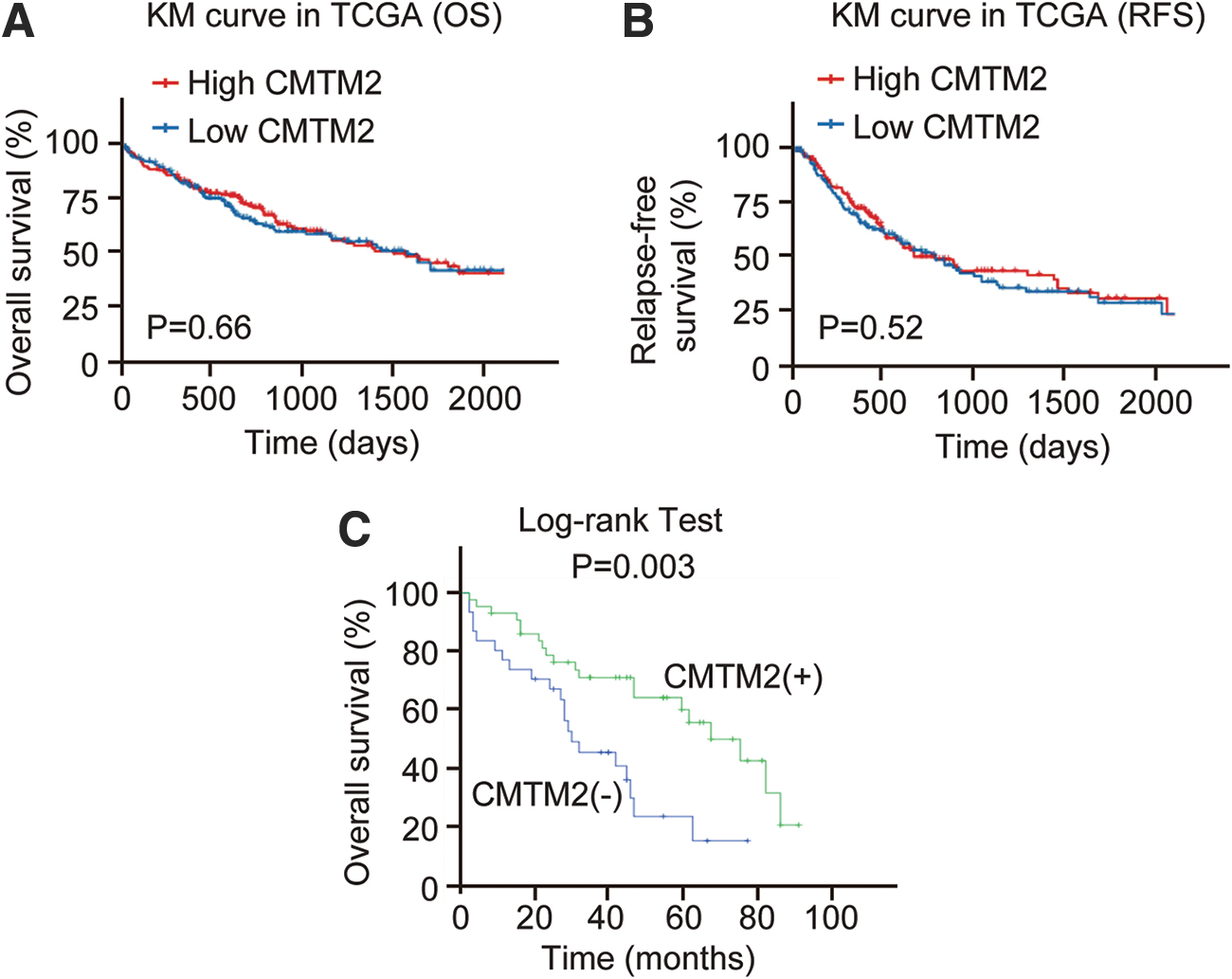
The relationship between CMTM2 expression and the prognosis of HCC patients.
COX Regression Analysis for Overall Survival of Hepatocellular Carcinoma Patients After Surgery
Bold values indicate significance.
Adjusted for age and gender.
Discussion
In this study, CMTM2 was found downregulated in HCC tissues by bioinformatics analysis and IHC assay. Moreover, CMTM2 expression was significantly correlated with tumor grade and vascular invasion of HCC patients, and tumor grade was an independent risk factor for CMTM2 expression after multivariate logistic regression analysis. The OS time of HCC patients was significantly different between CMTM2-positive and -negative expression groups from IHC result, and negative expression of CMTM2 was related to a poor prognosis of HCC patients.
The CMTM family is a new family first discovered and cloned by Chinese researchers. They are widely expressed in various systems of the body and function in immune, reproductive, and blood systems. Recently, they are found closely related to tumorigenesis and tumor development (Delic et al., 2015; Lu et al., 2016). They can regulate the cell cycle and participate in some signaling pathways of tumorigenesis, then affect tumor development and prognosis (Wang et al., 2009; Li et al., 2014). The discovery about synergistic protection of PD-L1 by CMTM6 and CMTM4 is particularly important, providing a potentially novel role of CMTM family in tumor immunotherapy through PD-1/PD-L1 signaling pathway (Imamovic and Vranic, 2017; Mezzadra et al., 2017). However, current studies are still relatively limited, and the specific molecular mechanisms of CMTM family in tumors are still not clear enough.
CMTM2 is widely expressed in human testes and spermatozoa (Shi et al., 2005). CMTM2 is found in the cell membrane of spermatogenic cells in testicular tissues, while locates near the posterior nuclear area in spermatozoa, which is the fusion site between sperm plasma membrane and egg plasma membrane (Zhang et al., 2017). The location and expression profile of CMTM2 suggest that an important role of CMTM2 might play in male reproduction. Some studies have found that there was a significant relationship between CMTM2 expression and spermatogenesis through animal models. The deregulation of CMTM2 expression could cause spermatogenesis disorders, and spermatogenesis disorders could also result in the mRNA expression level of CMTM2 decreased significantly (O'Hara and Smith, 2015). In the testicular tissues of patients with sertoli cell only syndrome, there was even no expression of CMTM2 (Zhang et al., 2005). In addition, varicocele can decrease the expression of CMTM2 through damaging the quantity and quality of spermatozoa (Zhang et al., 2016).
There are also some studies reporting the potential role of CMTM2 in tumors. Choi et al. (2018) identified a significant novel mutation at CMTM2 in diffuse-type gastric cancer (DGC) and CMTM2 expression could predict the prognostic outcomes of DGC. Mays et al. (2016) found that CMTM2 was highly expressed in tissues of patients with metastatic SACC. Fang et al. (2012) showed that CMTM2 exhibited hypermethylation in its promoter regions in colorectal cancer. The results of our study are consistent with these studies, suggesting a close relationship between CMTM2 expression and clinicopathological features of patients with tumors.
In addition, we found that some IHC results were not consistent with the bioinformatics analysis. The IHC results were analyzed under microscope by two professional pathologists, which might cause discrepancy with the results from the TCGA and GEO databases due to potential bias from different observers. We assume other reasons might be of different HCC patients from different places and races, and the small sample is also a main influence factor for the result. Further studies of CMTM2 in HCC need to be elucidated from bigger population sample, cell lines, and animal models.
Conclusion
In conclusion, CMTM2 is downregulated in HCC tissues and correlated with the prognosis of HCC patients, suggesting a potential tumor suppressor role of CMTM2 in HCC progression.
Footnotes
Disclosure Statement
No competing financial interests exist.
Funding Information
This work was supported by the National Natural Science Foundation of China (81860586, 81860602), Natural Science Foundation of Guangxi Province (2018GXNSFAA281054, 2018GXNSFBA281216) and Key Science and Technology Research and Development Program Project of Guangxi (AB17292074).
Supplementary Material
Supplementary Table S1
References
Supplementary Material
Please find the following supplemental material available below.
For Open Access articles published under a Creative Commons License, all supplemental material carries the same license as the article it is associated with.
For non-Open Access articles published, all supplemental material carries a non-exclusive license, and permission requests for re-use of supplemental material or any part of supplemental material shall be sent directly to the copyright owner as specified in the copyright notice associated with the article.
