
Abstract
Epithelial ovarian cancer (EOC) treatment strategies mainly focused on surgery combined with chemotherapy. Recent targeted therapy techniques emerge as milestone and could be used for management of ovarian cancer (OC) progression with more efficacy. The aim is to evaluate the therapeutic and diagnostic potential of microRNA (miRNA) in management of EOC using in silico and quantitative real-time PCR (qRT-PCR) expression analysis. We performed functional enrichment and miRNA-Target genes expression analysis in 48 EOC and 22 normal tissue samples using qRT-PCR and correlated with miRNA expression data in matched samples to evaluate the diagnostic and therapeutic potential of miRNA in OC management. In silico functional enrichment analysis revealed miRNA association with disease. Target genes of miRNAs participate in several biologically important pathways leading to cancer progression. Targets of miRNA-205 and miRNA-34a were significantly downregulated, and upregulated, respectively, in EOC. Moreover, significant negative correlation between relative expression of miRNA-205 and target genes (BCL2, ZEB1, E2F1, and TP53) was observed with r = −0.813; r = −0.755; r = −0.559; and r = −0.767, respectively. Similarly, miRNA-34a also showed higher negative correlation with target genes (MDM4, MAPK3, BRCA1, AREG) with r = −0.840; r = −0.870; r = −0.622; and r = −0.623, respectively. In addition, receiver operating characteristics analysis of combined miRNA panel, miRNA-205-Target gene panel, and miRNA-34a-Target gene panel exhibited higher diagnostics value with area under the curve (AUC) of 92.7 (p < 0.0001), 94.8 (p < 0.0001), and 98.3 (p < 0.0001), respectively. Negative Correlation between miRNA and target genes expression data in matched samples highlights therapeutic potential of miRNA in EOC management. Moreover, combined diagnostic potential of miRNA-target gene panel could predict risk of EOC with higher AUC, sensitivity, and specificity.
Introduction
Ovarian cancer (OC) is the 7th most common and 18th most deadly cancer-causing death in women worldwide (Shabir and Gill, 2020). The asymptomatic characteristic of OC leads to the presentation of most patients at the advance-stage of disease, thus, increasing morbidity. Due to OC, around 1 out of 78 women are affected in their life span, while morbidity accounts for 1 out of 108 women affected with OC (Holschneider and Berek, 2000; Karst and Drapkin, 2010). According to the American Cancer Society, around 19,880 new cases and 12,810 deaths are estimated due to OC in 2022 (Torre et al, 2018). According to cancer statistics data, India recorded 26,834 new cases (crude rate of 4.4), with morbidity of 19,549 (crude rate of 3.2) raking second in Asian countries (Razi et al, 2016).
Moreover, National Cancer Registry Program of India denoted
The most effective option for the treatment of epithelial ovarian cancer (EOC) is debulking surgery with the combination of chemotherapy. However, the final 5-year relative survival rate is only 49.1%, primarily due to EOC resistance to chemotherapy and recurrence of the disease (Caswell-Jin et al, 2018; Gadducci et al, 2019). The best treatment can only be provided when the incidence of the disease is diagnosed in the early stages, but that is not attained since most of the
Researchers are growing in investigating role of microRNAs (miRNAs) in disease progression since they are involved in essential processes such as cell differentiation, growth, and apoptosis. miRNAs are a family of small single-stranded noncoding RNAs, which are 22–24 nucleotides in length. The expression levels of miRNAs exhibiting tumor suppressor activity are significantly downregulated, whereas miRNAs which act as oncogenes have higher expression levels in
Epigenetic events occurring before the onset of disease, lead to overexpression of oncogenic miRNA and downregulation of tumor suppressor miRNA due to hypo/hypermethylation events (Loginov et al, 2015; Lopez-Serra and Esteller, 2012). Downstream effect of miRNA dysregulation results in abnormal expression of tumor suppressor genes (PTEN, SMAD4, TCF21, TP53, and ZEB1) and oncogenes (MDM4, MAPK3, AREG, and BRCA1) leading to carcinogenesis (Chu et al, 2018; Hu et al, 2016; Kong and Wang, 2020; Mandke et al, 2012; Nie et al, 2015; Niu et al, 2015; Tung et al, 2017). Thus, identifying miRNA-based dysregulation of target genes and their biological importance would render miRNA therapeutic role in
The present study aims to evaluate the impact of epigenetic status of miRNA in
Materials and Methodology
Study design
This study involves evaluation of therapeutic potential miRNA in EOC management. We selected candidate miRNA based on our previous study. Selected miRNA was further evaluated in-silico for their association specifically with
Patients and clinical features
A total of 70 EOC tissue samples (48 EOC and 22 normal) were recruited for this study (Supplementary Data). All samples were collected from Department of Surgical Oncology, King's George medical University, Lucknow, with dually signed informed consent form by patient. Study was performed according to Declaration of Helsinki and approved by the Institute Ethics Committee (Ref. No: IEC/2019–20/01). Further, samples were segregated according to their age, FIGO stage, histology, and metastatic nature of the samples. The segregated samples included 18 combined FIGO stage I + II (37.5%), and 30 stage III+IV (62.5%) and, similarly, 32 serous (66.0%), 6 mucinous (12.5%), 5 clear cell (10.4%), and 5 endometrioid (10.4%). In addition, samples were segregated as 30 metastatic samples (62.5%) and 18 nonmetastatic cases (37.5%). Around 58.3% of patients were aged above 45 years while 41.6% of patients were below 45 years (Table 1).
Represents the Clinical Features of Patients Recruited in this Study
Screening of candidate miRNA
Several miRNAs have been identified in EOC with reported hypo- and hypermethylation at gene level in several individual studies. Similarly, in our previous study, we reported hypo- and hypermethylation of several miRNA genes based on MeDIP NGS sequencing and further correlated them with miRNA expression in tissue and matched tissue samples (Kumar et al, 2022; Kumar et al, 2021). As an outcome of these studies, miRNA-205 and miRNA-34a exhibited highest correlation with miRNA-gene methylation and dysregulated expression in EOC compared to normal samples. Therefore, these miRNAs may qualify as potential candidate miRNA for further investigating their role in miRNA-based therapeutics in EOC management.
Bioinformatics analysis of the therapeutic role of miRNA in OC
miRNA-disease association enrichment analysis
Selected candidate miRNA was evaluated for association with disease using an online tool (
Target prediction and functional enrichment analysis
Candidate miRNA putative target genes were predicted using seven online tools (miRbase, miRDB, miRTarBase, Tools4miRs, miRnet, Diana-microT, miRwalk). Only miRNA-Target genes overlapping in all seven databases were selected for further analysis. miRNA-Target enrichment network was built using
Quantification of miRNA-target genes
mRNA samples were isolated from 48 EOC and 22 normal samples. All mRNAs from OC and normal samples were converted into cDNA using QuantiTech® Reverse Transcription Kit (Cat. No. 205313; Qiagen, India) following manufacturer's instructions. In brief, 1 μg of mRNA was treated with gDNA Wipeout solution by incubating at 42°C for 2 min, followed by the addition of QuantScript Reverse Transcriptase, QuantScript RT Buffer, and RT Primer mix (Cat. No. 205313; Qiagen) and incubated at 42°C for 15 min followed by heat inactivation at 95°C for 3 min. Next, all mRNA samples were diluted to bring final concentration of 10 ng/μL and stored at −20°C till further processing.
Further, qRT-PCR-based quantification of each target gene was performed in triplicates using SYBR Green Method (Cat. No. F416L; ThermoFisher, India) with the respective primer set and cycling condition. In brief, for 10 μL of reaction DyNamo Color Flash SYBR Green 5.0 μL, ROX dye 0.2 μL, forward primer 1.0 μL, reverse primer 1.0 μL, template 0.5, and 2.3 μL of DNase free water was added followed by respective qRT-PCR cycling condition for the primer set of each gene are depicted in Table 2.
Represent MicroRNA-Target Genes Primer Sequences with Optimized Annealing Temperature and Quantitative Real-Time PCR Cycling Condition for Each Gene
miRNA, micro
Statistical analysis
The miRNA-target genes' relative expression was evaluated using the LIVAK method (2−ΔΔCT). The Mann–Whitney U test was used to discover the statistical significance of differences between two groups of variables. Further, the correlation between miRNA expression and target gene expression was evaluated using Pearson's Coefficient Correlation analysis. The Association of miRNA and target genes with disease progression was evaluated using binary logistic regression analysis and predicted probability values obtained from the regression model were used to evaluate the combined diagnostic potential of the miRNA panel and gene panel using Receiver Operating Characteristics curve analysis.
All statistical analyses were executed in SPSS® (Version 26; IBM Chicago, Chicago, IL) and graphs were plotted in GraphPad Prism (Version 8.0). All statistical tests were two-sided, and a p-value <0.05 was considered statistically significant.
Results
Selection of candidate miRNA
In our previous study, MeDIP-NGS sequencing-based differentially methylated region associated with miRNA-205 and miRNA-34a genes promoter region was evaluated and found to have significant hypomethylation and hypermethylation on miRNA-205 and miRNA-34a genes with log2FC value of −0.21 (p = 0.002) and 0.71 (p = 0.032), respectively. In addition, our previous expression analysis of these miRNA showed to have significant dysregulation in EOC compared to normal tissue samples and were significantly correlated with methylation status miRNA-genes obtained from MeDIP-NGS sequencing (Kumar et al, 2022; Kumar et al, 2021). Therefore, based on our previous analysis, we selected these two miRNAs (miRNA-205 and miRNA-34a) for evaluation of their therapeutic role in
Candidate miRNA-disease association and miRNA-target enrichment analysis
In the first step toward identifying the potential role of miRNA in
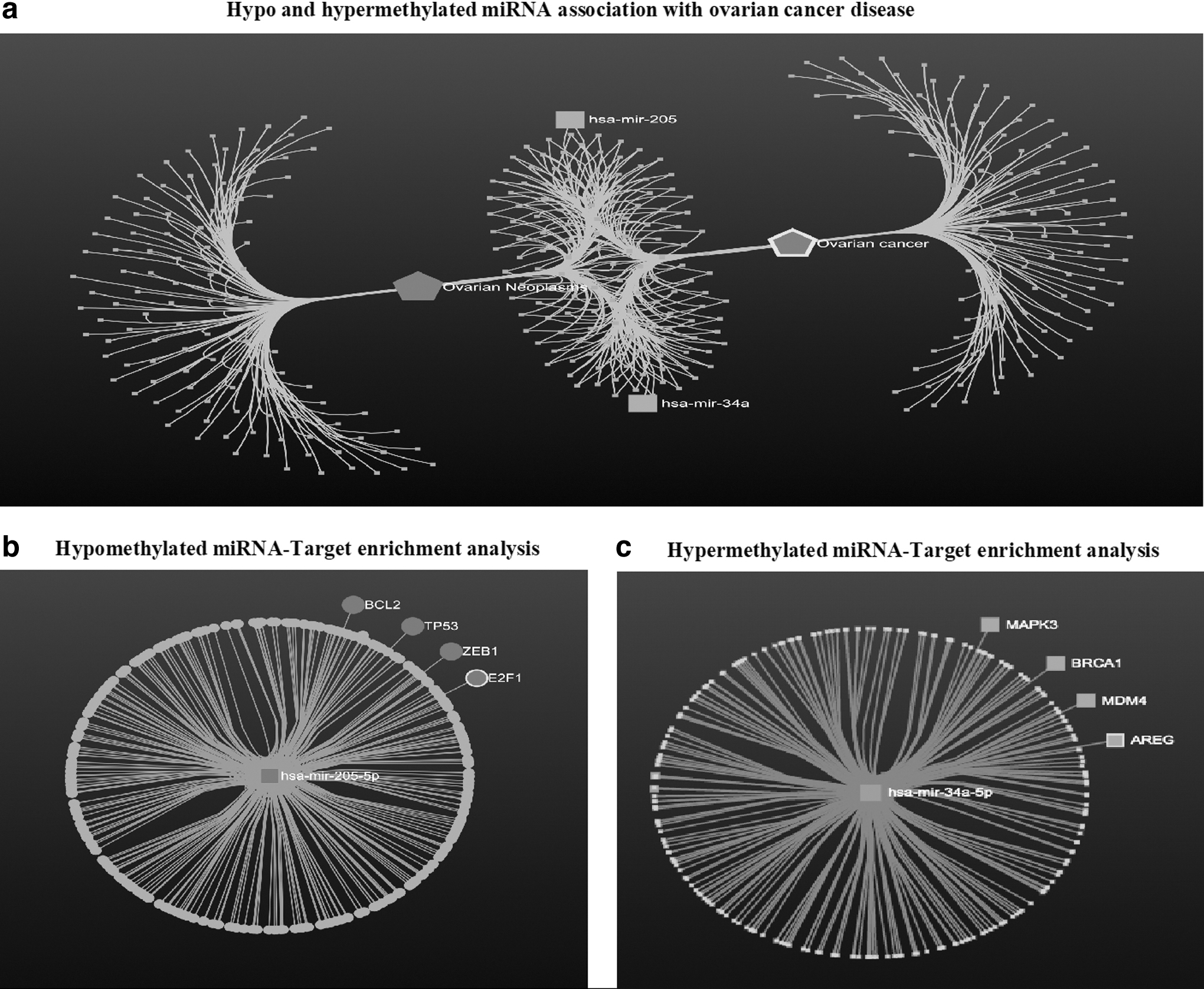
Represents miRNA-disease enrichment and miRNA-Target gene enrichment analysis of candidate miRNA.
Further, we used seven different miRNA databases (miRbase, miRDB, miRTarBase, Tools4miRs, miRnet, Diana-microT, miRwalk) to evaluate miRNA target molecules, more than thousands of genes were significantly targeted by individual candidate hypomethylated and hypermethylated miRNAs, however, we only selected overlapping target genes involved in
In our study, most significantly enriched miRNA-205 target genes were BCL2, ZEB1, E2F1, TP53, WNT5A, WAC, TET3, PANK1, HMGA2, PFN2, LRRFIP1, ZNF217, RAB23, POU2F2, RASSF8, RSF1, CDK14, GAS1, STAT3, MYO10, THBS2, ZMIZ1, TSC22D2, MYC, NOT, and AGO (Fig. 1b) and most significant miRNA34a-target genes were MDM4, MAPK3, BRCA1, AREG, VEGFA, AGO2, CXCL5, MYC, NOTCH2, SMAD2, RASSF2, CDK1, MBD4, MYCN, TRPS1, SOX2, MYC, MAPK8, MAPK1, BCL2L2, E2F3, MYCBP2, AGO3, CDK6, FAM135A, TBP, MYB, BCL11A, HIPK3, and MYO5C (Fig. 1c). Moreover, MDM4, MAPK3, BRCA1, and AREG were selected as the most significant downstream targets of miRNA-34a while BCL2, ZEB1, E2F1, and TP53 for miRNA-205 with a higher degree of betweenness, shortest path value, and p-value.
Functional enrichment analysis
After identifying putative candidate hypomethylated and hypermethylated downstream miRNA-target genes, we tried to analyze (1) the functional role of these target genes in cancer progression and (2) the therapeutic potential of candidate miRNAs. Therefore, we used DAVID software for functional annotation (Gene Ontology and KEGG analysis) of predicted miRNA-Target genes. We evaluated highly enriched top 10 Gene Ontology terms for biological process (BP), Molecular Function (MF), and Cellular Component (CC) of hypomethylated and hypermethylated candidate miRNA-Targets. Most of the Gene Ontology terms of hyper- and hypomethylated miRNA-Target genes were found to play a pivotal role in cancer progression and are presented in Supplementary Tables S1 and S2.
Further, we assessed KEGG enrichment analysis of selected candidate hyper- and hypomethylated miRNA-target genes to assess their involvement in biologically important pathways. Our hypermethylated miRNA targets were highly enriched in the small cell lung cancer, apoptosis, pathways in cancer, P53 signaling pathways, and several other cancers with a high enrichment score. Similarly, hypomethylated miRNA-target genes were significantly enriched in the cell cycle, pathway in cancer, PI3K-Akt signaling pathways, miRNA in cancer, and other important cancer pathways with higher enrichment scores (Supplementary Table S3).
Quantification of miRNA-target genes in OC
Further, we performed qRT-PCR-based relative expression analysis of miRNA-Target genes in EOC tissue samples compared to control tissue samples. The relative expression of miRNA-34a target genes MDM4, MAPK3, BRCA1, and AREG was significantly upregulated in EOC samples compared to healthy control with an increase in fold change of 7.22 (p < 0.0001), 6.39 (p < 0.0001), 4.69 (p < 0.0001), and 7.87 (p < 0.0001), respectively (Fig. 2a). Similarly, the relative expression of miRNA-205 target genes BCL2, ZEB1, E2F1, and TP53 was significantly downregulated in EOC samples compared to healthy control with a drop-in fold change of 4.09 (p < 0.0001), 0.94 (p < 0.0001), 3.1 (p < 0.0001), and 3.48 (p < 0.0001)], respectively (Fig. 2b).

Represents relative expression of enriched target genes of miRNA-205 and miRNA-34a in matched EOC tissue samples (48) and normal tissue samples (22).
Correlation between miRNA and target gene expression
Our in silico analysis of miRNA disease association, miRNA-target enrichment analysis, and functional enrichment analysis revealed several potential downstream target genes involve in
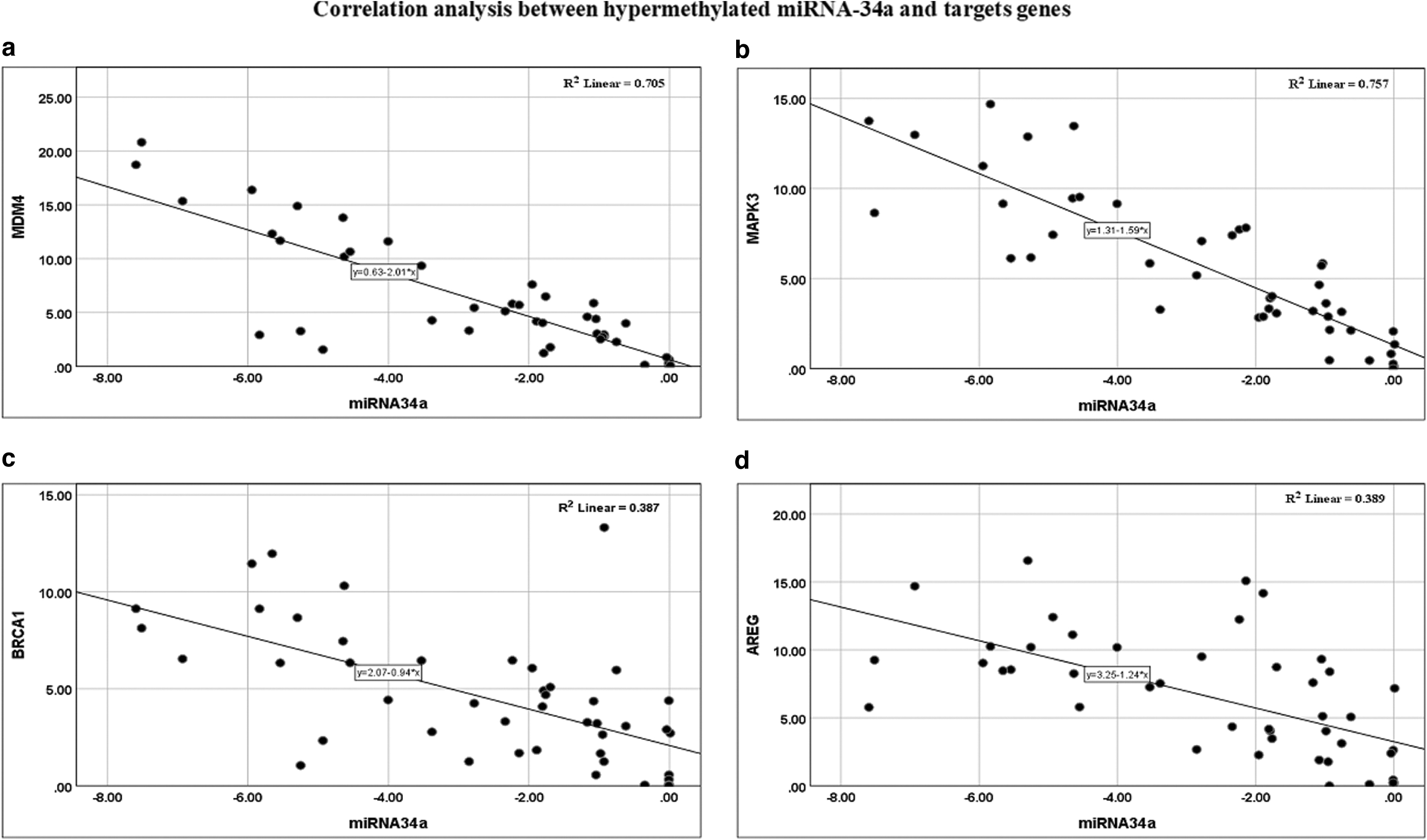
Represents correlation between dysregulated miRNA-34a expression with target gene expression in matched tissue samples. Upregulated expression of MDM4
Represents Correlation Analysis of MicroRNA-34 and MicroRNA-205 Expression with Corresponding Target Genes Expression in Matched Tissue Samples
miRNA-34a has significant negative correlation with MDM4, MAPK3, AREG, BRCA1, while age does not show any significant correlation.
miRNA-205 showed strong negative correlation with BCL2, ZEB1, E2F1, and TP53, while, age does not show significant correlation with any tested parameters. r = regression coefficient, p = probability value.
Similarly, upregulated miRNA-205 was negatively correlated with downstream target genes BCL2 (r = −0.813; p < 0.0001), ZEB1 (r = −0.755; p < 0.0001), E2F1 (r = −0.559; p < 0.001), and TP53 (r = −0.767; p < 0.0001), respectively (Fig. 4) (Table 3b).

Represents correlation between dysregulated expression of miRNA-205 with target gene expression in matched tissue samples. Downregulated expression of BCL2
Diagnostic potential miRNA panel and target gene panel
Further, to evaluate the diagnostic potential of miRNA and their downstream target genes, we conducted multivariate binary logistic regression analysis on a combined miRNA panel (miRNA-205+miRNA-34a), hypermethylated miRNA-34a target gene panel (MDM4+MAPK3+AREG+BRCA1), and hypomethylated miRNA-205 target gene panel (BCL2+ZEB1+E2F1+TP53). The expression data of miRNA were used from the previous study (Kumar et al, 2022; Kumar et al, 2021). The expression of the miRNA panel and hyper- and hypomethylated target genes panel were significantly associated with disease progression (Supplementary Table S4).
Furthermore, predicted probability values obtained from multivariate regression model was used to assess the diagnostic potential of each panel using receiver operating characteristics analysis. The combined diagnostic potential of miRNA-34a and miRNA-205 was evaluated by area under the curve (AUC) at 95% confidence interval (CI), p-value was 82.7% (0.715–0.940; p < 0.0001) with sensitivity = 91.3% and specificity = 78% at cutoff value = 0.523 (Fig. 5a). Combined diagnostic potential of miRNA-34a target genes (MDM4+MAPK3+AREG+BRCA1) was evaluated by AUC at 95% CI, p-value was 98.3% (0.960–1.002; p < 0.0001) with sensitivity = 93.5% and specificity = 90.9% at cutoff value = 0.523 (Fig. 5b).
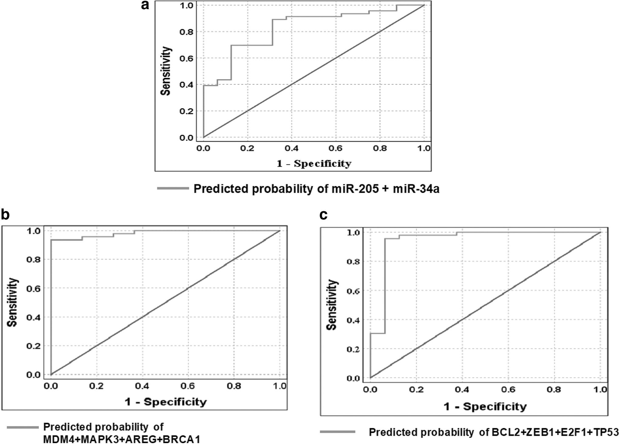
ROC curve analysis of miRNA panel, miR-205-associated target gene panel, and miR-34a-associated target gene panel toward diagnosis of EOC.
Similarly, combined diagnostic potential of miRNA-205 target genes (BCL2+ZEB1+E2F1+TP53) was evaluated by AUC at 95% CI, was 94.8% (95% CI = 0.862–1.204; p < 0.0001) with sensitivity = 95.7% and specificity = 93.7% at cutoff value = 0.871 (Fig. 5c). Combined diagnostic ability of miR-34a and miR-205 downstream target genes has better predictive value for detection of EOC. The detailed AUC, sensitivity, specificity and cutoff values of each panel is given in Table 4.
Represents Receiver Operating Characteristics Curve Analysis for Predicting Risk Epithelial Ovarian Cancer Using Relative Expression Level of MicroRNA Panel and MicroRNA-Target Gene Panel from Tissue Samples
AUC, area under the curve; CI, confidence interval; CV, optimal cutoff value; EOC, epithelial ovarian cancer; SEN, sensitivity; SPE, specificity.
Discussion
In this context, our previous analysis on miRNA-205 and miRNA-34a expression and correlation with epigenetic changes on their gene was in line with recent articles (Kumar et al, 2022; Kumar et al, 2021). Therefore, we extended our analysis toward identifying therapeutic potential of miRNA-205 and miRNA-34a in
The first step toward identifying the therapeutic potential of miRNA, we assessed miRNA-disease enrichment, miRNA-Target enrichment, and functional enrichment analysis of candidate hypo-/hypermethylated miRNA. Our analysis revealed that several tumor suppressor genes and oncogenes involved in cancer progression pathways were targeted by candidate hypomethylated (miR-205) and hypermethylated miRNA (miR-34a). Most significant target gene of miR-205 was BCL2, TP53, ZEB1, and E2F1, while, MDM4, MAPK3, BRCA1, and AREG were significantly targeted by miR-34a.
Wang et al (2015) revealed miR-205 upregulation leading to downregulation of BCL2 in cell line, moreover, miRNA-205 targets BCL2 in cell line was confirmed through luciferase assay, qRT-PCR, and immunohistochemical assay. Similarly, miR-205 enhances the motility of
Similarly, downregulated expression of miRNA-34a was correlated with overexpression of direct target AREG in
In addition, we explored downstream functional analysis (Gene Ontology and Kyoto Encyclopedia of Genes and Genomes) of selected candidate miRNA-Target genes exclusively in EOC. Functional enrichment analysis of hypomethylated and hypermethylated miRNA-Target genes using Gene Ontology and KEGG analysis showed significant involvement of miRNA-Targets in different biologically important BP {[Hypomethylated BP (GO:0010628, GO:0006357, GO:0008285); Hypermethylated BP (GO:0008219, GO:0006915, GO:0010941, GO:0012501)); MF (Hypomethylated MF (GO:0005524, GO:0000978, GO:0008134, GO:0019899); Hypermethylated MF (GO:0044877, GO:0051087, GO:0004861,GO:0042802)); CC (Hypomethylated CC (GO:0005654, GO:0005694, GO:0043234, GO:0044427); Hypermethylated CC (GO:0005634, GO:0097136, GO:0043233, GO:0005654)]}, and different cancer-related pathways (Supplementary Table S1).
Further, expression analysis of miRNA-205 and miRNA-34a target genes in EOC and normal samples showed significant downregulation of miRNA-205 target gene (BCL2, TP53, ZEB1, and E2F1) in
Conclusion
Overall, our analysis revealed miRNA-205 and miRNA-34a disease association, therapeutic potential, and diagnostic ability. Moreover, identified miRNAs-Target genes showed tumor suppressors and oncogenic roles in several cancer-associated pathways and involved in cancer progression, thus revealing miRNAs therapeutic potential for
However, this study is an only preliminary report highlighting epigenetic regulation of miRNA, target identification, expression analysis, evaluation of their diagnostic potential, and correlation of miRNA and target gene expression. Therefore, additional high-throughput analysis is required at cellular and molecular level to validate the obtained result in a larger cohort of samples and further claiming the therapeutic role of candidate miRNA and there are target molecules in
Footnotes
Acknowledgments
We want to thank the Central instrumentation facility of MNNIT Allahabad, Prayagraj, for facilitating Real-Time PCR. We further extend our gratitude to Department of Gynecology and Obstetrics, Swaroop Rani Nehru Hospital Allahabad, Prayagraj, for providing normal tissue samples recruited for this study.
Authors' Contributions
V.K. performed investigation, writing-original draft preparation, data curation, and software; A.P. performed investigation, writing draft, and software; A.A. data curation and writing draft; P.G. draft formatting and data curation; D.B. software and data curation; M.S. helped in resources, conceptualization, methodology, validation, and supervision of this study.
Ethics Approval and Consent to Participate
The study was conducted according to the guidelines of the Declaration of Helsinki, and approved by the Institutional Review Board of Motilal Nehru National Institute of Technology Allahabad (Ref. No: IEC/2019–20/01). Moreover, informed consent was obtained from all patients involved in the study.
Availability of Data and Materials
The datasets used and/or analyzed during the current study are included in this article and can also be made available from corresponding author on reasonable request.
Disclosure Statement
No competing financial interests exist.
Funding Information
This research received specific grant from Indian Council of Medical Research, Government of India, Grant number 5/13/58/2015/NCD-III.
Supplementary Material
Supplementary Data
Supplementary Table S1
Supplementary Table S2
Supplementary Table S3
Supplementary Table S4
References
Supplementary Material
Please find the following supplemental material available below.
For Open Access articles published under a Creative Commons License, all supplemental material carries the same license as the article it is associated with.
For non-Open Access articles published, all supplemental material carries a non-exclusive license, and permission requests for re-use of supplemental material or any part of supplemental material shall be sent directly to the copyright owner as specified in the copyright notice associated with the article.