
Abstract
Abstract
There has been a growing trend toward the addition of surface modifiers, surfactants, or capping agents to the precursors of titanium dioxide nanoparticles to tailor their morphology, structure, and size. In this study, surface-modified, manganese-doped titanium dioxide nanoparticles were successfully synthesized under mild hydrothermal conditions (pressure is autogenous; temperature, 250°C; time, 18 h). Using different molar ratios, we applied caprylic acid to change the surface chemistry and photocatalytic efficiency of manganese-doped titanium dioxide nanoparticles. Reagent grade titanium dioxide and manganese dioxide were the precursors, and concentrated hydrogen chloride was used as a solvent. Synthesized nanoparticles were characterized using powder x-ray diffraction, transmission electron microscopy, BET surface area, and ultraviolet-visible spectroscopy. Photodegradation studies of methylene blue were then carried out using ultraviolet and sunlight irradiation to compare the photocatalytic efficiency of the products. Results of chemical oxygen demand and transmission percentage revealed a higher photocatalytic efficiency of the surface-modified, manganese-doped titanium dioxide nanoparticles compared to reagent grade titanium dioxide.
Introduction
There are a number of methods for synthesizing nanoparticles, including sol-gels, microemulsions, homogeneous hydrolysis, the use of supercritical fluid processes, and hydrothermal techniques (Mishra and Srivastava, 2008; Hu et al., 2003; Stengl et al., 2008; Aymonier et al., 2007; Byrappa and Adschiri, 2007). Unfortunately, while conventional methods yield highly agglomerated particles, there is little control over their morphology and crystal size. In most cases, the particles require further treatment. Although hydrothermal techniques are effective in obtaining high quality nanoparticles because of highly controlled diffusion during the synthesis process, particle size and morphology cannot be controlled. The application of a suitable surfactant or surface modifier or capping agent with optimum concentration can overcome these imperfections. For example, Deng et al. (2011) synthesized manganese-doped TiO2 (Mn-TiO2), using a sol-gel method and applied the product for the photodegradation of methylene blue (MB) under visible light. In other research, toluene gas was degraded using Mn-TiO2 nanoparticles under UV irradiation. It was found that the Mn-TiO2 nanoparticles were more efficient than pure TiO2 powder (Jothiramalingam and Wang, 2007). For our study, different concentrations of caprylic acid were applied as a surfactant and manganese dioxide (MnO2) was used as a dopant to narrow the band gap energy of TiO2 nanoparticles.
One major advantage of using dyes as model pollutants is the simplicity in monitoring the extent of their degradation, which can be done using spectrophotometry. The breaking of bonds in a dye molecule and its subsequent decolorization by a photocatalyst simulates what a photocatalyst can do when it comes to degradation of pollutants. The photodegradation of MB was carried out using aqueous dispersion of surface-modified hydrophilic Mn-TiO2 nanoparticles under UV and sunlight irradiation.
Experimental Studies
Preparation of surface-modified Mn-TiO2 nanoparticles
Mn-TiO2 hybrid nanoparticles were synthesized under mild hydrothermal conditions (T=250°C; pressure is autogeneous). To do so, reagent grade TiO2 (Loba Chemie) was used as the starting material, and MnO2 (2 mol% and 5 mol%) was added into it. Concentrated HCl (10 mL) was added as a mineralizer to the precursors. At the same time, different concentrations (0.8 M, 1.0 M, 1.2 M, and 1.4 M) of caprylic acid (Sisco Research Lab PVT, Ltd.), were added to the mixture, which was then stirred vigorously for a few minutes. The final compound was transferred to a 25 mL Teflon liner (Vfill=10 mL), which was later placed inside a General Purpose autoclave. The assembled autoclave was kept in an oven with a programmable temperature controller for 8–18 hrs. The temperature was kept at 250°C. At the end of experimental run time, the autoclave was cooled to room temperature. The product in the Teflon liner was transferred to a clean beaker, washed with double distilled water, and allowed to settle. The surplus solution was removed using a syringe, and the remnants were centrifuged using a Kubota KN-70 at 1500 rpm for 20 min. The product was recovered and dried in a hot air oven at 40–50°C for a few hours. The dried particles were subjected to systematic characterization and photocatalytic studies.
Characterization of the surface-modified Mn-TiO2 nanoparticles
The fabricated products were characterized using different analytical techniques. Powder X-ray diffraction patterns (XRD) (Bruker, D8 Advance, with Cu Kα, λ=1.5420 Å radiation; voltage, 40 mV; current, 30 mA; scan speed, 5 min−1) were recorded. The data were collected in the 2θ range 5–100°. UV-Vis spectrophotometry was used to study the optical properties of the products. Transmission electron microscope (TEM) images of the Mn-TiO2 hybrid nanoparticles were recorded, using a JEM 2000FX II (JEOL. Ltd.). The BET surface area was measured using a Belsorp mini II (Bel Japan, Inc.)
Photreactor and experimental procedure
Photoreaction experiments were carried out using a cylindrical flow photoreactor that was designed and fabricated in our laboratory. A 6 W, low-pressure mercury lamp with an emission peak at 264 nm was placed inside a quartz sleeve and positioned at the center our photoreactor. This was surrounded by a circulating water jacket, meant to control the temperature during reaction. The same reactor was used without the UV lamp (i.e., sunlight was used) for visible light irradiation. All photodegradation experiments using sunlight were carried out from 12:00 p.m. to 3:00 p.m. The reaction suspension was prepared by adding different amounts (0.1–1 g) of photocatalyst powder per liter of dye solution. A small aquarium aerator was used as an oxygen source, and air was continuously pumped into the reactor through air diffusers to fully fluidize the Mn-TiO2 hybrid nanoparticles during photoreaction. The hybrid nanoparticles were thoroughly mixed with the MB solution and then placed in darkness for 30 min. to ensure a balance of adsorption and desorption. The suspensions were sampled at specific intervals to monitor the changes in MB concentration. Sampled suspensions were centrifuged using a Kubota KN-70 at 1500 rpm for 30 min. to remove the Mn-TiO2 hybrid nanoparticles and then analyzed by UV-Vis spectrophotometry.
Results and Discussion
Characterization of surface-modified Mn-TiO2 nanoparticles
Powder XRD results indicated that the crystal symmetry of the synthesized hybrid nanoparticles well matched the I41/amd space group. XRD data revealed a slight change in the lattice parameters of the Mn-TiO2 nanoparticles (TiO2-x, where x=2 mol% MnO2 or 5 mol% MnO2) at a-axis and c-axis when compared to reagent-grade TiO2. This confirms the existence of Mn atoms in the TiO2 nanoparticles. As Rizwan et al. (2008) have noted, the ionic radii of Mn ions (53 pm) are bigger than those of Ti (42 pm), and, on doping, are certain to make the cell parameter bigger than that of reagent-grade TiO2. The powder XRD pattern of (2 mol%; 5 mol%) Mn-TiO2 hybrid nanoparticles shows five primary peaks: 25.30°, 37.80°, 48.04°, 53.88° and 62.68° for 2 mol% dopant, and 25.33°, 37.80°, 48.05°, 54.24° and 62.66° for 5 mol% dopant (see Fig. 1). These can be attributed to different diffraction planes of TiO2 (anatase). The appearance of a few small peaks in the case of 5 mol% doping indicates that the structure of the TiO2 nanoparticles may change as the concentration of dopant increases. However, as shown in Fig. 1, no extra peaks were observed in the case of surface-modified, undoped TiO2 nanoparticles. Therefore, it can be concluded that the change in the structure of TiO2 is because of doping and the concentration of dopant. The powder XRD data indicate that the cell volume of Mn-TiO2 hybrid nanoparticles has slightly increased with 2 mol% and 5 mol% Mn doping (see Table 1) (Howard et al., 1992).
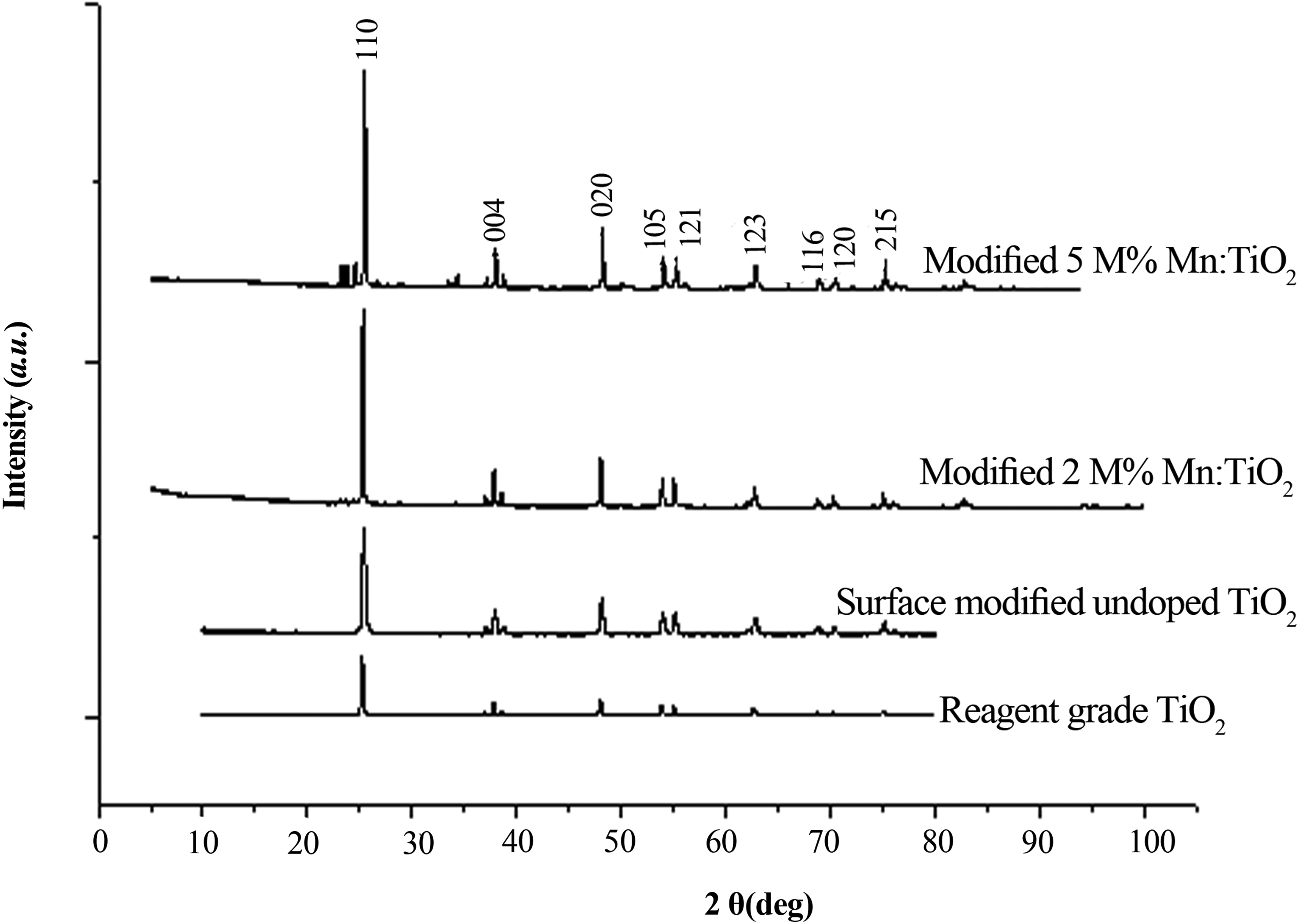
Powder X-ray diffraction pattern of Mn-TiO2 nanoparticles modified using 1.0 M caprylic acid.
The average size of the surface-modified Mn-TiO2 nanoparticles was determined from the XRD patterns of TiO2 nanoparticles by applying the Scherrer equation:
where k is the particle shape factor and taken as 0.89, λ is the wavelength of CuKa1 radiation (0.1542 nm), β is the calibrated half-intensity width (FWHM) of the selected diffraction peak (degrees), and θ is the Bragg angle (half of the peak position angle). The average nanoparticle size was obtained from the maximum intensity of anatase at 25.3° and 25.33° (Behnajady et al., 2008). Using this equation, the average size of the surface-modified Mn-TiO2 hybrid nanoparticles fabricated under mild hydrothermal conditions was 65 nm and 84 nm for 2 mol% and 5 mol% doping, respectively. These results are in agreement with the powder XRD data and TEM images.
Fig. 2 shows characteristic TEM images of 2 mol% and 5 mol% Mn-TiO2 nanoparticles modified with 0.8 M and 1.4 M caprylic acid. A thin organic coverage of the surface modifier on the synthesized nanoparticles is clearly evident. The agglomeration was less when a higher concentration of the surface modifier was used. The surface modification led to control of the direction of growth as well as particle size, thereby, preventing agglomeration. The surface modifier can not only affect the dispensability of the synthesized Mn-TiO2 nanoparticles but also change their growth habit. The stains observed in TEM images are due to the effect of both surface modifier and dopant application. The characteristic BET surface area of the surface-modified Mn-TiO2 nanoparticles (1.0 M caprylic acid; 5 mol% MnO2) was found to be 23.5 m2/g.
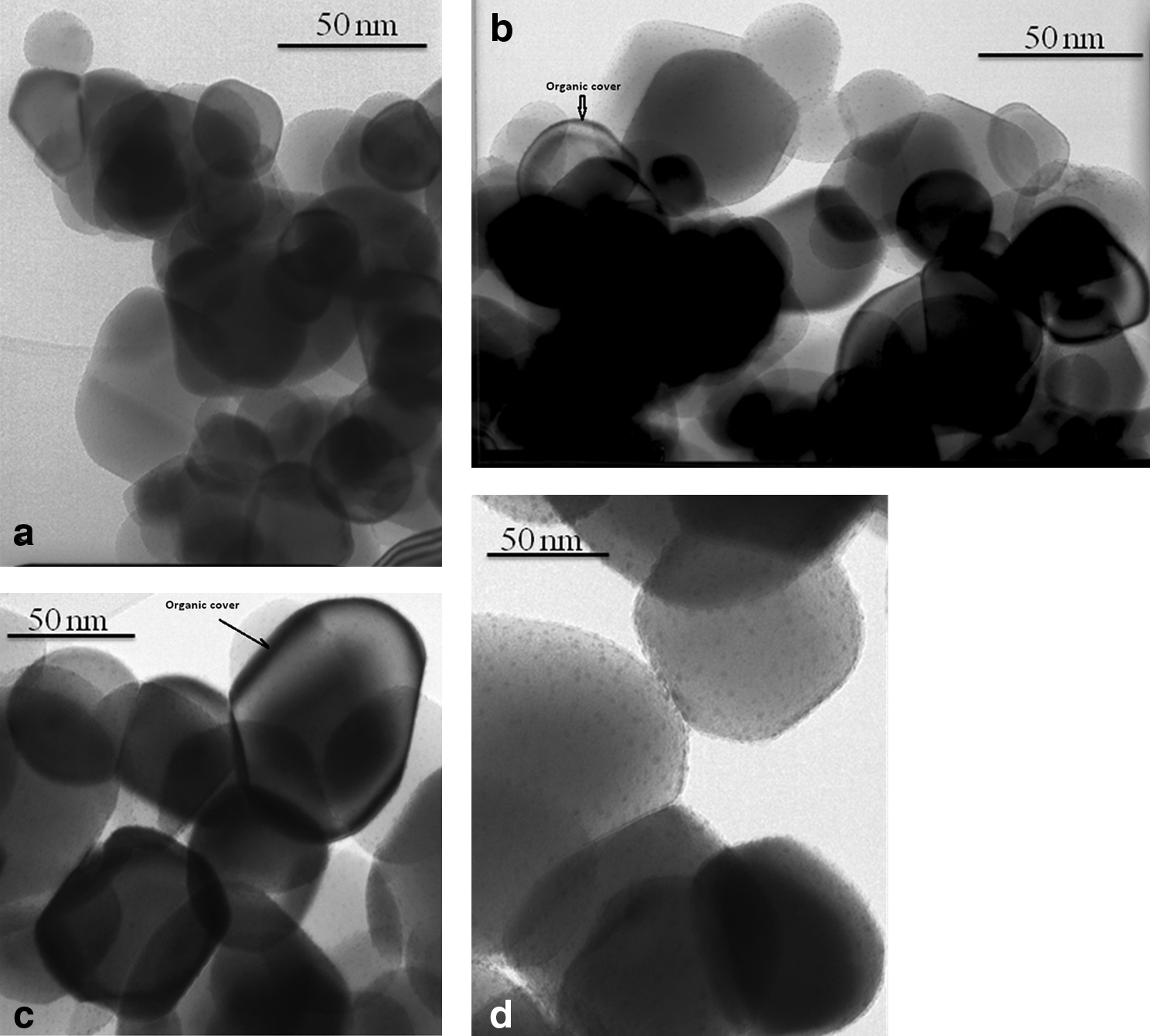
Characteristic TEM images of surface-modified Mn-TiO2 nanoparticles:
Band gap energy of surface-modified Mn-TiO2 nanoparticles
The effect of doping was studied by UV-Vis spectroscopy in the range of 200 nm to 600 nm. Fig 3 shows that reagent grade TiO2 and surface-modified, undoped TiO2 nanoparticles had no absorption in the visible light region (λ>400 nm). The surface-modified Mn-TiO2 hybrid nanoparticles showed a remarkable absorption band shift toward the longer wavelength region, which indicates a decrease of the band gap energy. In addition, the band gap of the three samples were measured at room temperature and calculated using the equation Eg=hc/λ. In this equation, Eg is the band gap energy (eV), h is Plank's constant, c is the speed of light (m/s), and λ is the wavelength (nm). The E g of reagent grade TiO2 nanoparticles, 2 mol% Mn-TiO2 hybrid nanoparticles, and 5 mol% Mn-TiO2 hybrid nanoparticles was 3.32, 3.12, and 3.01 respectively.
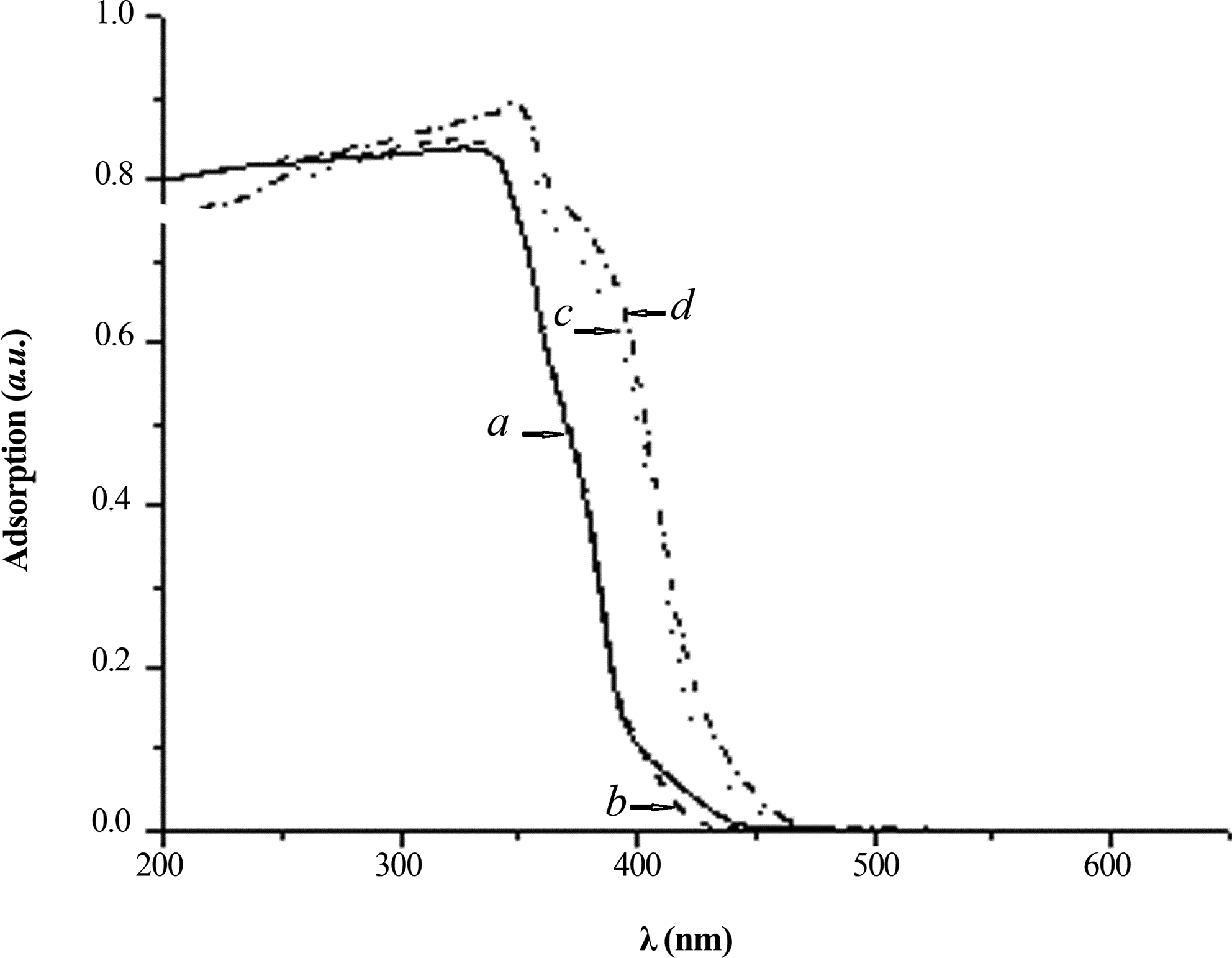
Effect of doping on the band gap energy TiO2 nanoparticles.
Photodegradation of MB
MB (Aldrich) absorption was measured at 404 nm (λmax) of the remaining solutions. The absorption was then converted to the relative concentration of the photodegraded dyes (ln C/C0, where C and C0 represent the concentration after reaction and the initial concentration, based on a standard curve and the displayed linear behavior between relative concentration and absorption at this wavelength, respectively). For comparison, the photocatalysis of MB on reagent-grade TiO2 powders (Loba Chemie) was conducted under the same operating conditions.
Effect of surface-modified Mn-TiO2 nanoparticles on photocatalytic degradation of MB
In a slurry photocatalytic process, catalyst dosage is an important parameter. Photocatalytic degradation of 10 mg/L MB was carried out with modified, tungsten-doped (2 mol% and 5 mol%) TiO2 hybrid nanoparticles and a reagent-grade TiO2 catalyst loading of 0–1.0 g/L under sunlight irradiation. The degradation of MB as a function of the fabricated TiO2 nanoparticles and pure TiO2 dosage is presented in Fig. 4a.

Effect of catalyst dosage on Methylene Blue (MB) photodegradation efficiency under
As shown in Fig. 4b, in the absence of TiO2, the removal percentage of MB was almost zero. The addition of nanoparticles enhanced the removal of MB. The removal rate of the contaminants increased as the concentration of the dyes thickened. In the case of reagent grade TiO2, there was no difference in the extent of degradation based on the light source. As shown in Fig. 4, MB degradation also occurred in visible light, which serves to confirm the effect of doping on MB photodegradation. An optimal result was achieved at a TiO2 dosage of 0.8 g/L. With a further increase in the catalyst dosage, removal decreases.
In analyzing the kinetic data of photodegradiation mediated by semiconductors or nanoparticles like TiO2, the data are fitted to a simple rate expression of Langmuir–Hinshelwood (L-H) form (Kim et al., 2007):
where C0 is the initial concentration of dye, KAd is the pseudo–first-order L-H-type adsorption coefficient, and K is the reaction rate constant. The integration of Equation (2) yields Equation (3):
where t is the reaction time. The integrated form of Equation (3) for a low initial concentration of MB of this study can be written as follows:
where kapp is the apparent reaction rate constant. Rate constant kapp has been chosen as the basic kinetic parameter for different systems, because it is independent of used concentration and can be determined from the slope of the curve obtained from Fig. 4 (Wang et al., 2008).
Effect of light source and its duration on the photocatalytic degradation of MB
Figure 5 shows a comparison of the photodegradation of MB in the presence of surface-modified (2 mol%; 5 mol%) Mn-TiO2 hybrid nanoparticles at different times under sunlight and UV irradiation. The photodegradation ratio increased as the sunlight irradiation time increased, attaining about 96.9% and 94.2%, respectively, within 3.0 hours. In the case of UV irradiation, the efficiency was 76.35% and 72.5%, respectively. The results indicate that the photocatalytic degradation reaction is a pseudo–first-order kinetic reaction. Moreover, up to a certain concentration (pseudo–first-order), there is a direct relationship between the concentration of surface modifier applied and photodegradation efficiency. However, we have not presented any data regarding the stability of caprylic acid with regard to both washing away and oxidation (Shahmoradi et al., 2010a, 2010b).
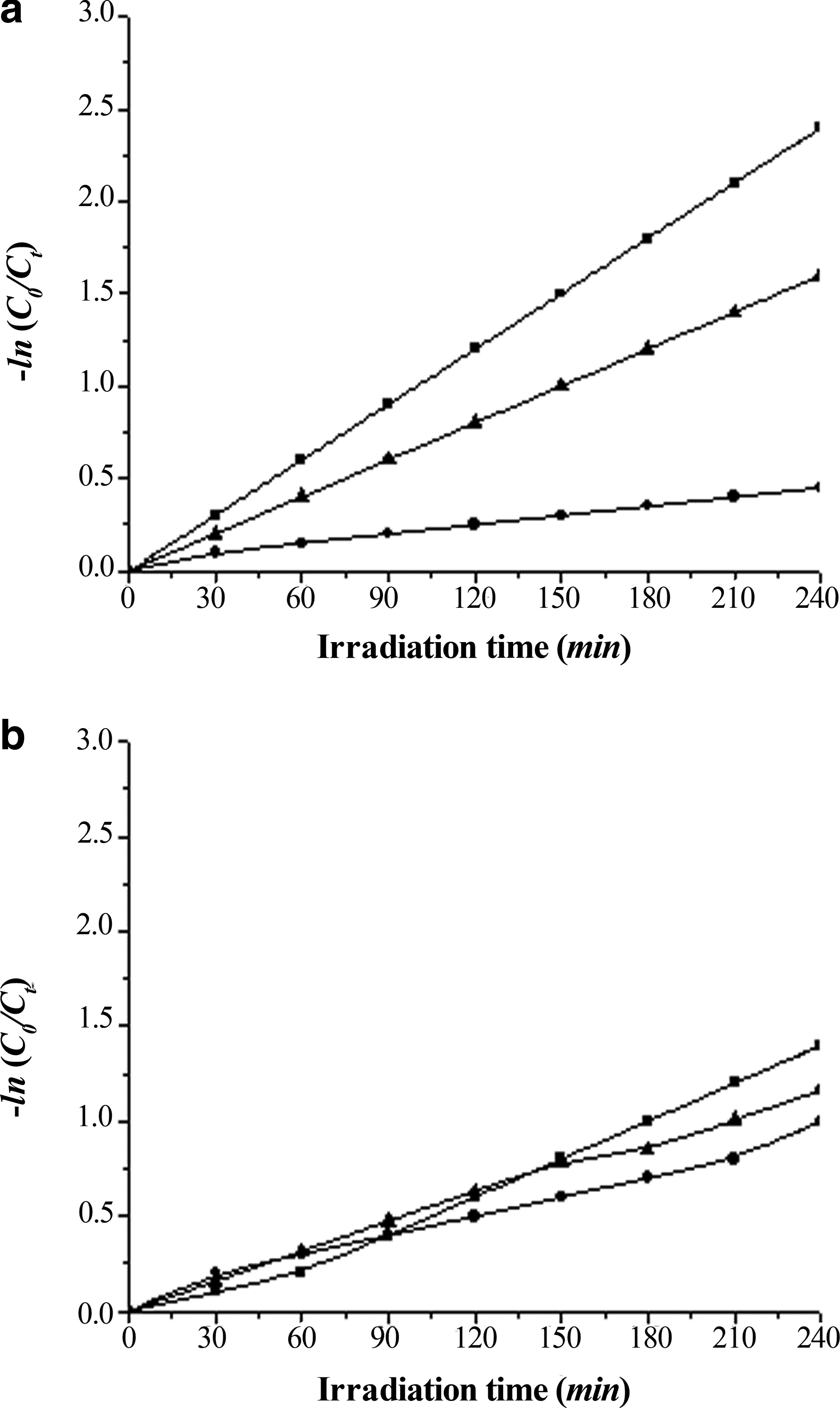
Effect of irradiation time on the photodegradation of MB under
Conclusion
Mn-TiO2 nanoparticles were successfully modified under mild hydrothermal conditions. Caprylic acid was used as a surface modifier because it has low toxicity, low density, and a low melting temperature. It is also eco-friendly. Surface modification and doping changed the morphology and size of the synthesized nanoparticles. Modification and doping also changed the surface charges and increased the stability of the nanoparticles, which is necessary to achieve higher photodegradation efficiency. Among the important factors that may affect photodegradation are initial concentration and type of pollutant. Simultaneous surface modification and doping significantly shifted the band gap energy to that portion of the visible region where photodegradation efficiency is more comparable to that of the UV region.
Footnotes
Author Disclosure Statement
No competing financial interests exist.