
Abstract
Abstract
Vanadium (V)-doped WO3/TiO2 microporous film on a titanium (Ti) plate was prepared by using the micro-arc oxidation process. Incorporating V and tungsten (W) species into the TiO2 lattice in one step yields band gap narrowing and strong visible-light (Vis) photoactivity, dominated mostly by incorporation of WO3 and V2O5 oxides, and possibly W x Ti1−xO2. Photoactivity under ultraviolet light (UV) irradiation was likely attributable to its WO3-loaded microporous structure, which favored cationic molecule adsorption on its active sites. Although post annealing enhanced film crystallinity, photocatalytic ability was decreased largely because of changes in doping levels. This suggests that the as-oxidized microporous film is feasible for photocatalytic cationic dye degradation under UV and Vis irradiation.
Introduction
T
It has been observed that a heterostructured catalyst can provide a potential driving force for the separation of photogenerated charge carriers, provided that the band edges of the constituent catalysts are properly positioned. Among the various heterostructured oxides, TiO2/WO3 has shown promising photocatalytic applications, mostly because of increased surface acidity (He et al., 2009; Yu et al., 2010) and the proper coupling of the WO x species with TiO2 for enhancing charge-separation efficiency (Li et al., 2001). For instance, the conduction band (CB) edge of TiO2 (−0.19 eV vs. NHE) is more negative than that of WO x (0.3 eV) and the valance band (VB) edge of TiO2 (3.01 eV) is more positive than that of WO x (3.5 eV). Under UV illumination, the photogenerated electrons and holes will transfer to the CB of WO x and to the VB of TiO2, respectively (Higashimoto et al., 2006; Chen et al., 2008). Similarly, the heterostructured V2O5/TiO2 favors the photogenerated electron transfer to the CB of V2O5, due to a proper CB edge of 0.48 eV of V2O5 oxide (Dong et al., 2012). As a result, the recombination of the photogenerated carriers in the heterostructured catalyst is suppressed, potentially resulting in higher photocatalytic ability. Additionally, a highly specific surface area reportedly favors photocatalytic activities of the porous anodized TiO2 oxides (Liu et al., 2009).
Micro-arc oxidation (MAO), also known as plasma electrolytic oxidation, is regarded as a fast electrochemical process that has recently been used for preparing porous TiO2 layers coupled with vanadia or tungsten oxide (Bayati et al., 2010a, 2010c; Yeh et al., 2012). One of the potential advantages of the MAO process is the possibility of incorporating anionic and/or cationic ions into the TiO2 layer on a Ti substrate, by controlling the composition and concentration of the electrolyte (Li et al., 2009). The main mechanism in the MAO process involves dielectric breakdown, which causes spark discharge, micro arcing, and anodic oxidation in potentiostatic condition. Under this condition, small luminescent sparks are observed to move rapidly across the surface of the oxide film, facilitating its continued growth. Therefore, porous oxide films continuously form on the anode surface (Yerokhin et al., 1999; Khan et al., 2008; Li et al., 2009). In general, the current density is normally in the region of 0.5–1.0 A/cm2 during the MAO process (Yerokhin et al., 1999).
Addition of fluoride into the electrolyte has reportedly increased the electrolyte conductivity, leading to change of the MAO microstructure and properties (Mu and Han, 2008; He et al., 2009). The content of W doped into TiO2 lattices is affected by the fluorine ion (F−) concentration in the electrolyte; the higher content of W generally provides better photocatalytic activity. Note that F-doping does not cause any change in the optical absorption of TiO2 (Minero et al., 2000; Li et al., 2005) and little change in the surface morphology of MAO films (He et al., 2009). In this study, NaF was added into the electrolyte solution to enhance the MAO properties and hence the photocatalytic activities as well.
We previously determined that, by applying the MAO process at a low applied voltage of 300 V, the loaded WO3 favored the formation of the well-crystallized anatase with a trace of rutile TiO2 phase (Yeh et al., 2012). The aim of this study was to elucidate the structural and photocatalytic characteristics of the V-WO3/TiO2 layers prepared at a high applied voltage of 380 V. The effects of annealing are also discussed.
Experimental Protocol
Microporous V-WO3/TiO2 films were synthesized on Ti sheet substrates (25 mm×75 mm×1 mm) by using an in-house MAO system (MIRDC) that consisted of a high-voltage DC power supply, anode, and cathode. A stainless steel container with stirring and cooling systems was used as an electrolyte bath. The Ti sheet substrate was connected as anode, whereas the stainless steel container was used as cathode. The electrolyte solution (pH=8) comprised Na2WO4 (15 g/L), NaVO3 (5 g/L), and NaF (2 g/L) solutions. The Ti sheet substrates were oxidized using a bipolar pulse current mode, in which the positive voltage, negative voltage, and duty cycle were 380, −10, and 0.2 V, respectively, for a period of 10 min during the MAO process. The electrolyte temperature was maintained at ∼40°C±2°C by the stirring and cooling systems. The as-oxidized MAO (denoted as V-WTO) samples were rinsed with distilled water and then dried in air at room temperature. Some of the as-oxidized V-WTO samples were then annealed at 873 K for 6 h in atmosphere, and were denoted as Vh-WTO herein. For comparison, N,C-codoped TiO2 (N,C-TiO2) films were deposited on ITO glass substrate by using a magnetron sputtering technique for a duration of 7.2 h (Wu and Hung, 2009). The thicknesses of the V-WTO and N,C-TiO2 films were 1.4 and 1.8 μm, respectively.
The crystal structures of the samples were analyzed using a high-resolution X-ray diffractometer (XRD; Rigaku ATX-E). The microstructures and thicknesses of the films were examined using a scanning electron microscope (SEM; JEOL JSM-6700F). A transmission electron microscope (HRTEM; Philips Tecnai 20) equipped with an energy dispersive X-ray spectrometer (EDS) was employed for composition analysis. X-ray photoelectron spectroscopy (XPS) analysis was performed using a high-resolution XPS spectrometer (ULVAC-PHI Quantera SXM/Auger AES 650). The binding energy of XPS spectra was calibrated using the C1s energy of 284.6 eV. The diffused reflectance of the films was measured using a UV–vis–NIR spectrometer (Hitachi U-4100) equipped with an integration sphere. The band gap energy of each sample was calculated using the formula stated by Bayati et al. (2010c).
The photocatalytic activities of the samples were evaluated by immersing the samples with a size of 25 mm×65 mm into 30 mL of aqueous methylene blue (MB) solution with an initial 10 mg/L (C0) concentration. Prior to every MB degradation test, the samples were soaked in another beaker of aqueous MB solution for 3 h in darkness for equilibrium surface adsorption. Aqueous MB solutions without samples were also illuminated to generate a blank value and found slightly degraded (<10%) under 2 h of UV radiation (Yeh et al., 2012). Two light sources, ultraviolet (UV) lamps (λ=365 nm) and blue-light-emitting diodes (BLEDs; 420<λ<530 nm), were used to provide irradiated light intensities of 2.7 and 10.5 mW/cm2, respectively. The degradation rate constant (k) of MB over film was assumed to be a first-order reaction for the evaluation, where the rate constant is expressed from the linear fitting of k=−ln(C/C0)/t and the degraded MB concentration C (mg/L) at reaction time t (h). The reaction times were set at 0.5-h intervals from 0.5 to 2 h for the UV tests. The experimental details were described in related reports (Wu and Hung, 2009; Yeh et al., 2012).
Results and Discussion
Microstructures
As shown in Fig. 1, the V-WTO diffraction peaks are ascribed to the TiO2 anatase (101) phase (2θ=25.3°) located at broad and yet to be identified peaks ranging from 2θ=20°–35°, and two weak Ti peaks at 2θ=38.3° and 2θ=40.1°. Moreover, no other peak associated with another phase is found in the probed range. A slight diffraction angle shift, caused by different ion radii of Ti to V and W, has often been observed in the XRD patterns of the anatase TiO2 (101) phase as W or V doped on titania (Bayati et al., 2010a, 2010c). As seen in the inset of Fig. 1, a slightly low angle shift of the diffraction (101) plane of 2θ=25.32° of TiO2 phase was observed in the V-WTO, but not in the Vh-WTO. A diffraction angle shift with a broad peak is possibly because of the WO x monolayer and V2O5, or the amorphous phases highly dispersed on the TiO2 lattice (Sajjad et al., 2009). This suggests that the as-prepared V-WTO is likely doped with W, V, and/or other ions, which have a significant effect on its optical property. The amorphous phases are commonly found in the MAO samples prepared at a low applied voltage (Bayati et al., 2010c). The annealed Vh-WTO exhibits various well-crystallized TiO2 anatase phases along with crystalline WO3, V2O5, and a trace of TiO2 rutile phase. An intense diffraction peak at 2θ=23.67° of the crystalline WO3 (020) plane is observed as shown in the inset of Fig. 1. The other two diffraction peaks at 2θ=29.1° and 2θ=33.5° are assigned to crystalline WO3 phases. Moreover, the diffraction peaks at 2θ=21.6° and 2θ=26.6° are the predominant (101) and (110) planes of crystalline V2O5, respectively. Only two weak peaks at 2θ=38.42° and 2θ=40.17° (JCPDS No. 44-1294) that possibly derive from the Ti substrate are found in both the V-WTO and Vh-WTO. These findings imply that the Vh-WTO is of the crystallized TiO2 anatase (101) phase incorporated with noticeable contents of crystalline V2O5 and WO3.
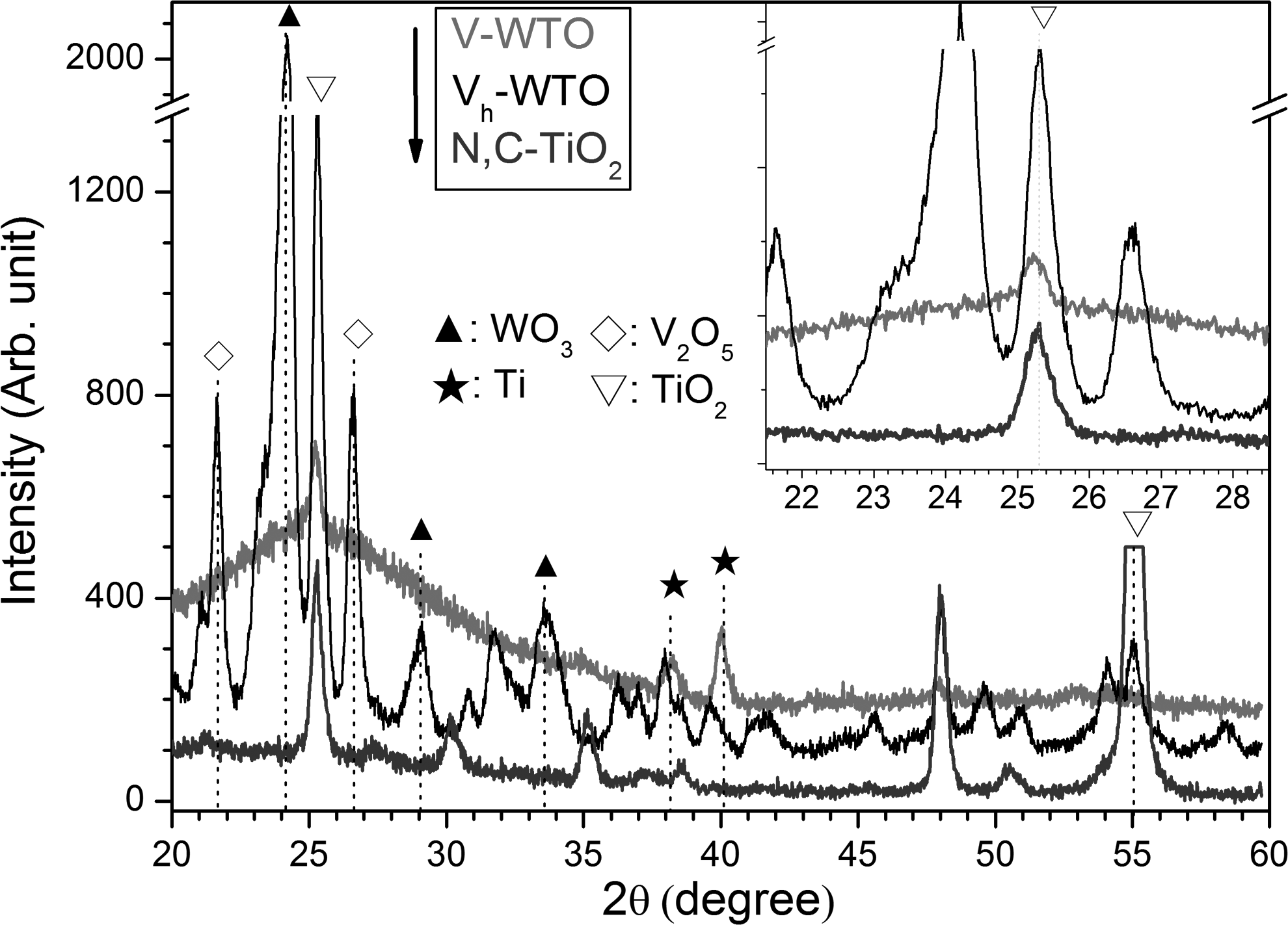
X-ray diffraction patterns of V-WTO and Vh-WTO samples along with N,C-TiO2 film.
A significant difference in morphological characteristics is shown in Fig. 2a and b. With randomly dispersed deep pores accompanied by smaller pores inside, the V-WTO surface appears to have less pore density. Such pores constitute the specific surface area of the film's lesser pore density, which is related to a film grown at a high applied voltage, whereas the pore size increases with increased electrolyte concentration (Bayati et al., 2010c). The typical WO3/TiO2 microporous film (Fig. 2b) that possessed some submicron pores and colloidal particles on a large cavity was fabricated in electrolyte, comprising Na2WO4 (15 g/L) and NaF (2 g/L), without V ions in the solutions at a low applied voltage of 300 V (Yeh et al., 2012). Thus, a high applied voltage of 380 V and a high concentration of NaVO3 (5 g/L) added in the electrolyte are mostly ascribed to this unique morphological feature of the V-WTO. As expected, the plain SEM image (Fig. 2c) of the annealed Vh-WTO sample shows no noticeable difference in porous appearance to the as-prepared V-WTO sample (Fig. 2a).
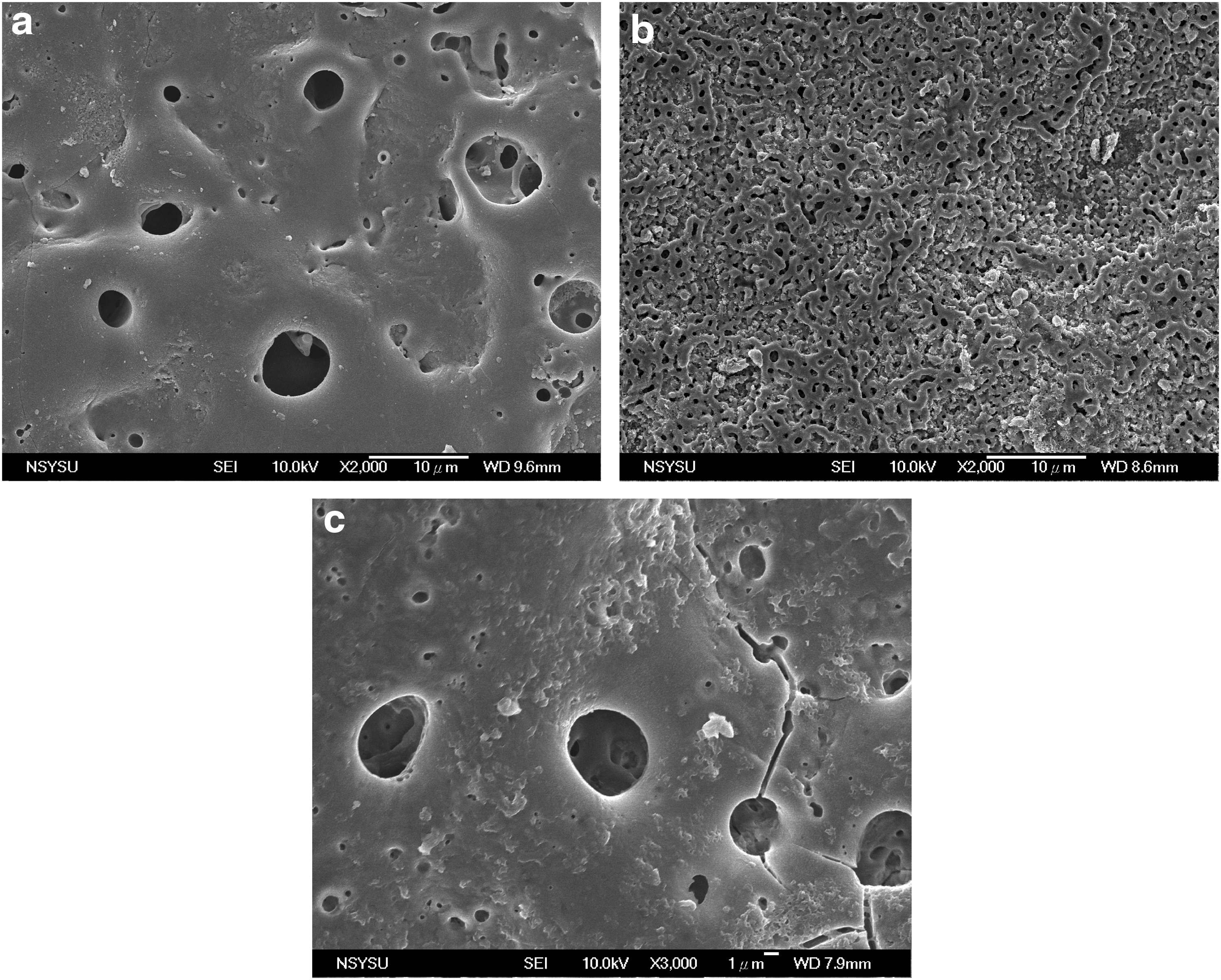
Scanning electron microscopy images of
XPS analysis
As conveyed in Fig. 3a, the N,C-TiO2 shows a slightly asymmetric Ti2p peak, whereas the V-WTO and Vh-WTO display reasonably asymmetric peaks. N,C-TiO2 was rationally resolved into two peaks at 458.8 and 464.4 eV, corresponding to Ti2p3/2 and Ti2p1/2, respectively (figure not shown). V-WTO and Vh-WTO were convoluted into two additional Ti2p peaks at 456.0±0.1 eV and 461.1±0.1 eV, assigned to Ti2+of TiO phase (McCafferty and Wightman, 1999).
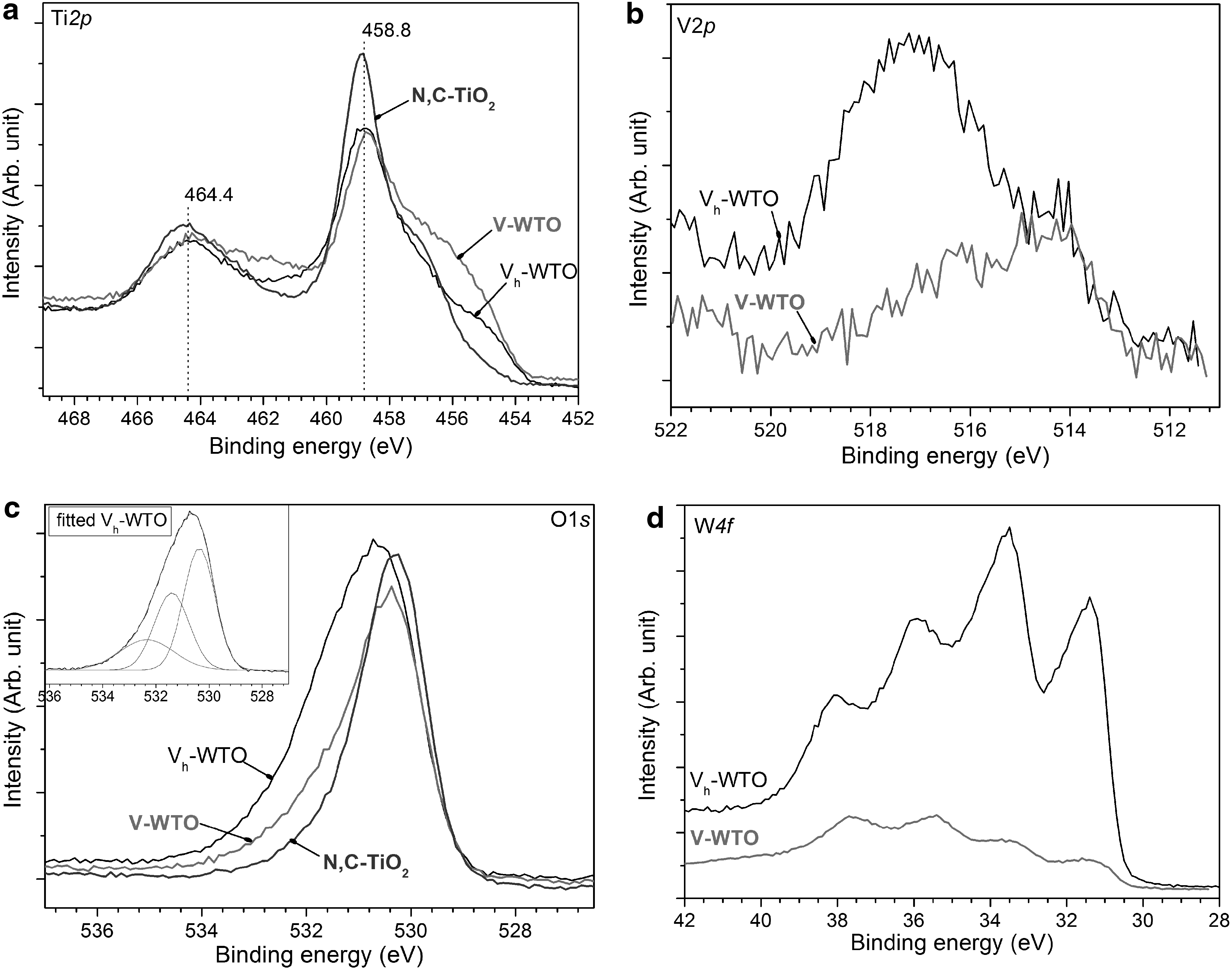
High-resolution X-ray photoelectron spectroscopy spectra of
The O1s spectrum of the Vh-WTO shown in Fig. 3b was resolved into three peaks, as shown in the inset. The first peak, at 531.6 eV, is ascribed to hydroxyl (OH) groups from adsorbed water on the surface. The second peak, at 530.3 eV, is associated with the oxygen bond (O2−) in TiO2. However, the O2− value (530.0±0.1 eV) recorded for the TiO2 reference materials shifted by ∼0.3 eV to a higher binding energy of 530.3 eV (McCafferty and Wightman, 1999). This may suggest a moderately different oxide state for the W and/or V ions in the Vh-WTO sample. The third peak, at 532.5 eV, is not directly associated with titania, but reportedly corresponds to O ions at a tungsten content of 22 at.% (Kubacka et al., 2010). Moreover, a shift and tail of the O1s spectrum toward the high binding energy is clearly observed in the Vh-WTO, as compared with the N,C-TiO2 sample. Thus, the incorporation of W and/or V ions in the TiO2 lattice is related to a binding energy shift at the O1s core level.
The major peak at 517.0 eV is assigned to V5+, which confirms the formation of V2O5, as shown in Fig. 3c. The binding energy at 517.0 eV is slightly lower than the bulk V2O5 value and the composite V2O5/TiO2, but is analogous to the reported value for V2O5/TiO2-SnO2. The weakly minor peak, at 514.2 eV, may correspond to V2+ (Zhou et al., 2010). This indicates that the V species, mostly in the form of V2O5, exist in the lattice of the Vh-WTO samples. The post annealing effect on the crystallization of V2O5 in the film is also apparent in the binding energy increase of Vh-WTO at ∼517.0 eV.
As shown in Fig. 3d, two peaks, at ∼38 and 36 eV of W4f5/2 and W4f7/2, respectively, are associated with the oxidation state (6+) of pure WO3 (Li et al., 2001). This indicates that both samples were loaded with a considerably high amount of WO3 phase. The two other peaks (at ∼33.5 and ∼31.5 eV) are possibly assigned to a different form of tungsten oxide (i.e., WO2), or nonstoichiometric tungsten oxides such as W12O39n−. In fact, because of the similarity in their ion radii, W4+ can substitute Ti4+ in the TiO2 lattice to form W x Ti1−xO2, which is a nonstiochiometric solid solution. Thus, W x Ti1−xO2 produces a tungsten impurity energy level (Li et al., 2001). Accordingly, the Vh-WTO W4f spectrum shows higher binding energy intensity than that of the V-WTO. This indicates that purer WO3 crystals were formed after annealing. The doping concentration of W ions allowed in the anatase TiO2 lattice is considerably higher than that of V ions, based on the TEM-EDS measurements (data not shown). Additionally, the content of fluorine is so limited to be detected by the TEM-EDS. This is in good agreement with the fact that the content of W ions doped into TiO2 lattices is affected by the fluorine ion (F−) concentration in the electrolyte; the higher content of W generally provides better photocatalytic activity (He et al., 2009). Therefore, the effect of W ions is implicitly the most critical factor in photocatalytic activity.
Absorption spectrum
Displaying broad absorbance across the 400–600 nm UV-Vis regions, Fig. 4 shows the diffused reflection spectra of the V-WTO and Vh-WTO microporous films. Thus, the shift toward the longer wavelengths markedly originates from the band gap narrowing of TiO2 by coupling with tungsten and vanadium oxides, and possibly W x Ti1−xO2. The respective band gap energies are estimated to be 2.41 and 2.25 eV for the Vh-WTO and V-WTO, in comparison with 2.95 eV for the N,C-TiO2 (Wu and Hung, 2009). These are close to the reported values of the V2O5/TiO2, which has V4+ transitions at ∼550 nm and V5+ excitations at ∼450 nm (Kubacka et al., 2009). As discussed in the XRD and XPS analyses, a lesser red shift of the Vh-WTO is closely related to the formation of V2O5 and WO3 after being heated at 873 K. Though, the crystalline bulk WO3 and V2O5 have band gap energies of 2.8 and 2.2 eV, respectively (Higashimoto et al., 2006), about 0.4 eV lower than the corresponding monolayered oxides (Londero and Schröder, 2010). The V-WTO is likely doped with W, V, and/or W x Ti1−xO2, which possibly results in a more red shift in visible-light region (Gu et al., 2008; Kubacka et al., 2009; Bayati et al., 2010b).
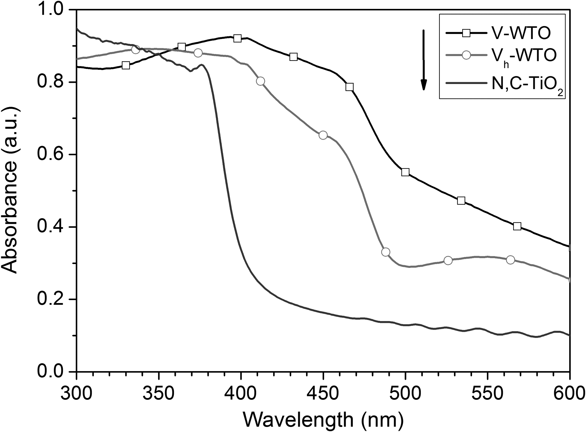
Diffused reflection spectra of microporous V-WTO and Vh-WTO along with N,C-TiO2 film.
Photocatalytic activities
Figure 5 shows the degraded MB concentrations of the samples under UV and BLED irradiation. The V-WTO exhibits degradation rate constants of 0.58 and 0.36 h−1, which are ∼66% and 53% higher than the planar N,C-TiO2 film with rate constants of 0.35 and 0.22 h−1 under UV and BLED irradiation, respectively. The photocatalytic activity of both V-WTO and Vh-WTO composite films is enhanced by its WO3 and V2O5 crystallites and its high specific surface area as well (Liu et al., 2009; Yeh et al., 2012). Because of its WO3-loaded acidic surface, microporous TiO2 film loaded with WO3 reportedly favors the adsorption of more MB (a cationic dye) molecules on its active sites, resulting in a higher degradation rate (He et al., 2009; Yu et al., 2010). The sponge-like porous samples can cause a charge carrier to reach the surface more easily than does a typical planar N,C-TiO2 (Liu et al., 2009). Moreover, the V2O5 and WO3 are mostly loaded onto the TiO2 matrix, which can generate more electron–hole pairs available for photocatalytic reactions under BLED irradiation, as indicated in the inset of Fig. 5. Thus, the doping of the V and W ions in a form of V2O5, WO3, and/or W x Ti1−xO2 is responsible for a strong visible-light photocatalytic activity that is superior to those associated with anionic dopants (i.e., N and C) in the TiO2 lattice (Kubacka et al., 2009; Wu and Hung, 2009).

Degraded methylene blue concentrations of microporous V-WTO and Vh-WTO along with N,C-TiO2 film under ultraviolet irradiation. Inset: The same samples under blue-light-emitting diode (BLED) irradiation.
In general, crystallinity is one of the most critical factors among several others, including surface area, crystal orientation, surface hydroxyl density, and phase composition, in determining photocatalytic activity (Fujishima et al., 2000; Toyoda et al., 2004; Inagaki et al., 2006; Yang et al., 2008). Surprisingly, the V-WTO shows a slightly higher photocatalytic activity than that of the annealed Vh-WTO. The photoactivity of these two types of V-WTO films tested is really negligible—a slight difference between 3.94 and 4.16 mg/L of degraded MB concentration, that is, k=0.58 and 0.53 h−1, under the same UV treatment conditions, as shown in Table 1. This is because that the annealing process diminished the WO x monolayer on the crystal surface, while enhanced the crystallinity of TiO2, WO3, and V2O5 in the V-WTO. The formation of WO3 crystallites, randomly distributed along TiO2 nanocrystals, rather than the highly adsorbing WO3 monolayer or amorphous WO x , reduces the MB adsorption capacity of WO3 (Kwon et al., 2000). Therefore, as shown in Fig. 5, a decrease in MB molecule adsorption is observed in the Vh-WTO. This implies that, with no need for post heat treatment, the V-WTO is suitable for photocatalytic degradation. Moreover, all the MAO samples lasted more than 80 test cycles, a total of 160 h, of MB degradation, and ultrasonic washing/cleaning and showed no signs of physical and photocatalytic deterioration.
BLEDs, blue-light-emitting diodes;
Conclusion
In this study, photocatalytic V-WO3/TiO2 microporous films with crystallized anatase TiO2 phase and network-framed structures were successfully obtained through MAO. The absorption edges of the films were shifted toward the visible wavelengths by incorporating the W and V species into TiO2 phases. The visible-light photocatalytic efficiency was dominated mostly by the incorporation of WO3 and V2O5 oxides, and possibly W x Ti1−xO2. In addition, the microporous film loaded with WO3 favored the adsorption of more MB molecules on its highly specific surface area. The MAO process, as-oxidized V-WO3/TiO2, is one of the most promising methods for efficiently manufacturing photocatalysts for dye degradation.
Footnotes
Acknowledgments
The authors would like to thank the National Science Council of the Republic of China, Taiwan, for financially supporting this research under Contract No. NSC 98-2221-E-022-004-MY2. The authors wish to thank Jui-Ching Sun of National Kaohsiung Marine University for his sample preparations and experimental tests.
Author Disclosure Statement
No competing financial interests exist.