
Abstract
Abstract
A confluence of water and silver-containing products or compounds results in unintentional or intentional discharge of silver species (including silver nanoparticles [AgNPs], free or organic matter-bound Ag+, Ag-halides, or soluble Ag-complexes) into visible light-illuminated waters. To date, little is known about the transformations of Ag species and mechanisms of AgNP formation in light-illuminated waters consisting of components such as bacteria, extracellular polymeric substances (EPS), natural organic matter (NOM), chloride, and/or nitrate. In this work, we demonstrate that in the presence of combinations of water components the transformation of ionic Ag to “new” AgNPs occurs via heterogeneous nucleation followed by (1) light-induced growth mediated by surface plasmon resonance activity of AgNPs, and (2) Ostwald ripening in the absence of light and chloride anions. Further, oxidative dissolution of AgNPs at near-neutral pH (6.8) releases Ag ions, which either complex with the components or form “new” AgNPs depending on solution chemistry. AgNP nucleation and growth on bacterial cells (e.g., Pseudomonas aeruginosa) may cause cell lysis. This study highlights that light-illuminated waters comprising common aquatic components are highly favorable for the AgNP formation over dissolution under neutral pH conditions, in which the presence of chloride accelerates AgNP formation and the reducing capacity of EPS/chloride medium may be greater than NOM (humics and fulvics). Findings of this study suggest that AgNP formation and dissolution may be a continual process and AgNPs thus formed may be stabilized by organic matter, which possibly leads to persistence of the nanoparticles in sunlit waters.
Introduction
W
The overall goal of this study was to demonstrate that Ag persists as “new” AgNPs formed from dissolved Ag in light-illuminated oxic surface waters. For this study, Pseudomonas aeruginosa was used as a model bacterium, as these bacteria produce copious amounts of EPS in a short duration and are commonly found in freshwaters, marine waters, and recreational waters systems (Khan et al., 2010). Commercially available humic and fulvic acids were mixed in a 1:1 (w/w) ratio to represent the major components of NOM. Separate experiments were also conducted with synthesized polyvinylpyrrolidone (PVP)-AgNPs in selected media to discern the AgNP dissolution behavior and possible transformations of released Ag ions. The results obtained from the earlier sets of experiments were evaluated to reveal the possible cyclic nature of AgNP formation and dissolution, and possible nanoparticle persistence and bacterial lysis in light-illuminated waters. Enhanced darkfield-hyperspectral imaging (HSI) microscopy, UV-Visible spectrophotometry (UV-Vis), transmission electron microscopy (TEM), scanning electron microscopy (SEM), X-ray photoelectron spectroscopy (XPS), and inductively coupled plasma-mass spectrometry (ICP-MS) were used for the characterization and analysis of AgNPs.
Materials and Methods
Chemicals and stock suspensions
Silver nitrate (AgNO3), sodium nitrate (NaNO3), sodium chloride (NaCl), and humic and fulvic acids were purchased from Sigma-Aldrich (St. Louis, MO). Tryptone, Bacto agar, and yeast extract were purchased from Difco (Detroit, MI). Stock suspension of PVP-coated AgNPs (mean diameter ∼25 nm) was synthesized according to published procedures (Ma et al., 2012). Stock suspensions of 100 mg/L AgNO3, 100 mg/L NOM (humic acids and fulvic acids were mixed at 1:1 w/w), and 8 g/L NaCl were prepared in sterile ultrapure water. Bacterial growth medium consisted of 10 g/L tryptone, 1 g/L yeast extract, and 8 g/L NaCl (aka LB w/NaCl). Similarly, LB w/NaNO3 medium was prepared. All media were prepared in ultrapure water (Barnstead NANOpure Diamond), and growth media were autoclaved before use.
Bacterial culture and test media
P. aeruginosa (ATCC # 47085) culture was grown in LB w/NaCl medium that was incubated at 37°C with shaking at 150 rpm for 18 h overnight. Supernatant (free of bacteria and cell debris) was prepared by subjecting 10 mL aliquots of overnight culture to sequential centrifugation, once at 6,000 g for 5 min and twice at 12,000 g for 30 min at 4°C. The collected supernatant, comprising secretions of P. aeruginosa (or EPS) and the spent LB w/NaCl medium, was used as EPS medium (this is referred to here as EPS w/NaCl). Similarly, EPS w/NaNO3 was prepared from P. aeruginosa culture that was grown in LB w/NaNO3 medium (to study the effect of chloride, NaNO3 was substituted for NaCl in the growth media). For the purpose of this study, various test media were prepared and are referred to here as follows: (1) overnight grown P. aeruginosa cultures in LB broth with NaCl are identified as “P. aeruginosa+EPS w/NaCl”; (2) after removing the bacteria through centrifugation, EPS medium is identified as “EPS w/NaCl”; (3) overnight grown P. aeruginosa cultures in LB broth with NaNO3 are identified as “P. aeruginosa+EPS w/NaNO3”; (4) the P. aeruginosa+EPS w/NaNO3 medium without bacteria is identified as “EPS w/NaNO3”; (5) LB broth with NaCl is identified as “LB w/NaCl” (this is the growth media); (6) LB broth with NaNO3 is identified as “LB w/NaNO3”; (7) LB broth without NaCl is identified as “LB wo/NaCl”; and (8) 100 mg/L NOM medium [1:1 (w/w) humic and fulvic acids that contains no chloride or bacteria] is identified as NOM medium. These media were stored at 4°C until use.
Experimental protocol
A typical test suspension consisted of 100 μL of 100 mg/L AgNO3, 100 μL of test medium, and 800 μL of ultrapure water (total volume 1 mL). Two sets of 1 mL test suspensions comprising Ag ions at 10 mg/L were prepared, in which one set was exposed to visible light (light source and flux information is provided in the Supplementary Data; Supplementary Data are available online at www.liebertpub.com/ees) and the other was kept in the dark (no visible light) for 90 min. Further, the experiments in the dark were continued for 14 days, as no observable changes occurred within the first 90 min. We acknowledge that 10 mg/L Ag is not an environmentally relevant concentration, but it enables the real-time visualization of Ag ion transformations in the test media with or without light [via darkfield HSI microscopy] and, thus, provides information on the transformation mechanisms. The absorbance spectra in wavelength range of 300–800 nm were collected for samples at predetermined time intervals using a UV-Vis spectrophotometer (Varian Cary 300). In addition, a set of experiments with selected media in the absence of light was carried out using PVP-AgNPs at a near-neutral pH of 6.8 and room temperature for a period of 4 days to assess the kinetics of AgNP dissolution.
Characterization and analysis
Enhanced darkfield HSI microscopy (aka CytoViva HSI Microscopy) was used for imaging samples in wet-form to observe and analyze Ag ion transformations in real-time (Badireddy et al., 2012). Hyperspectral image scans on sample droplets consisted of 512 lines rastered at a step size of 10 nm at a spectral resolution of 1.5 nm in the visible and near-infrared wavelengths (400–1,000 nm). TEM (FEI Tecnai G2 Twin, Hillsboro, OR) was used to image samples dried on the Formvar side of a Carbon Type A Formvar-coated copper grid (Ted Pella, Redding, CA) at an accelerating voltage of 160 kV. SEM (FEI XL30 SEM-FEG, Hillsboro, OR) was performed on gold-sputtered samples that were fixed with 2.5% glutaraldehyde and sequentially dehydrated and washed, respectively, with graded ethanol and 1× phosphate-buffered saline. Inductively coupled plasma-mass spectroscopy (ICP-MS; 7700 Agilent Technologies, Santa Clara, CA) was used for measuring Ag ion concentration in the test suspensions (Lowry et al., 2012). X-ray photoelectron spectroscopy (XPS) was used to collect the high-resolution scans (step size 0.1 eV) of the samples that were air dried on a silicon wafer for verifying the presence of zerovalent silver (Ag0). Total organic carbon analyzer (TOC 5050A; Shimadzu, Kyoto, Japan) was used for measuring the NOM concentration. Oxidation-reduction potential (ORP) measurements were made on all test suspensions against a silver/silver chloride electrode in the presence and absence of light using an Orion ORP probe (Thermo Scientific, Waltham, MA) (see Supplementary Data for more details). Note that no attempt was made to determine which specific components of EPS or bacteria or NOM were responsible for the Ag ion transformations; however, the experiments were designed to demonstrate the resulting effect of the combination of water components on Ag ions and nanoparticles in the visible light-illuminated waters.
Results
Ag ion reduction in P. aeruginosa+EPS w/NaCl+visible light
In P. aeruginosa+EPS w/NaCl medium, a broad UV-Vis absorbance peak associated with surface plasmon resonance (SPR) of AgNPs evolved at 396 nm initially, and then it gradually shifted toward 436 nm during 90 min of irradiation with visible light (Fig. 1a). UV-Vis spectra showed that after a short-lag period, the growth of AgNPs occurred rapidly in the medium (Fig. 1a and Supplementary Fig. S1). An increase in the absorbance of SPR peak and the asymmetric shape of the peak indicates a growing number of individual AgNPs as well as the formation of AgNP aggregates. At the end of 90 min, the average diameter of AgNPs was 25.5±2.5 nm (TEM image analysis was done using Image J) (Fig. 1b), and the identity of AgNP in the sample was confirmed by XPS (Fig. 1c).

Evolution of UV-Vis spectra of silver nanoparticles (AgNPs) in Pseudomonas aeruginosa+extracellular polymeric substances (EPS) w/sodium chloride (NaCl) medium exposed to Ag ions and visible light for a period of 0–90 min (the green colored spectrum corresponds to the sample at 90 min) is shown in
Further, the visual evidence for the presence of Ag nano-particulates (AgNPs and AgCl) in the medium exposed to 90 min of visible light is shown Figure 2. A representative darkfield image of P. aeruginosa in EPS w/NaCl medium without Ag ions (control) is shown in Figure 2a. After exposure to Ag ions and visible light for 30 min, numerous brightly colored Ag nano-particulates were observed to form in the bulk medium as well as on the bacterial cell walls; while the bacteria were catastrophically damaged as shown in Figure 2b, c, e, and f (also see Supplementary Figs. S2 and S3). SEM images suggest that AgNPs may have formed from Ag ions through nucleation and growth on the cell walls (Fig. 2d) and poles of the bacteria (Fig. 2e). The presence of colored particles is an indication of the SPR-activated AgNPs and the polydisperse nature of the nanoparticles formed. Real-time imaging and HSI analysis of the samples confirmed that AgNPs were, indeed, present on the cell walls as well as in the bulk medium (Supplementary Fig. S3 shows AgNPs on the cell walls).

CytoViva darkfield image (magnification: 40×) of P. aeruginosa+EPS w/NaCl media before the addition of Ag ions
The characteristic evolution of SPR absorbance in Figure 1a and in other test media (Supplementary Fig. S1) was nearly similar, suggesting that the mechanism of AgNP formation from Ag ions in all the media is also probably the same in the presence of visible light. The average diameters of AgNPs observed in the various media after 90 min of irradiation are summarized in Table 1. In NOM medium (a mixture of humics and fulvics that contains no chloride or bacteria), two distinct peaks at 400 and 500 nm in the absorbance band between 365 and 650 nm may be attributed to the formation of individual AgNPs and aggregates (Supplementary Fig. S1h), which is likely mediated by the NOM molecules bridging (or linking) individual nanoparticles and forming larger aggregates (Supplementary Fig. S4). Further, as shown in Figure 3, AgNPs with the largest average diameter were observed in NOM medium (31.5±5.1 nm); whereas the smallest ones were in EPS w/NaNO3 (13.5±0.8 nm) (Table 1). At the end of 90 min, it was observed that the UV-Vis absorbance maximum of each medium was different, which may imply that the rate of AgNP formation in each medium was also different (Table 1).
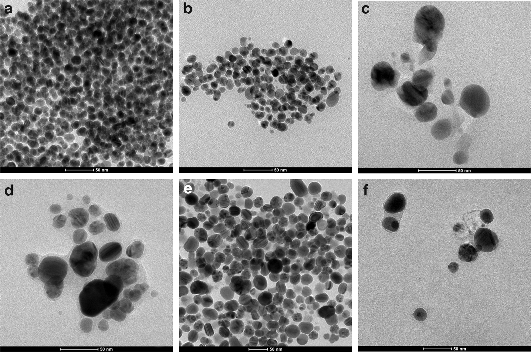
TEM images of spherical AgNPs formed in: EPS w/NaCl
LB is a mixture of 10 g/L tryptone and 1 g/L yeast extract [i.e., LB without (wo/) NaCl or NaNO3]. LB w/NaCl is a growth medium used for culturing P. aeruginosa; P. aeruginosa+EPS w/NaCl is a bacterial culture grown overnight (see Materials and Methods section); P. aeruginosa+LB w/NaCl corresponds to washed bacteria (extracted from P. aeruginosa+EPS w/NaCl) resuspended in fresh LB w/NaCl. EPS w/NaCl is simply P. aeruginosa+EPS w/NaCl without P. aeruginosa, which were removed by centrifugation. NOM medium is a mixture of humic and fulvic acids that contains no chloride, nitrate, or bacteria.
AgNP, silver nanoparticle; EPS, extracellular polymeric substances; NOM, natural organic matter; NaNO3, sodium nitrate; NaCl, sodium chloride.
Mechanism of AgNP formation in the presence and absence of visible light
Figure 4a shows the evolution of UV-Vis absorbance at 415 nm with time in each medium. The trace of absorbance over time was sigmoidal, starting with a slow induction phase. The data were described by a sigmoidal function that was analogous to a modified form of Hills equation (see Supplementary Data for details):
where A is the absorbance (arbitrary units), A1 and A2 are the left and right asymptotes (arbitrary units), respectively, t50% is the time (minute) at which 50% of the nanoparticle population is produced, and n is the binding coefficient describing cooperativity of Ag ions and AgNPs. Equation (1) provides a way to quantify the binding of Ag ions to SPR-activated Ag0 cluster (or AgNP) induced by visible light. Ag ion binding to AgNP is (1) positively cooperative when n>1, (2) negatively cooperative when n<1, and (3) noncooperative when n=1. The value of n for each medium was determined using Equation (1) (Fig. 4a and Table 1). The results in Figure 4a show that the values of n are greater than 1 for all the media in the presence of visible light, suggesting that the binding of Ag ions to SPR-activated AgNPs is positively cooperative, which followed a sigmoidal trend. Further, the n values were nearly twofold higher for the media w/NaCl compared to the media without NaCl, which suggests that the presence of chloride in the media enhances the AgNP formation. The n values for NOM, LB wo/NaCl, and LB w/NaNO3 were statistically indistinguishable, which may suggest possible molecular-level similarities between NOM and LB medium (with nitrate or without NaCl) and nitrate likely played a negligible role in Ag ion binding to AgNPs during 90 min of exposure to visible light. In the absence of light, no SPR peaks were recorded for the test suspension during the first 90 min.
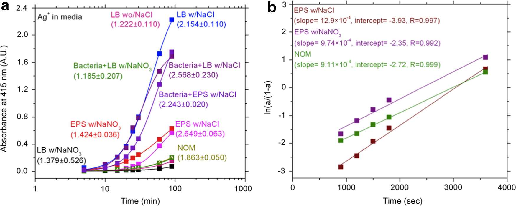
Sigmoidal evolution of absorbance at 415 nm with time in various media during 90 min of exposure to visible light is shown in
The progress of Ag ion binding to Ag particles via a reduction reaction could be quantitatively described by the following linear relationship with time (Esumi et al., 2000):
where a is the ratio of absorbance at time t (At) and the absorbance maximum (Amax), kobs is the observed binding constant (s−1), and t is time in seconds. The results in Figure 4b and Supplementary Fig. S5 show linear relationships (ln (a/(1−a))) as a function of time with reasonably high R2 values, suggesting that a light-induced heterogeneous nucleation and growth mechanism is likely responsible for the formation of AgNPs in the presence of visible light. The kobs value describing the binding process in each medium was determined from the slope of the Equation (2) and summarized in Table 1. In comparison to NOM and the media with NaNO3 (or without NaCl), the kobs of media w/NaCl were of greater, which implies that the presence chloride anion in the media likely enhances the AgNP formation rate, which is consistent with the two-fold increase in the binding coefficients (n) (Table 1).
Chloride anion enhances formation of AgNPs in the presence of visible light
Evidence for AgNP formation in AgCl(s) solutions exposed to visible light is shown in Figure 5. In the initial UV-Vis spectrum of AgCl suspension, a broad absorbance over the entire wavelength range of 300–800 nm was observed, with no characteristic AgNP SPR band (Fig. 5a). However, upon exposure to visible light, a broad SPR band began appearing at ∼415 nm. This peak gradually red-shifted, and then finally vanished after 20 min of exposure. The presence of the SPR band is indicative of transformation of AgCl to AgNP, and a gradual decrease in absorbance and disappearance of the SPR band was attributed to the aggregation of Ag nano-particulates (AgNP-AgCl) in the suspension. A color change from cloudy white to gray was observed at the end of 90 min of irradiation, which also qualitatively indicates the transformation of AgCl to AgNPs (Fig. 5e placed as inset in Fig. 5a). Both HSI analysis and TEM images confirmed the presence of AgNPs in AgCl suspensions. For instance, the brightly colored rings around the AgCl aggregates or the nanoparticles interspersed among aggregates are indicative of SPR activity of AgNPs (Fig. 5b). In the absence of visible light, the AgCl aggregates remained relatively free of smaller nanoparticles (Fig. 5c). However, after exposure to visible light for 90 min, numerous smaller nanoparticles coating the surface of AgCl aggregates were observed (Fig. 5d). Further evidence for the presence of AgNPs in AgCl suspension was provided by the hyperspectral fingerprint analysis, which confirmed that spectral peaks of colored nanoparticles were exactly matching with those of AgNPs in P. aerugionosa+EPS w/NaCl and engineered PVP-AgNPs (Fig. 6). Thus, it would appear that AgCl which is formed in a medium w/NaCl is further transformed to form AgNPs in the presence of visible light. Thus, the results in Figures 4 and 5 confirm that the presence of chloride enhances the AgNP formation in the presence of visible light.

Evolution and disappearance of a characteristic peak associated with AgNP surface plasmon resonance (SPR) in AgCl(s) suspension during exposure to visible light for 30 min is shown in
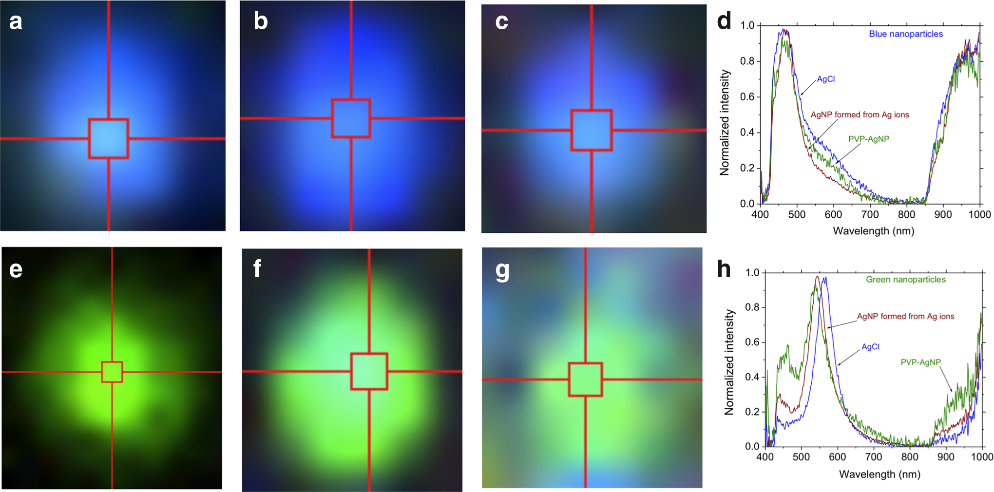
Enhanced darkfield hyperspectral images (magnification: 400×) of individual AgNPs formed in AgCl suspension
Dissolution of AgNPs in the test media
Selected experiments were designed to specifically study the oxidative dissolution of engineered AgNPs (PVP-AgNP) at pH 6.8 in the following media: (1) ultrapure water in the absence and presence of visible light, and (2) LB w/NaCl, EPS w/NaCl, simulated seawater media (i.e., 0.7 M NaCl in ultrapure water), and NOM in the absence of light. The dissolution trends of PVP-AgNPs in the selected media for the duration of 24 h are shown in Figure 7, and TEM images of samples obtained from each medium on day 4 are shown in Figure 8. In the presence or absence of visible light, AgNP dissolution rates were similar in ultrapure water. Further, the dissolution rates in ultrapure water were also similar to those observed in LB w/NaCl and NOM media. However, the dissolution rate is higher in the EPS w/NaCl medium and highest in the seawater medium. In addition, a TEM examination of the samples obtained on day 4 (Fig. 8a–f) revealed that (1) in ultrapure water [Fig. 8a (dark) and 8b (light)] and in NOM medium (Fig. 8c), PVP-AgNPs remained free of precipitates likely due to the absence of light and chloride; (2) in LB w/NaCl media, Ag ions released from AgNPs likely complexed with chloride and formed AgCl precipitates on AgNP surfaces and in their vicinity (Fig. 8d); (3) in seawater medium, Ag ions released from AgNPs likely formed precipitates of Ag-chloro complexes, which is observed as dispersed gray matter surrounding AgNPs (Fig. 8e); and (4) in EPS w/NaCl medium, Ag ions released from AgNPs were likely reduced to smaller “new” AgNPs by the organic components of the medium (Fig. 8f and Supplementary Fig. S7). The earlier results and observations suggest that dissolved Ag ions may be transformed to (1) AgNPs and AgCl in EPS w/NaCl medium, which is consistent with the results from dark experiments shown in Supplementary Figure S8, and (2) soluble Ag-chloro complexes in the seawater medium.
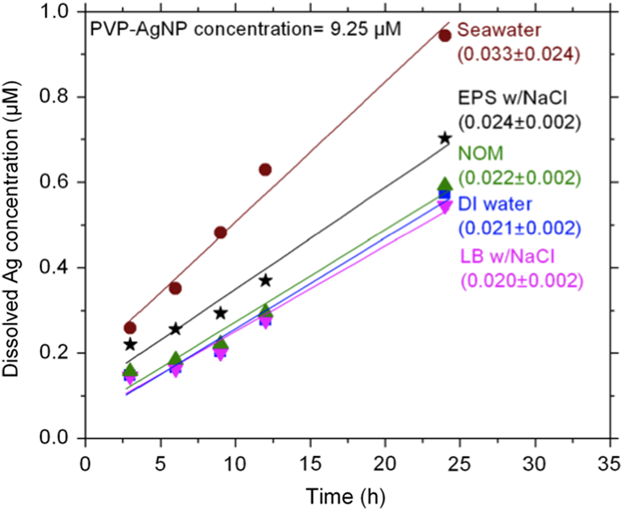
Dissolution behavior of PVP-AgNPs in seawater, EPS w/NaCl, NOM, DI water, and LB w/NaCl media over a 24 h duration in the absence of visible light are shown here. The nanoparticle dissolution rates (μM/h) are shown in the parentheses in the figure.

TEM images of PVP-AgNPs after dissolution for 4 days in: DI water in the absence of light
Discussion
Silver in surface waters exists in colloidal and particulate forms, free ions, and complexes with organic and inorganic matter (e.g., EPS, bacteria, NOM, and chloride, and sulfide) (Chinnapongse et al., 2011). The speciation of Ag in aquatic systems depends on the redox potentials and availability of ligands (organic molecules, Cl−, and S2−) (Adams and Kramer, 1998; Levard et al., 2012). Ag has been demonstrated to be prevalent in the form of Ag-chloro complexes as opposed to Ag-sulfide complexes, because sulfide concentration is low in surface waters. Therefore, for this study, the emphasis was placed on the interaction of Ag ions with chloride anions. This study investigates the resulting effect of Ag ion (or AgNP) interactions with a combination of components of water (EPS, bacteria, NOM, nitrate, and chloride) in the presence and absence of visible light.
Our experiments revealed that a rapid reduction of Ag ions (within minutes) to AgNPs was mediated by the water components in the presence of visible light (Fig. 4). Conclusive evidence for AgNP formation in the media was obtained through UV-Vis spectrophotometry, HSI microscopy, TEM, SEM, and XPS analysis. Since Ag ions can exist freely, bound to organic matter, or as soluble complexes in freshwaters and seawaters, any exposure to visible light could readily transform Ag ions to AgNPs via a reduction reaction (Fabrega et al., 2011). In addition, chloride anions in the aqueous media can substantially enhance the AgNP formation rates under visible light since AgCl also undergoes a photo-reduction reaction and forms “new” AgNPs (Fig. 5); a process similar to silver halide-based photography (Wang et al., 2011). Control experiments in the absence of visible light and chloride showed that AgNP formation was rather slow (order of days), which was revealed by a gradual increase in SPR-absorbance (at 415 nm) over a period of 14 days (Supplementary Fig. S8). However, the AgNP formation was minimal and the solution remained colorless (aside from the complete lysis of bacteria) for the duration of 14 days in the presence of chloride but without light. These findings suggest that in the absence of visible light, a significant portion of Ag ions may remain bound to and form precipitates with chloride. However, organic matter (bacteria, EPS, and/or NOM) may reduce Ag ions to Ag0 nuclei (Ishibashi et al., 1990; Maurer et al., 2012; Kang et al., 2014) in the absence of chloride due to the differences in ORPs [Eq. (a), Table 2, and Supplementary Fig. S9]. For example, the reducing capacity of EPS w/NaCl was substantially greater than NOM medium in the absence of light and, further, NOM medium was a stronger reducing agent with light than without light (Supplementary Fig. S8). Ag0 nuclei formed are thermodynamically unstable. In a process to minimize surface energy, Ag0 nuclei thus formed as an aggregate to optimal-sized Ag particles, which may be stabilized by a layer of organic matter in the medium. After this stage, larger particles grow at the expense of smaller ones through the Ostwald ripening process (Hoang et al., 2002) in which the smaller Ag particles via oxidation dissolve in the medium by releasing Ag ions, which then migrate to the surface of larger AgNPs where they undergo reduction via binding [Eq. (b), Table 2]. Such a reduction of Ag ions at the silver surface lowers the surface energy by increasing the size of the particles, which is consistent with the findings from the previous studies (Meisel, 1979; Henglein and Meisel, 1998; Pillai and Kamat, 2004) [Eq. (4)].
It should be noted that neither AgNO3 nor the test media (colorless suspensions) exhibited any UV-Vis absorbance in the 300–800 nm wavelength range. AgNP formation and dissolution processes are summarized in Table 2 and described here: Initially, Ag0 nuclei formation occurs in the media, due to (1) differences in redox potential [Eq. (a)] (Supplementary Fig. S8), (2) photo-thermal process [Eq. (c)] and photo-reduction of AgCl [Eq. (d)], and (3) photosensitized O2•− [Eq. (e)] facilitated by OM, which includes EPS and NOM. Although bulk O2•− concentration was low (see Supplementary Fig. S10), it was likely sufficient near the photosensitizer (i.e., EPS and/or NOM) to initiate Ag0 nuclei formation (Jones et al., 2011). These nuclei while aggregating to an optimal size, via Ostwald ripening [Eq. (b)], began absorbing significant amounts of visible light due to plasmon excitation, which likely enhanced reduction potential at the surface of the particles and subsequently resulted in rapid growth of AgNPs in the media [Eq. (f)]. This implies that SPR-activated Ag particles, covered by organic components of the media, served as heterogeneous nucleation sites favoring the formation and growth of AgNPs, via reduction of Ag ions onto their crystalline faces (i.e., binding of Ag+ to surface of Ag particles) (Xue et al., 2008). Spherical AgNPs thus formed were stabilized by OM components of the media (AgNP diameters are shown in Table 1). Thus, light-induced heterogeneous nucleation, Ostwald ripening, and SPR of Ag particles play a key role in the AgNP formation in the presence of light. The apparent differences in the lag period (nucleation) before AgNP growth in individual media could be attributed to factors, such as differences in ORP potential (Supplementary Fig. S9), chloride content, and O2•− generation rates (Supplementary Fig. S10). No attempt was made to compare the chemical nature of EPS, LB, or NOM medium, but based on previous studies it is likely that they share similar photoactive groups that mediate the reduction of metal ions via photosensitized O2•− production (Rose and Waite, 2005).
Real-time HSI microscopy observations of the interactions of Ag ions with the components of P. aeruginosa+EPS w/NaCl medium revealed that P. aeruginosa lost their swimming ability after a few seconds of exposure to Ag ions (observed in real-time using darkfield HSI microscopy). During 30 min of exposure to Ag ions, Ag0 nucleation and growth occurred on the cell walls and poles, and in the bulk medium (Fig. 2d–f and Supplementary Fig. S2) resulting in considerable cell damage at those regions as evidenced by hyperspectral, SEM, and TEM images (Figs. 2 and 6, and Supplementary Fig. S2). Ag0 nucleation and AgNP growth on the bacterial cell walls could be considered one of the mechanisms through which cell lysis occurs in the visible light.
When AgNPs are discharged through human activity and/or natural processes into the surface waters, they undergo oxidative dissolution, through which the release of Ag ions is favored in the presence of dissolved oxygen and excess protons [Table 2, Eq. (g)] (Liu and Hurt, 2010). As expected, the PVP-AgNPs were oxidized in the test media releasing Ag ions for the duration of the experiment (Fig. 7). Visible light had a negligible effect on PVP-AgNP dissolution rate in ultrapure water (pH 6.8), as organic matter and/or chloride were absent. However, the organic components in NOM and LB w/NaCl media showed negligible influence on the Ag dissolution rate as evidenced from similar dissolution rates in ultrapure water (light and dark), LB w/NaCl, and NOM media (Table 1). The PVP-AgNP oxidation was higher in EPS w/NaCl and highest in seawater medium (Fig. 7) when compared with other media. The nanoparticle oxidation in EPS w/NaCl medium was higher, as EPS medium was highly reducing for Ag ions causing the formation of smaller “new” AgNPs around the PVP-AgNPs (Fig. 8f and Supplementary Fig. S7), and therefore likely favored increased dissolution. In addition, the dissolution was highest in seawater medium possibly due to the formation of soluble Ag-chloro complexes (Liu et al., 2010; Levard et al., 2013).
In surface waters, Ag ions are regarded as equilibrium species whereas AgNPs are unstable and nonpersistent species (Liu and Hurt, 2010; Liu et al., 2010; Zhang et al., 2011). Contrary to the common belief, the results of this study show that the formation of AgNPs is a continual process, through which “new” AgNPs form spontaneously in waters under visible light illumination. However, in the absence of light, the nanoparticles dissolve through oxidation releasing Ag ions, which then transform into “new” AgNPs when re-exposed to visible light. The most likely pathways involved during Ag ions reduction to AgNPs (or formation) followed by re-oxidation (or dissolution) to form Ag ions are shown in Figure 9. Given the abundance of bacteria and its EPS in visible light-illuminated waters, their role in forming and dissolving metallic nanoparticles and the associated toxicity effects should be taken into consideration when assessing the environmental fate, persistence, and toxicity of metallic nanoparticles, especially in sunlit waters.

Pathways through which “new” AgNPs (Nano-Ag0) could form from dissolved Ag (Ag+), which may be associated with environmental water components such as chloride, EPS or soluble microbial products (SMP), bacteria, and NOM (humic and fulvic acids), in the presence of visible light are shown here.
Conclusions
When discharged into visible light-illuminated waters, Ag species (AgNPs, free or organic matter bound-Ag+, Ag-halides, or soluble Ag-complexes) are continually re-cycled and form “new” AgNPs that may be stabilized by environmental organic matter (e.g., EPS and NOM), which, in turn, may contribute to their persistence. EPS medium appears to be a stronger reducing agent than NOM medium, with regard to the reduction of Ag ions to AgNPs. Organic components reduce Ag ions to Ag0 nuclei due to redox potential differences, and Ag0 nuclei thus formed may then grow into larger nanoparticles through (1) Ostwald ripening and light-induced SPR of AgNPs, or (2) Ostwald ripening process in the absence of light. The chloride in the media either enhances AgNP formation with visible light or forms Ag precipitates (or soluble-complexes) without light. Photosensitized superoxide radicals may also play an important role in Ag ion reduction. AgNPs formed in the visible-light illuminated waters stabilized by organic matter are likely to persist in surface waters, as the formation rates were higher (order of minutes) than dissolution rates (order of days). Bacterial cell walls may also serve as nucleation sites for Ag, through which Ag ions deposit and transform into AgNPs, via the mechanisms described earlier. As nanoparticles grow on the cell walls, they cause cell rupture. In addition to plasmonic activity of AgNPs, redox and photosensitizing properties of the media may play a major role in the metallic nanoparticle formation and persistence in sunlit waters.
Footnotes
Acknowledgments
This work was funded through the Center for the Environmental Implications of Nanotechnology (CEINT) by the NSF and the EPA under NSF Cooperative Agreement Number EF-0830093. Any opinions, findings, conclusions, or recommendations expressed in this material are those of the author(s) and do not necessarily reflect the views of the NSF or the EPA. This work has not been subjected to EPA review and no official endorsement should be inferred.
Author Disclosure Statement
No competing financial interests exist.
References
Supplementary Material
Please find the following supplemental material available below.
For Open Access articles published under a Creative Commons License, all supplemental material carries the same license as the article it is associated with.
For non-Open Access articles published, all supplemental material carries a non-exclusive license, and permission requests for re-use of supplemental material or any part of supplemental material shall be sent directly to the copyright owner as specified in the copyright notice associated with the article.