
Abstract
Abstract
Nosocomial infections and food spoilage, caused by different airborne bacteria, is of major concern and titania nanostructure coated surfaces are effective in reducing pathogenic bacteria present in the indoor air. In the present study comparative photocatalytic disinfection ability of pure titania nanotube (TNT) and 1% iron-doped TNT coated wood surface, to disinfect airborne bacteria, has been examined. Prepared nanotubes (both pure and iron doped) were characterized by scanning electron microscopy, energy dispersive spectroscopy, X-ray diffraction, Brunauer–Edward–Teller surface area measurement, and band gap energy calculations. Nanotubes were coated on wood surfaces using a novel and green method of coating using a natural resin as the binding agent. Coated wood surfaces were found effective in reducing important airborne bacterial species Pseudomonas aeruginosa (Gram negative) and Staphylococcus aureus (Gram positive) present in indoor air. With a 70% reduction in the population of P. aeruginosa being observed when pure TNT coated wood surfaces were exposed to fluorescent light for 2 h. In the case of 1% iron-doped TNT coated wood surfaces, P. aeruginosa species were completely degraded within 75 min of exposure to fluorescent light. Similar trends were observed with S. aureus, with 80% removal in the case of pure TNT coated wood surface after 2 h, and 100% removal within 90 min, when 1% iron-doped TNT coatings were placed under fluorescent light.
Introduction
A
A standard method to reduce airborne bacterial infections is ultraviolet germicidal irradiation (Reed, 2010) whereby the hospital air can be easily disinfected by using UV lamps of uniform size (Shah, 2009). Chlorine Dioxide (ClO2) can also be used to reduce residual bacterial and fungal concentrations present in the environment (Hsu et al., 2012). Such methods of disinfection, however, have many demerits like chlorination is chemically intensive. Adding to the problem some bacterial species may become resistant to chlorination treatment and chlorination itself may produce chlorination byproducts (Pontius and American, 1990; Huang et al., 2005).
Photocatalytic disinfection of bacteria is currently gaining importance because of its effectiveness against highly virulent and antibiotic resistant strains of bacteria, especially in hospital environments, that demands newer disinfection methods (Cheng et al., 2009). The first report on utilization of photocatalytic oxidation to disinfect the microorganisms (Escherichia coli) emerged in 1993 (Ireland et al., 1993). Since then, bacterial inactivation, using TiO2, has been mostly carried out in aqueous media, by a number of researchers, but only a few studies are available that deal with such photocatalytic disinfection of air microbial flora (Vorontsov et al., 2010).
TiO2 is one of the best photocatalysts and most widely used all over the world (Sökmen et al., 2001) because it is cost effective, highly efficient, stable to light, and chemically inert (Jo and Kim, 2009; Ilyas et al., 2011). TiO2 acts as a photocatalyst when UV or visible light strikes on its surface, which generates peroxide and superoxide radicals responsible for bactericidal action and degradation of organic compounds (Hoffmann et al., 1995).
Importance of disinfection of benchtop surfaces, particularly in the healthcare units and microbiology laboratories is, however, essential. Compared to the conventional methods of (Gamage and Zhang, 2010) photocatalytic oxidation on such surfaces coated with titania nanoparticles, provides an alternative solution to the problem as pathogenic bacteria like E. coli, Staphylococcus aureus, and Enterococcus faecium can be easily killed by titania coated surfaces (Kühn et al., 2003). TiO2 coated on glass and tiles, was found effective for killing of bacteria (Amin et al., 2009; Yang et al., 2012), and self-cleaning (Nishimoto et al., 2008). Similarly, 1% Ag-TiO2 nanoparticles, water, and ethanol-based coating on substrates like glass and plastic venetian blinds kill 93% to 100% bacteria (Khan et al., 2013). Since TiO2 has been approved by US Food and Drug Administration for use in human food, food packing materials, medicines, and cosmetics (Chawengkijwanich and Hayata, 2008) and because it is nontoxic in nature (Nonami et al., 1998), it can also be used as a photocatalyst in food industries and pharmaceuticals to reduce airborne bacteria, where the use of cleaning chemicals or biocides is prohibited or are ineffective (Skorb et al., 2008).
Bactericidal activity of TiO2 increases as the surface area increases due to the increase in interfacial electron transfer rate (Vorontsov et al., 2010). Vorontsov and coworkers showed that 90% to 99.9% inactivation of certain viruses and bacteria can take place when titania nanoparticles are exposed to soft UV rays for 30 min and platinum-doped titania nanoparticles showed better bacterial disinfection than the pure titania nanoparticles (Kozlova et al., 2010). Similarly, it has been reported that silver-doped titania nanotubes enhance the photocatalytic activity of nanotubes to disinfect bacterial species by absorbing visible light and minimizing electron–hole recombination during photoexcitation (Grandcolas et al., 2011).
In the present study pure and 1% iron-doped titania nanotubes (TNTs) were prepared by the hydrothermal method followed by their coating on wood surfaces by using gum acacia as a binding agent. The resulting coated wood surface was then evaluated for its effectiveness against bacterial disinfection, by studying its ability to inactivate Pseudomonas aeruginosa and S. aureus under visible and ultraviolet light.
Material and Methods
Reagents and materials
General purpose reagent (GPR, TiO2), sodium hydroxide pellets, hydrochloric acid, ferric oxide, nutrient agar, and nutrient broth were obtained from Sigma Aldrich. Gum acacia was obtained from the local market. Bacterial cultures of P. aeruginosa and S. aureus were grown in the laboratory using standard protocols. Distilled water was used during the whole exercise and all chemicals of analytical grade were used without further purification.
For photocatalytic experiments, a fluorescent lamp (commonly used for room illumination), with a wavelength range of 500–600 nm, was used as a light source. UV lamp having peak intensity at 365 nm was also used to study the photocatalytic activity in the UV region.
Synthesis of nanotubes
Synthesis of precursor material for nanotubes
Titania nanoparticles were used as a precursor for the formation of pure TNTs. To prepare the precursor, GPR was mixed in distilled water and stirred for 24 h on a magnetic plate. The resulting slurry was placed in an oven for 12 h at 105°C for drying. After drying and crushing, the powder was placed in a muffle furnace for 6 h at 450°C for calcination.
Synthesis of pure TNTs
Pure TNTs were synthesized by the hydrothermal method (Asapu et al., 2011) with some modifications. For this purpose, 2.5 g prepared titania nanoparticles were added to 100 mL of 10 M NaOH solution with 2 h of stirring followed by 1 h of sonication. The resulting suspension was placed in a Teflon-lined steel autoclave. The autoclave was maintained at a temperature of 135°C for 24 h, with continuous stirring at 300 rpm. The treated solution was allowed to cool to room temperature, and washed with 0.1 M HCl, and distilled water repeatedly, until the pH was nearly neutral. The sample was dried at 105°C for 24 h. Finally the sample was calcined in a muffle furnace at 500°C for 1 h to obtain TNTs in anatase phase.
Synthesis of iron-doped TNTs
Iron-doped TNTs were prepared by using the hydrothermal method (Nguyen and Nguyen, 2011). 1.5 g of prepared titania nanoparticles and 32 mg of ferric oxide was first stirred for 2 h and then mixed ultrasonically in 100 mL of 10 M NaOH for 1 h. The resulting solution was placed in a Teflon-lined autoclave at 135°C for 24 h with continuous stirring at 300 rpm. Following this, the solution was allowed to cool at room temperature and was washed with 0.1 M HCl and distilled water repeatedly, until the pH become neutral. The sample was then dried in an oven at 105°C, for 24 h. Finally, the sample was calcined in a muffle furnace at 500°C for 1 h to obtain nanotubes in the anatase phase.
Characterization of pure and doped nanotubes
The morphology of the nanotubes was observed using scanning electron microscopy (SEM) whereas the elemental analysis of the material was carried out using energy dispersive spectroscopy (EDS) attached with SEM. Particle size and crystalline phase was identified with the help of X-ray diffraction (XRD). Brunauer–Edward–Teller (BET) analyzer was used to calculate the specific surface area of pure and iron-doped TNTs. For BET analysis TNT samples were first degassed at 130°C for 4 h under vacuum (Akarsu et al., 2006). Band gap energies of TNTs were calculated by using UV–Vis Spectrophotometer in the absorbance mode. For this purpose, suspensions of pure and iron-doped TNTs were prepared by adding 0.1 g of sample in 10 mL ethanol (Reddy et al., 2002) with 1 h of sonication. Band Gap energies were then calculated by using Planck's equation (E=hc/λ).
Nanotubes coating on wood surfaces
Preparation of coating agent
Coating/Binding agent was prepared by adding 250 g crushed and sieved gum acacia (a natural resin) in 250 mL of methyl alcohol and heating the mixture at 175°C for 10 min with constant stirring with the help of a glass rod.
Preparation of nanotube-coated surfaces
Nanotubes (pure and iron doped) were dispersed ultrasonically in the binding agent for 15 min. Wood pieces of 10×10 cm2 were prepared from a wood log of Beyar tree (used in furniture also) and a single layer of pure binding agent (a common wood furniture polishing material) was coated on these wood pieces. Then the layer of the binding agent, containing pure and doped TNT, was deposited on separate wood pieces and allowed to dry at ambient temperature.
Bacterial culture preparation
One milliliter of pure bacterial cultures of P. aeruginosa and S. aureus were inoculated in 100 mL of nutrient broth and Luria Bertani broth (Sigma Aldrich) separately and incubated in a shaker incubator at 37°C for 24 and 16 h, respectively. The density of the bacterial suspensions was estimated using optical density at 600 nm and the range of 0.8–1.0 was chosen as optimal density for subsequent experimental testing. The cell suspension used for bacterial testing was ∼134×105 CFU/mL. Standard plate count technique was used to calculate the number of colonies throughout the experimental testing. Colony range of 25–250 was taken as a standard during viable count procedure.
Bacterial disinfection efficiency test of nanotube-coated surfaces
The overnight grown bacterial cultures were transferred into a spray bottle (autoclaved at 121°C for 15 min) and then sprayed evenly on nanotube-coated wood surfaces (pure and iron doped) and simple wood surface coated with pure binding agent only, acting as a control surface. The wood surfaces were exposed to fluorescent light for 2 h under laminar flow hood, and swab samples, by evenly streaking sterile swab sticks on the entire coated surface, were taken at intervals of 0, 15, 30, 45, 60, 75, 90, 105, and 120 min. Bacterial samples, collected on the swab sticks, were immersed in 0.85% saline solution and serial dilutions were made. One milliliter of the final dilution was poured on to the surface of agar plates and spread evenly with the help of a glass spreader (entire procedure was performed in a laminar flow hood). Following this, the agar plates were placed in an incubator at 37°C for 24 h. Colonies were counted after 24 h with the help of a Stuart SC6 colony counter to check the disinfection efficiency of the prepared surfaces. The same procedure was followed where coated wood surfaces were exposed to UV light.
Results and Discussions
Characterization of nanotubes
SEM analysis
SEM was used to observe the morphology and diameter of the nanotubes. Figure 1 shows the images of Pure TNTs by JEOL JSM-6460 at 10,000 and 30,000 magnifications. SEM images confirm that tubular structures have been formed with diameters ranging from 38 to 73 nm for pure TNTs.
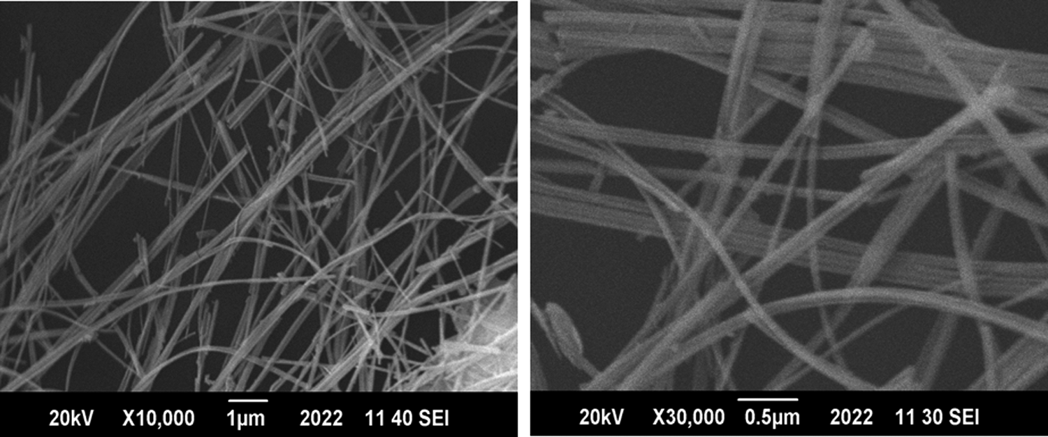
Scanning electron microscopy (SEM) images of pure titania nanotubes (TNTs) at ×10,000 and ×30,000.
Figure 2 displays the SEM images of iron-doped TNTs at 5,000 and 30,000 magnifications, again showing that very fine tubular structures have been formed with diameters ranging from 18 to 42 nm. These tubular structures exhibit high surface area as compared with nanoparticles and are expected to have higher photocatalytic activity. From images of doped nanotubes, it can be further inferred that iron doping has helped in reducing diameters of the tubes compared to the pure TNTs.
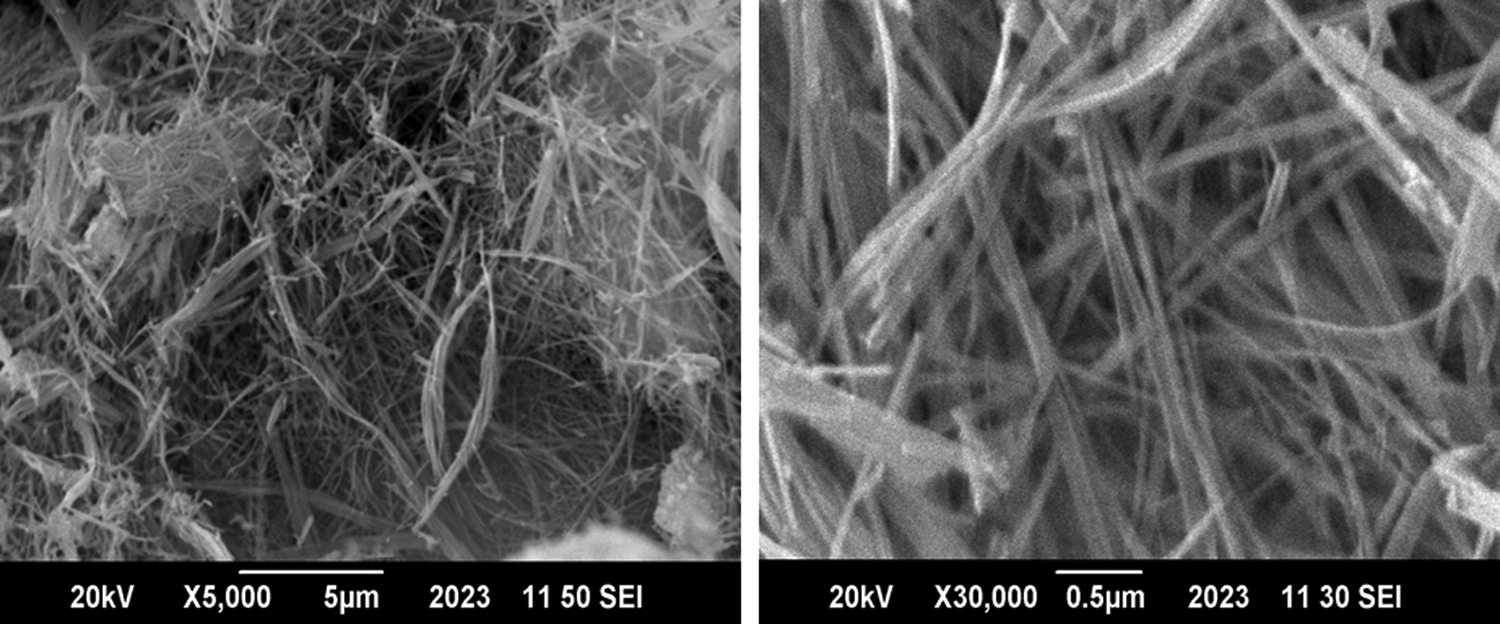
SEM images of 1% iron-doped TNTs at ×5,000 and ×30,000.
EDS analysis
Figure 3 shows that the major constituents of pure TNTs are titanium and oxygen that is, ∼61% and 39%, respectively (as determined by EDS). EDS results of iron-doped TNTs in Fig. 3 show that the product is composed of titanium (50.94%), oxygen (46.84%), iron (1.06%), and sodium (1.2%). The source of sodium is NaOH, which was used during the synthesis process as small quantity of sodium was left over, even after several washings. EDS results confirmed that 1% iron doping of nanotubes has been successfully achieved.

Elemental compositions of pure and 1% iron-doped TNTs.
XRD analysis
XRD characterization for both pure TNTs and 1% iron-doped TNTs were carried out using Cu-Kα radiations at an angle of 2θ from 10° to 80°. In case of pure TNT, peaks can be seen at 25°, 27.5°, 32°, 45.5°, 48°, 54°, 55°, 56.5°, 63°, 66°, and 76° in Fig. 4. These peaks are characteristics of TiO2 in the anatase phase confirming that the synthesized pure TNTs are highly crystalline in nature. The main reason behind this crystallinity is calcination of nanotubes at 500°C for 1 h.

X-ray diffraction intensity plot for pure and doped TNTs.
In the case of 1% iron-doped TNTs, Fig. 4 shows peaks at the same position as in the case of pure TNTs, but with some broadening due to iron doping. The doped TNTs are, however, still highly crystalline in nature attributing this characteristic to calcination of prepared nanotubes in the muffle furnace for 1 h.
BET analysis
BET analysis of pure TNTs (Fig. 5) showed that at a relative pressure of 0.25, the maximum amount of liquid nitrogen adsorbed on the surface of nanotubes is about 80 cm3/g at standard temperature and pressure (STP). This corresponds to almost 320 m2/g surface area of pure TNTs that matches well with the surface area limits (300–350 m2/g) for pure nanotubes (Sekino, 2010).

Isotherm linear plot for N2 adsorption (pure and doped TNTs) at 77 K.
BET analysis of 1% iron-doped TNTs (Fig. 5), on the other hand, shows that at a relative pressure of 0.25, the maximum amount of liquid nitrogen adsorbed on the surface of nanotubes is about 65 cm3/g at STP. This corresponds to almost 230 m2/g surface area of doped TNTs.
Band gap energy calculation
Band gap energy for pure and doped TNTs were found out by using Planck's equation (Dharma et al., 2009) E=hc/λ where,
E=Energy
h=Planck's constant that is, 6.626×10⁻³⁴ J·s
c=Speed of light that is, 3.0×10⁸ m/s
λ=cutoff wavelength obtained from absorbance spectra of pure and doped nanotubes as indicated in Fig. 6
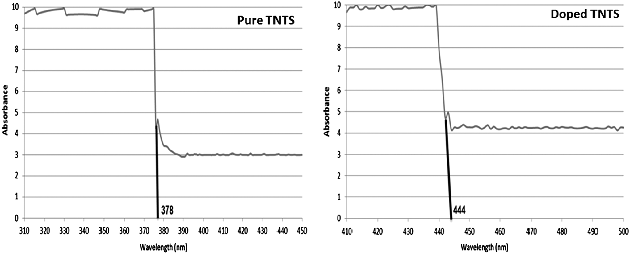
Absorption spectra for pure and doped TNTs in UV–Vis spectrophotometer.
In the case of pure TNTs, the cutoff wavelength came out to be 378 nm. By putting this value in the above equation
E=(6.626×10⁻³⁴) (3.0×10⁸)/378 nm=5.25×10⁻¹⁹ J
E=5.25×10⁻¹⁹ J/1.6×10−19 J=3.28 eV
Cutoff wavelength for 1% iron-doped TNTs came out to be 444 nm. By putting this value in the Planck's equation
E=(6.626×10⁻³⁴) (3.0×10⁸)/444 nm=4.477×10⁻¹⁹ J
E=4.477×10⁻¹⁹ J/1.6×10−19 J=2.80 eV
The results show that iron doping has significantly reduced the band gap energy for TNTs meaning that doped nanotubes can work more efficiently under normal fluorescent light than pure TNTs having higher band gap energies (close to 3.20 eV) that work effectively under UV light only.
Bacterial disinfection
To observe the inactivation of bacterial species as a function of time under fluorescent as well as UV light, fresh cultures of P. aeruginosa and S. aureus, grown overnight, were sprayed on wood surfaces that is uncoated (as a control surface), pure TNTs coated surfaces, and 1% iron-doped TNTs coated surfaces.
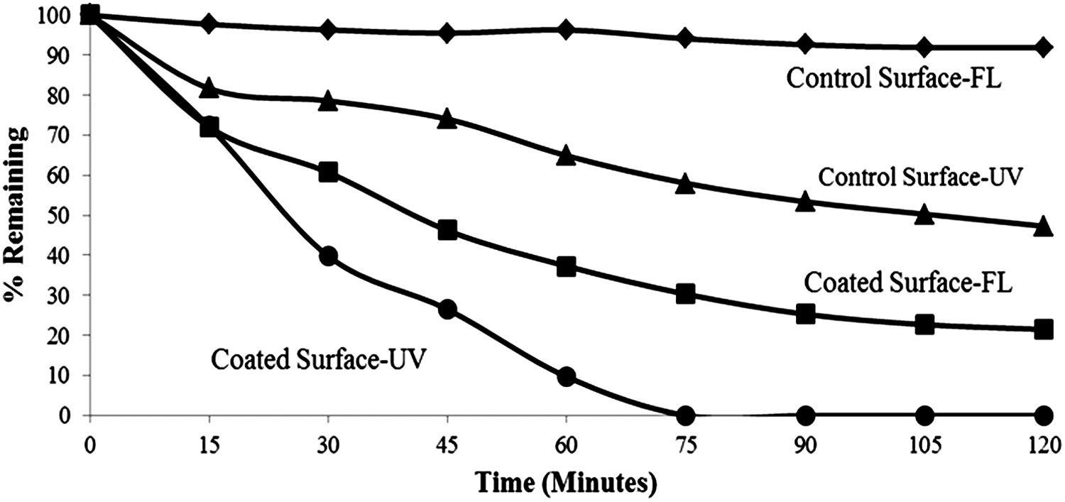
In the case of wood surface coated with pure TNTs exposed to normal fluorescent light, Fig. 7 the population of P. aeruginosa was reduced to almost 65% after 60 min. On the other hand, after 120 min of exposure to fluorescent light, almost 70% of the bacterial population was killed (cell count decreasing from 126×105 to 38×105 CFU/mL) illustrating that even after 120 min, almost 30% of the P. aeruginosa specie survived on pure TNT-coated wood surface. Incomplete disinfection of bacterial population is most probably due to the presence of only 2.5% UV in fluorescent light (NEMA, 1999) as pure TNTs work more efficiently in the presence of UV light due to large band gap energy requirements. When coated wood surface containing the culture of P. aeruginosa was exposed to UV light, complete disinfection was obtained after 105 min confirming that UV exposure has enhanced the bacterial disinfection rate as pure TNTs became highly functional under UV light.

P. aeruginosa disinfection by pure TNTs coated surface with respect to time.
Control wood surface (without any coating), did not show significant reduction in bacterial population in case of normal light whereas almost 40% killing of P. aeruginosa was observed under UV light after 2 h due to its germicidal effect.
Similar patterns were observed in the case of S. aureus when pure TNT-coated wood surface was exposed to fluorescent and UV light source. Figure 8 shows that S. aureus cultures exhibited 80% degradation (cell count decreasing from 236×105 to 50×105 CFU/mL) when coated surface was exposed to fluorescent light for 2 h. Incomplete inactivation of S. aureus can be attributed to its complex wall structure for being a gram negative species (Singleton, 2004) and large band gap energy requirements by pure TNTs (Linsebigler et al., 1995). On the other hand, inactivation rate increased under UV exposure with complete disinfection just within 75 min as high energy UV rays can easily generate electron–hole pairs in pure TNTs by satisfying its band gap energy requirements (Linsebigler et al., 1995).
Staphylococcus aureus disinfection by pure TNTs coated surface with respect to time.
Control surface showed negligible reduction in population of S. aureus under normal light whereas 50% degradation (cell count decreasing from 236×105 to 124×105 CFU/mL) under UV light after 2 h of exposure.
Doped nanotube coated surface when exposed to normal fluorescent light, under sterile laminar flow hood, showed far better disinfection of P. aeruginosa than pure TNT-coated wood surfaces. Figure 9 shows that 70% of the bacterial population was killed (cell count decreasing from 126×105 to 38×105 CFU/mL) just within the first 30 min of exposure. Almost complete sterilization was achieved just within 1 h of exposure as compared to pure TNTs wood surfaces where complete sterilization was not achieved even after 2 h. This is due to the fact that iron doping significantly reduced the band gap energy requirements (Grandcolas et al., 2011) for titania. Under UV exposure, bacterial degradation rate was enhanced and complete sterilization was achieved (cell count decreasing from 216×105 CFU/mL to 0) within 45 min of exposure. When visible light strikes the doped nanotubes, it produces electron–hole pairs, which react with water molecules present on the coated surface to form OH−, O2−, H2O2, and HO2 species (Huang et al., 1998). These species have great ability to degrade organic matter like bacteria.

P. aeruginosa disinfection by 1% iron-doped TNTs coated surface with respect to time.
Control wood surface (without any coating), showed a reduction of almost 50% (cell count decreasing from 222×105 to 110×105 CFU/mL) when exposed to UV light. Under normal fluorescent light, control surface showed negligible reduction in bacterial population.
For doped wood surface containing S. aureus when exposed to light, almost 90% of S. aureus population inactivated (cell count decreasing from 88×105 to 12×105 CFU/mL) in the first 60 min (Fig. 10). Whereas, complete killing of bacteria under fluorescent light was achieved after 105 min (showing more resistant than P. aeruginosa under similar conditions). When similar bacterial species were sprayed on doped nanotubes wood surface and exposed to UV light source, complete inactivation was achieved just within 45 min emphasizing that UV light enhanced the reaction kinetics.
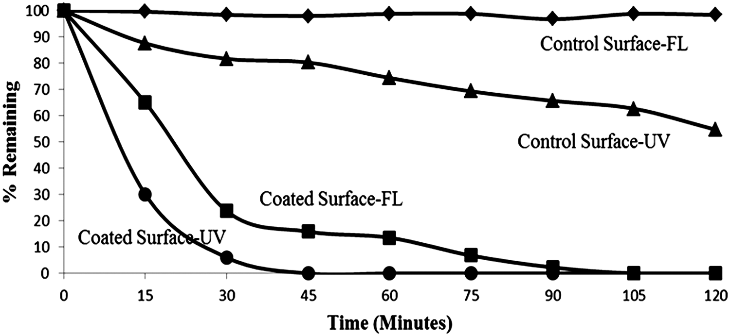
S. aureus disinfection by 1% iron-doped TNTs coated surface with respect to time.
Conclusion
Both pure and 1% iron-doped TNTs were synthesized by using the hydrothermal method along with ultrasonication. Nanotube-coated surfaces, even under fluorescent light, exhibited remarkable reduction in airborne bacterial population with 1% iron-doped TNT-coated wood surface being more effective in killing P. aeruginosa and S. aureus than pure TNT-coated wood surfaces, where bacteria remain viable even after 2 h of exposure to fluorescent light. The novel method of coating the wood surface with the nanotubes, using a natural resin (gum acacia), is not only cost effective, but also environment friendly as the resin does not require any processing except sieving and crushing.
Footnotes
Author Disclosure Statement
No competing financial interests exist.