
Abstract
Abstract
Sb2S3–TiO2 semiconductors heterojunction promote separation of electron–hole charges generated upon light irradiation. This enhancement in charge separation depends on the quality of interfacial contact between Sb2S3 and TiO2, their composition, and their morphological and surface properties. In this study, the Sb2S3–TiO2 nanocomposites were synthesized by two different methods (hydrothermal and mechanical mixing methods) with different percent compositions (5–20% of Sb2S3) and different calcination temperatures (200–600°C) to study their effects on improving the interfacial contact between both semiconductors. Among all synthesized samples, hydrothermally synthesized 10% Sb2S3–TiO2 nanocomposite calcined at 300°C showed the highest photocatalytic degradation of methyl orange (MO) dye solution. X-ray powder diffraction pattern showed that the anatase phase of TiO2 and the orthorhombic phase of Sb2S3 are retained in the nanocomposite. Scanning electron microscopy and energy-dispersive X-ray revealed formation of nanocomposite with purity. Transmission electron microscopy revealed that TiO2 is deposited better on Sb2S3 in hydrothermally synthesized nanocomposite than in mechanically mixed composite. However, 300°C calcinations of the composite helped improve the interaction between TiO2 and Sb2S3. Brunauer-Emmett-Teller (BET) surface area analysis showed the highest specific surface area for the hydrothermally synthesized 10% Sb2S3–TiO2 nanocomposite calcined at 300°C. Considering all results, a reasonable mechanism of photocatalysis of Sb2S3–TiO2 nanocomposite has been proposed. Furthermore, the chemical oxygen demand data showed substantial mineralization of MO dye by the 10% Sb2S3–TiO2 nanocomposite calcined at 300°C.
Introduction
I
Itzhaik et al. (2009) deposited Sb2S3 over TiO2 in the Extremely Thin Absorber cells as semiconductor sensitizers instead of using dyes, which show 3.3% efficiency. Tsujimoto et al. synthesized Sb2S3-based Extremely Thin Absorber solar cells and observed an increase in its efficiency up to 4.1% (Tsujimoto et al., 2012). Boix et al. presented the model of TiO2/Sb2S3/CuSCN semiconductor-sensitized photovoltaic cell with improved cell performance by focusing on their fundamental process of charge transport and recombination using CuSCN as hole transporter (Boix et al., 2012). Salunkhe et al. (2012) deposited Sb2S3 nanoparticles on mesoporous TiO2 and used as photoanode in the FTO/TiO2/Sb2S3/electrolyte/Pt device structure.
Li et al. synthesized pristine Sb2S3 by solid-state reaction and investigated its photocatalytic activity in visible light for degradation of methyl orange (MO). Its degradation rate was found to be much higher than those of P-25, CdS, and Bi2S3 under visible light irradiation (Li et al., 2008). Sun et al. (2008) synthesized Sb2S3 nanorods by a simple wet chemical method under refluxing condition and found good photocatalytic efficiency under visible light for degradation of MO and p-hydroxyazobenzene. They synthesized Sb2S3–TiO2 heterojunction catalyst using the same Sb2S3 nanorods and optimized the composition of Sb2S3 over TiO2 at 60% for efficient degradation of MO in visible light (Sun et al., 2012). Huang et al. (2011) synthesized Sb2S3 and Bi2S3 doped TiO2 by gel hydrothermal method and investigated its photocatalytic activity for degradation of 4-nitrophenol. This enhancement in charge separation depends on the quality of interfacial contact between Sb2S3 and TiO2, which may ultimately depend on the synthesis method, % composition, calcination temperature, and their morphological and surface properties, such as Brunauer-Emmett-Teller (BET) surface area.
In this view, in present investigation, an attempt has been made to reveal the key factors in synthesis of Sb2S3–TiO2 nanocomposite with enhanced photocatalytic efficiency. It has been observed that the effect of composition of each component in the composite, calcination of composite, and method of synthesis all together affect the photocatalytic degradation efficiency of MO dye solution in the presence of white light when the Sb2S3–TiO2 nanocomposite is used in a multilamp photoreactor.
Experiment
Materials
Titanium tetraisopropoxide (TTIP) was purchased from Alfa Aesar. Absolute ethanol, tartaric acid, and antimony trichloride were purchased from S D Fine-Chem Limited. Potassium thiocyanate was purchased from Fisher Scientific. The azo dye, methyl orange (MO dye) of Merck, was used as the model pollutant to study the photocatalytic efficiency of synthesized composite. Double-distilled water was used for preparation of solutions. All chemicals were AR grade and used without any further purification.
Synthesis of photocatalyst
Synthesis and characterization of TiO2 were described previously (Shitole et al., 2013). Sb2S3 particles were synthesized by a simple wet chemical method under refluxing conditions (Ota et al., 2007).
In hydrothermal synthesis of Sb2S3–TiO2 nanocomposite, the synthesized Sb2S3 particles were added to provide a weight ratio of Sb2S3 over TiO2 as 5%, 10%, and 20%. In a typical experiment, synthesized Sb2S3 particles were dispersed into double-distilled water and sonicated for 15 min. A predetermined amount of TTIP was mixed with ethanol in 1:5 ratio. After complete dispersion of Sb2S3 in water, TTIP:ethanol solution was added dropwise under sonication and kept overnight with vigorous stirring. On the next day, whole solution was transferred to the Teflon-lined stainless steel autoclave and placed in muffle furnace for hydrothermal treatment at 140°C for 24 h for effective interaction of Sb2S3 with TiO2 at high temperature and elevated pressure. After cooling the furnace to room temperature, autoclave was removed from the furnace, and the obtained composite was dried on a hot plate.
To study the effect of calcination on photocatalytic activity of the nanocomposite, the composite was calcined at various temperatures ranging from 200°C to 600°C for 2 h in muffle furnace. Furthermore, to study the effect of method of synthesis on the properties and photocatalytic activity of composite, Sb2S3–TiO2 nanocomposite was also synthesized by the mechanical mixing method. In hydrothermally synthesized composite, 10% Sb2S3–TiO2 calcined at 300°C for 2 h showed the best results for photocatalytic degradation of MO dye solution. Therefore, in mechanical mixing method, 90 mg of hydrothermally synthesized TiO2 and 10 mg of Sb2S3 synthesized by wet chemical method were mixed and grinded in a mortar and pestle until they became homogeneous. Then, the ground sample was calcined in muffle furnace at 300°C for 2 h.
Material characterization
Sb2S3 and Sb2S3–TiO2 nanocomposites were characterized by a range of analytical techniques. The X-ray powder diffraction (XRD) (Philips X'Pert PRO) patterns were recorded with Cu Kα radiation (α = 0.15406 nm) in the range of 10–80° 2θ at a scanning speed of 0.02°/s to determine the crystal structure. The morphology and structure of Sb2S3 and Sb2S3–TiO2 nanocomposites were examined by transmission electron microscopy (TEM) (TECNAI G2 20 TWIN; FEI) and scanning electron microscopy (SEM) (JEOL JSM-6360 A) along with energy-dispersive X-ray (EDX). The sample was subjected to thermal gravimetric analysis (TGA) (DTG-60H, simultaneous DTA–TG apparatus) to get information regarding thermal stability of the product. The surface areas of synthesized samples were measured by Thermo Scientific Surfer BET Surface Area Analyzer.
Photocatalytic degradation experiments
We developed the successful design for laboratory-made multilamp photoreactor to study the photocatalytic activity. The laboratory-made photoreactor (Fig. 1) consisted of quartz reaction vessel in the center surrounded by four white light lamps (T5-8W 6400K; Hybec). The reaction solution was constantly aerated using aerator pump, and reaction temperature was controlled at 30°C ± 1°C by an air- cooling system. The photocatalytic test was performed with 40 mg catalyst dose and 100 mL solution of 2.5 × 10⁻⁵ M MO dye solution. The aliquots of suspension were taken out after every 15 min and filtered using 0.2 μm pore size, 13-mm-diameter Millipore disc, and the undecomposed MO dye was determined using Shimadzu UV–visible spectrophotometer (UV-1800 PC). Chemical oxygen demand (COD) values were measured by standard dichromate method using Spectralab 2015M COD Digestor and CT-15 COD Auto-Titrator. Total organic carbon (TOC) analysis was performed using commercially available test kits (NANOCOLOR TOC 60) from Macherey-Nagel, Germany, using thermodigester (Spectroquant TR 320) and spectrophotometer (NANOCOLOR VIS; Macherey-Nagel) (Kale and Thakur, 2015).

Laboratory-made multilamp photoreactor.
Results and Discussion
Characterization
XRD patterns of the Sb2S3 in Fig. 2a show the peaks for 020 peak at 2θ = 15.65°, 120 peak at 2θ = 17.51°, 310 peak at (25.04), 121 (29.21), 221 (32.36), 240 (35.50), and 501 (46.79) crystal planes, and their relative intensities were matching with the standard spectrum (JCPDS No. 42–1393) of the orthorhombic phase of crystalline Sb2S3. Absence of unwanted peaks confirms the purity of the product. Figure 2b–f shows the XRD of Sb2S3–TiO2 nanocomposite from synthesized composite temperature to 600°C calcination temperature. The hydrothermally synthesized nanocomposite of Sb2S3–TiO2 shows the peaks for both the anatase phase of TiO2 and orthorhombic phase of Sb2S3 (Fig. 2b). The XRD also confirms the formation of crystalline and pure nanocomposite. The peak intensity increases as the nanocomposite calcination temperature increases from 200°C to 300°C. However, when the calcination temperature of nanocomposite increases to 400°C, the peaks for Sb2S3 are not observed, which is in accordance with the TGA data of Sb2S3 at 400°C, where the Sb2S3 oxidizes to Sb2O3. In the XRD pattern, the main peaks 25.42 (111) and 37.94 (141) for Sb2O3 (JCPDS No. 11–0689) might be shielded by the peaks of TiO2 and hence cannot be seen distinguishably. When composite was calcined at 600°C, Sb2O3 vaporizes, and some Sb2O3 gets converted to Sb2O4 and so very less Sb2O4 is present in the nanocomposite. Hence, the XRD of nanocomposite at 600°C shows the peaks only for TiO2 nanoparticles. Further characterization of the Sb2S3–TiO2 nanocomposite, such as SEM, TEM, and BET, was carried out only for sample calcined at 300°C.

XRD patterns of Sb2S3 and 10% Sb2S3–TiO2 nanocomposites calcined at 200°C, 300°C, 400°C, and 600°C and for TiO2. XRD, X-ray powder diffraction.
Figure 3a and b shows the SEM images and EDX of the synthesized Sb2S3. These images reveal the flower-like morphology along with the formation of rods, which may be advantageous for the photocatalysis applications. Ota et al. (2007) reported that as the duration of reaction increases, Sb2S3 induces the rod-like morphology. The EDX spectrum of synthesized Sb2S3 shows the peaks for Sb and S, which confirms the purity of the Sb2S3. Figure 3c depicts the SEM image of Sb2S3–TiO2 nanocomposite. Because of the more quantity of TiO2 over Sb2S3 (10% Sb2S3 content), most of the Sb2S3 are covered and hidden under TiO2 nanoparticles.

TEM images (Fig. 4) illustrate the morphologies of Sb2S3 and Sb2S3–TiO2 nanocomposites synthesized by hydrothermal and mechanical mixing methods. Figure 4a shows that Sb2S3 has a rod-like morphology. Figure 4b and c shows the TEM images of Sb2S3–TiO2 hydrothermally synthesized nanocomposites without calcination and at 300°C calcination temperature. It is evident from Fig. 4b that before calcination, TiO2 nanoparticles were dispersed around Sb2S3 rod, but after calcination at 300°C for 2 h, those dispersed TiO2 nanoparticles were embedded on the surface of Sb2S3 rod as in Fig. 4c, indicating improved bonding between TiO2 and Sb2S3 particles at higher calcinations. However, when the same composite was synthesized by mechanical mixing method, it shows the separate clusters of TiO2 and Sb2S3 particles (Fig. 4d). These results indicate that the hydrothermal mixing method disperses the TiO2 nanoparticles on the surface of Sb2S3 better than mechanical mixing method, and calcination supports for a strong bonding between them.

TEM images of
Table 1 shows the specific surface area of Sb2S3, TiO2, and 10 wt.% Sb2S3–TiO2 calcined at 300°C nanocomposite determined by BET analysis. Because Sb2S3 prevents the agglomeration of TiO2 nanoparticles, the surface area of nanocomposite increases in comparison with bare TiO2 and Sb2S3 nanoparticles.
BET, Brunauer-Emmett-Teller.
Figure 5a shows the TGA and DTA curves of synthesized Sb2S3. The TGA shows the first weight loss around 290–380°C and second weight loss around 415–440°C. DTA shows the peaks for the same weight losses. This indicates the oxidation of Sb2S3 to Sb2O3. There is a presence of both Sb2S3 and Sb2O3 after the first weight loss, and in the second weight loss, all Sb2S3 oxidize to Sb2O3. The third weight loss occurs around 473–563°C, which occurs owing to vaporization of Sb2O3 and continued oxidation of Sb2O3 to Sb2O4. Actually, the oxidation of Sb2O3 to Sb2O4 results in weight gain, but this weight gain was counteracted by the vaporization of Sb2O3 (Town et al., 1975; Kelley et al., 1987).
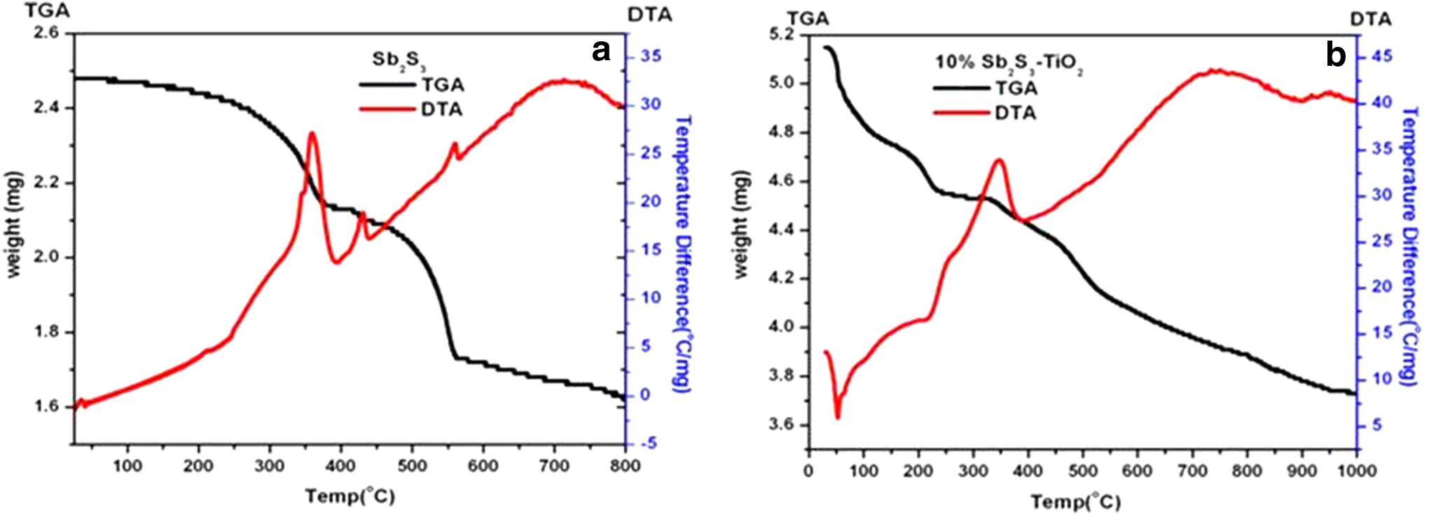
TG/DTA of
Figure 5b shows the TG/DTA of hydrothermally synthesized Sb2S3–TiO2 nanocomposite. TGA clearly reveals four weight losses. The first weight loss around 100°C was because of water evaporation, and the second weight loss around 200–350°C might be due to decomposition of organic matter. The third and fourth weight losses around 325–400°C and 450–550°C were because of oxidation and vaporization of Sb2S3 within the nanocomposite, respectively.
Photocatalytic activity study
Effect of composition
The effect of loading of Sb2S3 in the composite over TiO2 was studied to achieve the higher photocatalytic activity for degradation of targeted pollutant. It is a crucial step to control the composition ratio of Sb2S3:TiO2 to obtain the optimal synergistic effect between TiO2 and Sb2S3.
Figure 6a displays the concentration changes of the initial 100 mL 2.5 × 10⁻⁵ M MO dye solution at 463 nm as a function of UV light irradiation time in the presence of TiO2 and TiO2

Effect of calcination temperature
Efficient catalyst in weight ratio (10% Sb2S3–TiO2) was calcined between temperatures ranging from 200°C to 600°C for 2 h in muffle furnace and examined for its photocatalytic response for degradation of 2.5 × 10⁻⁵ M MO dye solution under UV light irradiation (Fig. 6b). Bare TiO2 degrades 64.15% MO dye from solution, while the 10% Sb2S3–TiO2 as prepared, calcined at 200°C, 300°C, 400°C, 500°C, and 600°C, degrades 73.09%, 86.31%, 95.31%, 70.53%, and 25.67% MO dye, respectively. 10% Sb2S3–TiO2 nanocomposite shows the best activity at 400°C calcination temperature, but as evident from XRD and TGA data, after 300°C, Sb2S3 from nanocomposite starts getting oxidized. Up to 300°C, the nanocomposite contains only TiO2 and Sb2S3, but above 300°C, along with TiO2 and Sb2S3, oxidized Sb2S3 was also observed because the Sb2S3 in nanocomposite is thermally unstable above that temperature. Hence, 300°C calcination temperature was finalized to prepare nanocomposite of TiO2 and Sb2S3 by mechanical mixing to compare its activity with hydrothermally synthesized 10% TiO2–Sb2S3 nanocomposite. It is also observed that at 300°C, a mixture of Sb2S3 and Sb2O3 along with TiO2 is more beneficial for photocatalytic degradation of MO dye solution, and interestingly above 400°C, Sb2O3–TiO2 has been found to be more efficient than Sb2S3–TiO2 nanocomposite (Liu et al., 2012).
Effect of method of synthesis
Nowadays, various types of composite synthesis methods have been developed. The method of synthesis largely affects the structure, morphology, and properties of composite. Hence, to study the effect of method of synthesis on the photocatalytic properties of nanocomposite becomes essential. Photocatalytic efficiencies of 10% Sb2S3–TiO2 nanocomposite synthesized by mechanical mixing (S1T Mech) and hydrothermal (S1T Hydro) methods were tested by degrading MO dye solution. The result shows that the S1T Hydro composite exhibits enhanced photocatalytic performance by degrading 86.31% MO dye solution than S1T Mech composite degrading only 53.61% MO dye solution under UV light irradiation (Fig. 6c).
It concludes that the synthesis method greatly affects the photocatalytic degradation efficiency.
Chemical oxygen demand
The mineralization of model pollutant by S1T Hydro calcined at 300°C (S1T Hydro 300) and bare TiO2 synthesized by the same method was studied by COD measurements. As shown in Table 2, after 2 h of photocatalysis reaction, there is a substantial decrease in the COD values of MO dye by S1T Hydro 300 nanocomposite than TiO2 alone.
COD, chemical oxygen demand; MO, methyl orange.
Total organic carbon
Mineralization of model pollutant by 10% Sb2S3–TiO2 hydrothermally prepared and calcined at 300°C and bare TiO2 synthesized by the same method was studied by TOC measurements. Figure 7 shows that 64% mineralization was achieved for Sb2S3–TiO2 nanocomposite compared to bare TiO2 (49%). Obtained results are a direct indication of enhanced degradation and mineralization efficiency of 10% Sb2S3–TiO2 hydrothermally prepared and calcined at 300°C nanocomposite compared with bare TiO2.
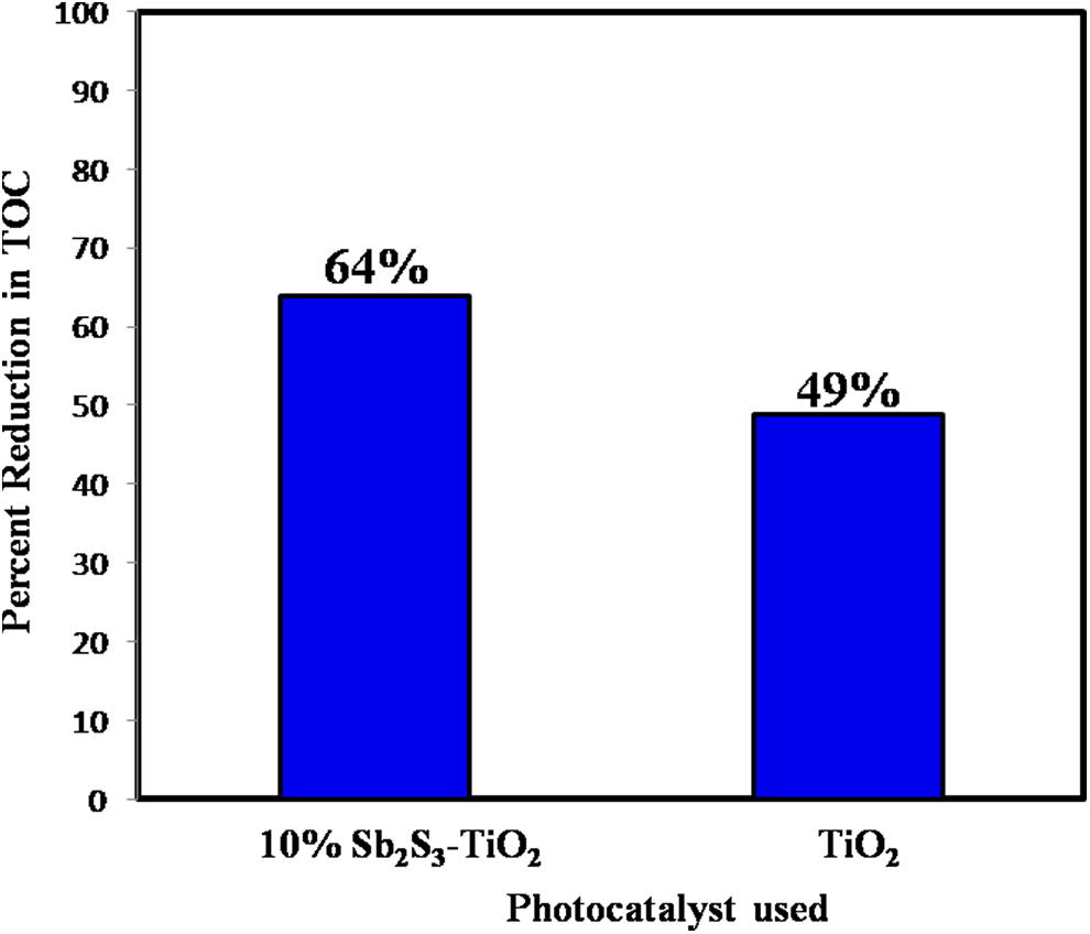
Percent reduction in TOC for 10% Sb2S3–TiO2 hydrothermally prepared and calcined at 300°C nanocomposite and bare TiO2 for degradation of MO dye. TOC, total organic carbon.
Proposed mechanism
A coupling of Sb2S3 with TiO2 increases the photocatalytic efficiency of pure TiO2. The conduction band of TiO2 is more anodic than Sb2S3 conduction band, which strengthens the driving forces of electron injection, as shown in Fig. 8. Hence, the TiO2 and Sb2S3 semiconductor heterojunction generates a beneficial role in improving charge separation and extends TiO2 response to a range of light irradiation.

Based on reported works (Huang et al., 2011), a mechanism was proposed for the photocatalytic degradation of organic pollutants using Sb2S3–TiO2 semiconductor heterojunction as a photocatalyst. Proposed mechanism is shown in Fig. 8b, with step-by-step equations. Upon irradiation, TiO2 and Sb2S3, get excited, and electrons are injected from the conduction band of Sb2S3 to that of TiO2, as shown in Eqs. (1) and (2). This electron reduces the molecular oxygen to superoxide radical anion and hydrogen peroxide [Eqs. (3) and (4)]. Holes from valence band of TiO2 oxidize H2O to hydroxyl radicals [Eq. (6)]. These hydroxyl radicals are powerful oxidizing agents and play an important role in the degradation of organic pollutants [Eq. (7)] (Huang et al., 2011; Sun et al., 2012).
Photocatalytic degradation of organic pollutant over Sb2S3–TiO2 nanocomposite.
Conclusion
In summary, Sb2S3–TiO2 nanocomposites were successfully synthesized by hydrothermal method. Furthermore, the effects of percent composition, calcination temperature, and synthesis method of Sb2S3–TiO2 nanocomposite on the photocatalytic activities were investigated in detail. Obtained results show enhanced degradation (86.31%) of MO dye, with efficient 10% Sb2S3–TiO2 nanocomposite synthesized by hydrothermal method calcined at 300°C compared to bare TiO2 (64.15%), which is even more than mechanically mixed nanocomposite (53.61%). The obtained results of enhanced degradation in case of hydrothermally synthesized composite clearly explain the effect of method of nanocomposite synthesis. Hydrothermally synthesized sample facilitates in establishing good interfacial contact between TiO2 and Sb2S3, thereby resulting in the effective separation of the photogenerated electron–hole pairs, leading to the faster rates of photocatalytic oxidation compared to bare TiO2 and mechanically mixed sample. Furthermore, the encouraging COD and TOC data throw light on the substantial mineralization of the pollutant tested, which is essential from the environmental point of view. Thus, the nanocomposites synthesized under present investigation by hydrothermal method with efficient percent composition and calcination temperature possess potential applications in environmental remediation as well as solar cell application.
Footnotes
Acknowledgment
The authors are thankful to the ISRO-UoP Cell, Savitribai Phule Pune University, for research funding.
Author Disclosure Statement
No competing financial interests exist.