
Abstract
Abstract
Because of it high solubility in water, m-cresol is an important class of water contaminants. Due to toxicity effects and endocrine disrupting properties of this phenolic compound, removal of it from water and wastewater has gained much attention. In the present work, m-cresol degradation has been studied in aqueous medium using silver nanoparticles (Ag NPs) synthesized using leaf extract of Nyctanthes arbor-tristis. N. arbor-tristis leaf extract was used as the reducing agent in fabrication of Ag NPs in different solvents (water, ethylene glycol, and ethyl acetate) using ultrasonic-assisted method. Ag NP prepared in different solvent shows different shape and size. Ag NPs prepared in water medium show different shape–triangular and spherical. Ag NPs tailored in ethylene glycol and ethyl acetate medium show spherical and elongated shape with different sizes. Particles as such prepared have diverse pioneer catalytic activities toward degradation of m-cresol. Ag NP prepared in water medium completely degrades m-cresol in 93 min, while Ag prepared in ethylene glycol and ethyl acetate medium took a longer time. Degradation process followed first order kinetics with rate constant value in the following order—kAg water > kAg ethylene glycol > kAg ethyl acetate. Superior catalytic efficacy of Ag fabricated in water is attributed to the synergetic effect of facet shape, exposing a highly active face and the size dependent surface activity of particles.
Introduction
P
These chemicals pose a serious threat to many ecosystems, water supplies, and human health because of their toxicity, endocrine disrupting abilities, and carcinogenic behavior (Gogate and Pandit, 2004). m-cresol is one of them, and it is commonly used in organic laboratory as solvent. m-cresol is a pollutant of great importance due to its toxicity and solubility. It is highly soluble in water (2.35 g/100 mL), so it is present in large amount in wastewater discharges from chemical industries (Wang et al., 1999). According to US health exposure limits, permissible amount of m-cresol is 5 ppm (22 mg/m3) and presence of 390 μg/L of cresol derivatives in surface water was reported (Helmet, 2007). So before discharging m-cresol and its derivatives to environment it should be treated. Various methods such as anaerobic digestion, advance oxidation process, hydroxylation of the aromatic ring, flocculation, precipitation, granular activated carbon, reverse osmosis, and so on are proposed by researchers for degradation of m-cresol (Chen et al., 2016).
In contrast, due to growing need to develop green routes for generating materials, biosynthesis of nanoparticles (NPs) has received tremendous attention. Use of plant, plant extract or product, microorganism, enzymes, and so on in synthesis processes is suggested as possible eco-friendly alternative routes to other conventional methods, which use lots of harmful chemicals. In this work, leaf extract of Nyctanthes arbor-tristis (NA) was used for the fabrication of silver NPs (Ag NPs). This plant, locally known as night jasmine, is rich in phenylethanoid derivatives and iridoid glycosides (Jesen et al., 2002). It is used in Ayurvedic medicine for the treatment of sciatica and arthritis (Pittaya et al., 2003). Extract of this plant possesses hepatoprotective, antileishmanial, antiviral, antifungal, analgesic, antipyretic, and ulcerogenic activities (Priya and Deepak, 2007). This leaf extract is rich in phytosterols, phenolics, tannins, phlobatannins, flavonoids, steroids, glycosides, saponins, and highly branched polysaccharide (75 kDa) containing esterified phenolic acids (Rathee et al., 2007; Shukla et al., 2012; Ghosh et al., 2015). These phytochemicals have the ability of reducing metal and providing robustness against agglomeration (Henam, 2015). Using this leaf extract of NA, Ag NPs of different shapes and sizes are fabricated through sonochemical method. Ultrasonic irradiation caused cavitation in the liquid medium where the formation, growth, and collapse of microbubbles occurred (Shi-Fan, 2015). Molecules undergo a reaction due to the application of the powerful ultrasound radiation. Sonochemistry has been used to synthesize various organic and inorganic materials (Tahmasian and Morsali, 2012). It has been indicated as a simple, clean, and convenient technique in chemical synthesis (Safaei-Ghomi and Akbarzadeh, 2015). Sonochemistry is based on a physical phenomenon called “acoustic cavitation.” Sonochemical process consists of three steps—creation of bubbles, growth, and collapse. In the first stage, the liquid undergoes series of compression–expansion (rarefaction) following the ultrasound wave encounter; the sudden drop in pressure forms small and oscillating bubbles of vapor. In second stage, the growth of the bubble occurs through the diffusion of solute vapor to the volume of the bubble. These bubbles expand in each cycle of applied ultrasound energy until they reach an unstable size. After reaching this unstable size they can collide or violently collapse to give the desired product. According to hot-spot mechanism, implosive collapse raises the temperature and pressure, which result in rupture of chemical bonds (Shafi et al., 2001). This collapse of bubbles occurs in less than a nanosecond and very high cooling rates are also obtained. Due to the fast kinetics, the growth of the nuclei is restricted and in each collapsing bubble few nucleation centers are formed, the growth of which is limited by the short collapse; as such the particles synthesized by this technique are in nanoscale.
In the present work, ultrasonic-assisted synthesis method is used as green synthetic approach for the synthesis of nanoscale Ag NPs in different solvents. These three solvents—water, ethylene glycol, and ethyl acetate have drastic variation in relative polarity and dielectric constant values (given in Table 1). Because of this variation it will be easy to understand the role of solvent in the synthesis of NPs. Ultrasonic radiation is used to maintain the size of Ag particles in nanoscale by preventing the growth of the particles. Ag NPs prepared using ultrasonic radiation are used as placebo for degradation of m-cresol.
Materials and Experiments
Materials
Ethylene glycol and acetyl acetate were obtained from HiMedia Company. Silver nitrate (AgNO3) of 99.9% pure was purchased from Sigma Pvt. Ltd. All the chemicals were of analytical grade, so without further purification they were used in synthesis process. Throughout the synthesis process double distilled water (DDW) was used. Leaf of NA was collected from local area of Imphal West, Nambol, Manipur and dried in a dark room. Syringe filter of Millex GV of 0.22 μm was used for filtering leaf extract.
Preparation of leaf extract
Leaf extract of NA was prepared in different solvent (water, ethylene glycol, and acetyl acetate). To 3 g of the dried leaf, 50 mL of the solvent was added and warmed at 40°C for 15 min with constant stirring. After cooling, the extract was centrifuged to settle down the unwanted and filtered using syringe filter of pore size 0.22 μm. This leaf extract was used for the fabrication of Ag NPs of different size and shapes.
Preparation of Ag NPs
To 25 mL of leaf extract, 0.5 g of AgNO3 was added and ultrasonic radiation was passed by sonicating the mixture for 60 min. Ultrasonic cleaner of 40 kHz frequency was used for the synthesis process. Formation of NPs is confirmed by the change of yellowish and greenish color of leaf extract to black and gray color. Particle formed was centrifuged and washed with ethanol twice, later dried at room temperature.
Growth kinetics of Ag NPs
Growth kinetic of Ag NPs was studied by monitoring the absorbance of reaction mixture of silver and leaf extract in ultraviolet-visible (UV-Vis) spectroscopy by scanning in range of 300–800 nm. While sonicating the reaction mixture, 3 mL of each reaction mixture was withdrawn and increase in the absorbance of Ag particles was measured at regular interval of time using UV-Vis spectroscopy.
Degradation of m-cresol
Degradation of m-cresol in aqueous medium was monitored with the help of UV-Vis spectrophotometer at λmax = 271 nm, a prominent characteristic peak of m-cresol. To 20 μL of 50 ppm m-cresol, 50 μL of Ag NPs prepared were added in a 3.5 mL capacity quartz cuvette. The total volume for the degradation reaction system was fixed at 3 mL by adding DDW. The progress of the reaction was monitored by noting the fall in absorbance at 271 nm at 25°C.
Characterization
UV-Vis spectroscopy
All UV-Vis spectra were recorded on Perkin Elmer UV-Vis spectrophotometer fitted with Peltier System. The absorption spectra of the prepared NPs were recorded by taking the aqueous dispersion of the NPs and scanned in the range of 200–800 nm.
Scanning electron microscopy
Scanning electron microscopy (SEM) images of the synthesized NPs were obtained using SEI quanta 250 model of SEM instrument. An adequate amount of Ag NPs were used for the measurement.
Transmission electron microscopy and selected area electron diffraction
Transmission electron microscopy (TEM) and selected area electron diffraction (SAED) micrographs of the particles were taken with JEM-2100 transmission electron microscope operating at 60–200KV STEPS. After preparation, the particles were centrifuged at 12,000 rpm for 10 min, washed with DDW thrice, and redispersed in it by sonication for 4–5 min. A drop of dilute aqueous dispersion of the Ag NPs was put on carbon-coated copper grids, and the grids were dried at room temperature. After complete drying of the grids, TEM and SAED patterns of the particles were taken.
X-ray diffraction analysis
X-ray diffraction (XRD) patterns of the samples were taken by Philips XRD instrument; properly dried powder samples were utilized to carry out the diffraction experiment. After preparation, the samples were washed effectively with DDW and finally with ethanol. The samples were then allowed to dry at room temperature.
Fourier transformed-infrared spectroscopy
IR-Perkin Elmer, Fourier transformed-infrared (FT-IR) system, Spectrum BX FT-IR instrument was utilized to measure FT-IR spectra of leaf extract and the synthesized NPs. FT-IR spectra of leaf extract of NA and the synthesized NPs were recorded in the range 400–4,000 cm−1.
Results and Discussion
Synthesis of Ag NPs using ultrasonic-assisted method is explained in Fig. 1. Change in the color of the leaf extracts from light yellow/green to black/gray after sonicating for 1 h confirms the formation of Ag NPs.
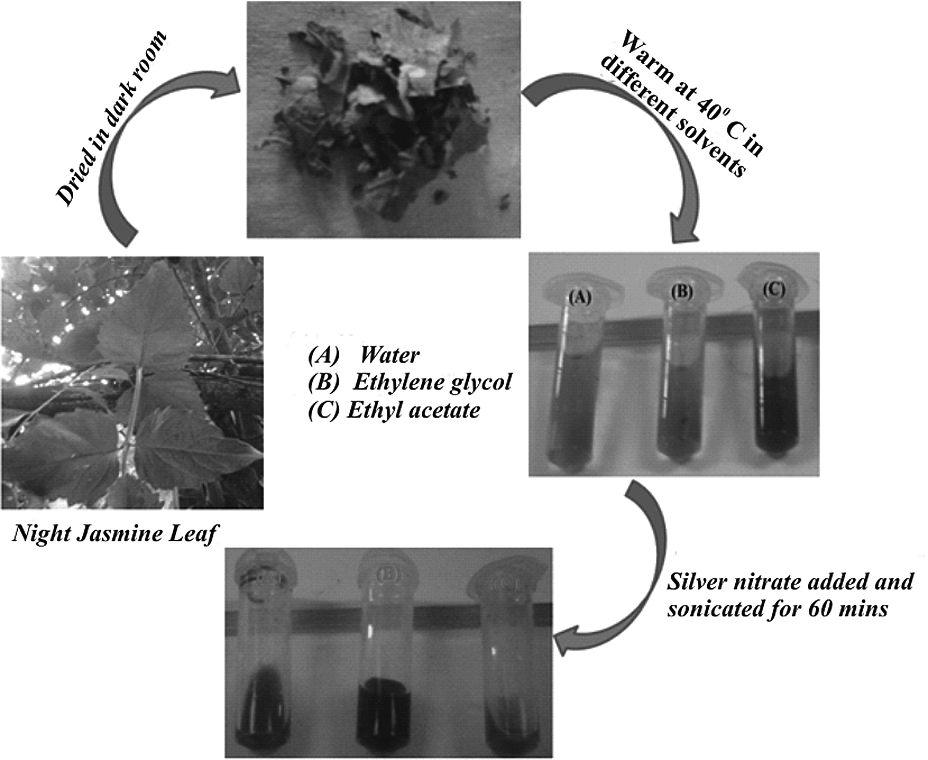
Synthesis of Ag NPs using leaf extract of night jasmine in different solvents. NP, nanoparticles.
Optical property of Ag NPs is studied using UV-Vis spectroscopy and found out that Ag NPs fabricated in different medium show unique surface plasma resonance band. Ag NPs obtained by reducing Ag ions by leaf extract in ethylene glycol (AgG) show a single but a broad peak at 452 nm, which may be due to the out of plane dipole plasmon resonance. Ag NPs prepared using water as solvent (AgW) show two plasmon resonance band at 346 and 454 nm; likewise, Ag NPs fabricated in ethyl acetate medium (AgE) also show two peaks one at 617 nm and another at 480 nm. Peak at 346 and 617 nm may be due to the out of plane quadruple resonance and inplane dipole plasmon resonance, respectively (Jin et al., 2001), as given in Fig. 2. In 1908, Mie shows that plasmon resonance depends explicitly on the particle size (Mie, 1908). Small spherical nanocrystals exhibit a single surface plasmon band, and anisotropic particles exhibit two or more bands, depending on shape of particles (Nikhil et al., 2001; Daniel and Didier, 2004; Mohamed et al., 2012). Larger metal particles can exhibit broad or additional bands in the UV-Vis range due to the excitation of plasmon resonances or quadrupole and higher multiple plasmon excitation (Kamat et al., 1998). However, in history it has been demonstrated both experimentally (Zeman and Schatz, 1987; Lisiecki et al., 1996; Van der Zande et al., 1997) and theoretically (Creighton and Eaton, 1991) that surface plasma resonances of the coinage metal NPs depend mostly on the particle shape than on sizes (Yu-Ying et al., 1997).
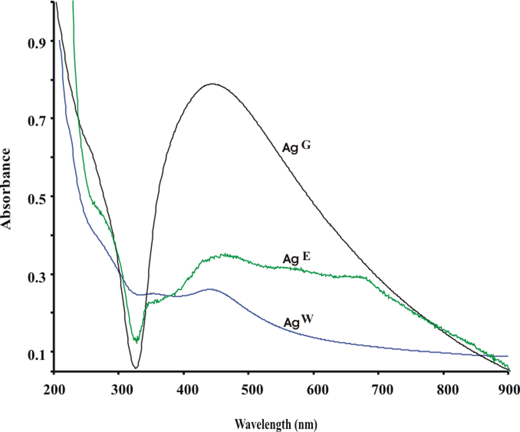
UV-Vis spectra of Ag NPs. UV-Vis, ultraviolet-visible.
Figure 3A–C highlights the TEM images of Ag NPs fabricated in water, ethylene glycol, and ethyl acetate medium. AgW NPs have triangular and spherical morphologies of different size indicating the polydispersity nature of the particles as given in Fig. 3A, which may be due to polar nature of solvent. Particles are faceted and average diameter of the particles fabricated is in the range of 10–54 nm and some larger particles are also observed. AgG NPs are spherical in shape and have an average diameter of 10–50 nm as shown in Fig. 4B. Ag particles fabricated using ethyl acetate as solvent are spherical, and some elongated oval shape particles are observed as given in Fig. 3C with average diameter in range of 9–45 nm. Ag particles fabricated in ethyl acetate are smaller in size, and possible reason of this observation is that dielectric value of ethyl acetate is very low so it hardly interacts with the ultrasonic radiation. Because of this ethyl acetate did not favor the growth and ripening of Ag crystals (Petroski et al., 1998). Ag NPs synthesized in ethylene glycol and ethyl acetate are more uniform in size compared to those prepared in water. Organic solvent containing alkyl group usually promotes the formation of spherical rods (Xu et al., 2009). Relative polarity of the three solvents follows the order water > ethylene glycol > ethyl acetate. Unlike polar solvent, organic solvent did not support the self-association of smaller particles to give structural NPs (Spiecker et al., 2003; Pradip et al., 2008). TEM analysis further confirmed that solvents have significant influence on the morphology of silver particles formed under ultrasonic irradiation. In contrast, SAED patterns show the well crystalline nature of the Ag NPs fabricated using this leaf extract of NA in different solvents. SEM images of the Ag NPs prepared using leaf extract in different solvents are given in Fig. 4; images highlight the aggregation of NPs due to the large surface energy of the particles.

Transmission electron microscopy images and selected area electron diffraction pattern of Ag NPs prepared in
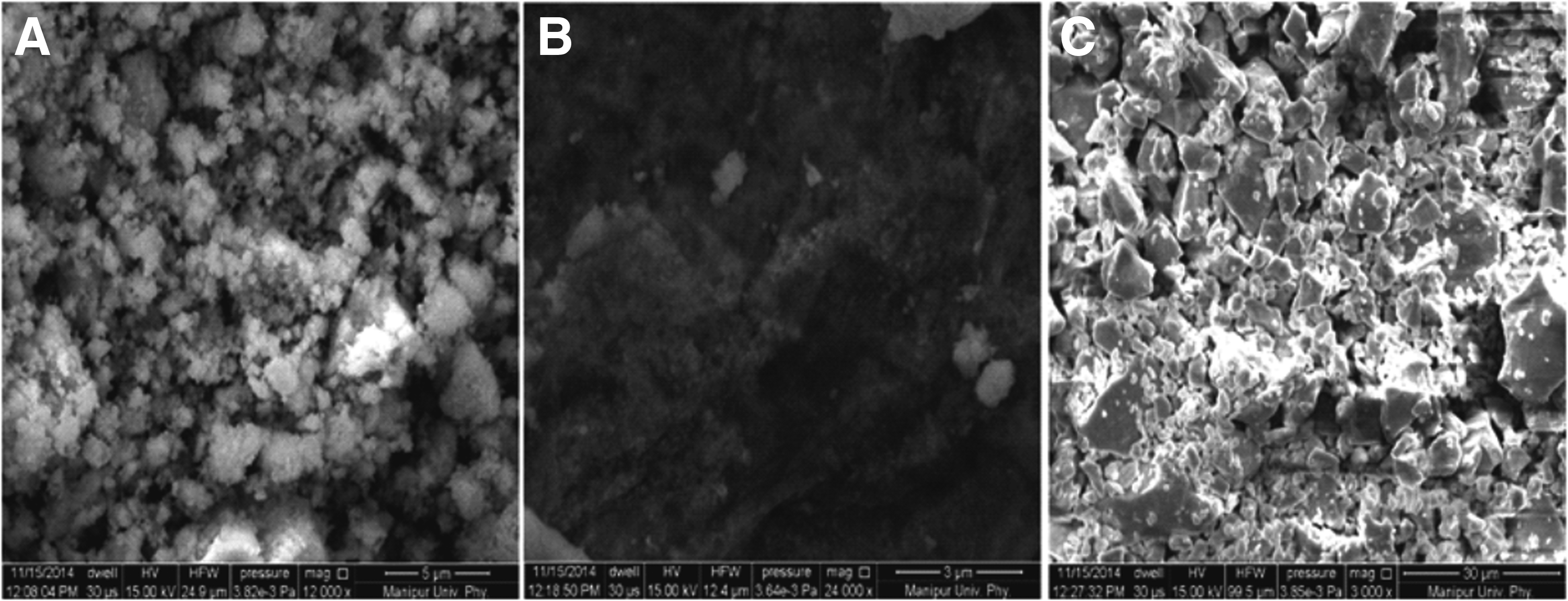
Scanning electron microscopy images of Ag NPs prepared in
Growth kinetic of Ag NPs in different solvent is given in Fig. 5. Particles of Ag grow spontaneously in water and ethylene glycol, but growth rate of Ag in ethyl acetate medium is very slow as given in Fig. 5D. Growth analysis of Ag NPs in different solvents clearly indicates that ethyl acetate did not favor the growth of NPs. The mechanism for the formation of different shape and morphology of Ag NPs may be due to variation in the growth rate at different planes of the particles and the particle growth competes with the coordinating action of the stabilizers (Petroski et al., 1998; He et al., 2002). Chemical components of leaf extract of NA may have interacted with different solvents in different manner and solvents also have different complexing abilities with Ag ions. IR spectra of leaf extract of NA in different solvents were recorded and observed that component of leaf extract interacted with solvents in different manner as given in Fig. 6A. IR spectra of leaf extract are composed of C-H stretching at 2,994 and 2,897 cm−1, C=C aromatic stretching at 1,559 cm−1, a broad and weak peak at 1,390 cm−1 is of C-O-H, and bands at 1,236, 1,059, and 1,010 cm−1 can be attributed to C-O stretching (Pavia et al., 2015). The peak located at around 2,357 cm−1 was attributed to the N-H stretching vibrations or the C=O stretching vibrations. N-H stretching of peptide linkages present in the proteins of the extract can be assigned to the broad band around 3,410 cm−1. Figure 6B presents the IR spectra of Ag NPs and it composed of IR stretching in region of 1,559 to 1,000 cm−1. These IR stretching can be assigned to the C=C, C-O-H, and C-O. From which it can be manifested that polyphenol, including tannin and flavonoids, and protein are present in metal sample and stabilized the particles from further changes like aggregation (Singh et al., 2014; Henam et al., 2015).
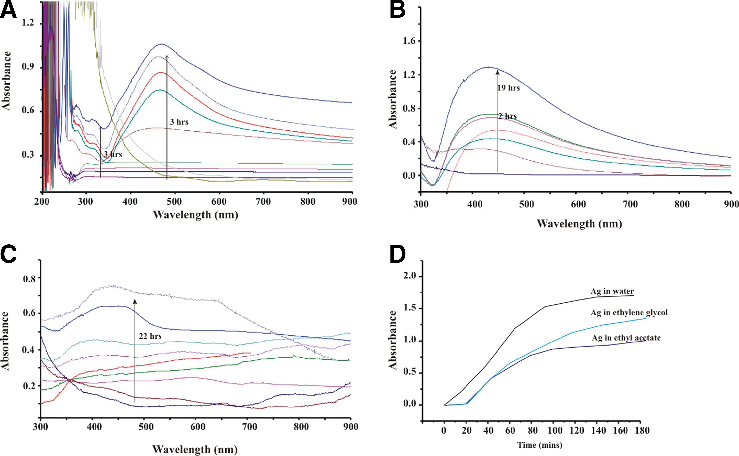
Growth kinetics of Ag NPs

IR spectra of
XRD further confirmed the crystalline nature of the Ag particles; diffraction pattern of Ag NPs prepared in water, ethylene glycol, and acetyl acetate medium is given in Fig. 7A–C, respectively. Particles of Ag NPs in water, ethylene glycol, and ethyl acetate show characteristic diffraction peak at 2θ values of 38.20, 38.35, and 38.25, respectively, showing face centered cubic crystal structure (Mittal et al., 2014; Vidhu and Philip, 2014) as given in Fig. 6. AgW NPs show additional peak of hexagonal phase as index in Fig. 7, and (110) plane of faced centered cubic (FCC) is more prominent than (111) plane. While AgE NPs show only one peak with average intensity, this proved that the growth of NPs is not favored in the ethyl acetate medium (Petroski et al., 1998).
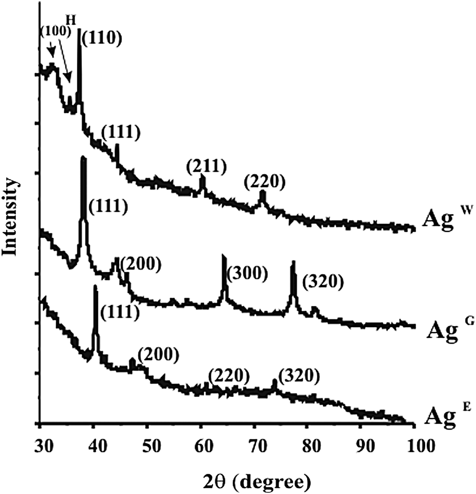
X-ray diffraction pattern of Ag NPs prepared in water, ethylene glycol, and ethyl acetate medium.
Catalytic activity of fabricated NPs of Ag was studied using m-cresol as role model organic pollutant. m-cresol shows absorbance peak at 271 nm; photodegradation and photocatalytic degradation of m-cresol were monitored at this wavelength (Yadollah et al., 2011). Photodegradation of m-cresol is given in Fig. 8A and it is also negligible. Ag NPs prepared in different medium have distinct catalytic potential. AgW NPs completely degraded m-cresol in 93 min as shown in Fig. 8B. AgG and AgE particles are able to reduce 96.2% and 60% of m-cresol in 146 and 180 min as given in Fig. 8C and D. Degradation of m-cresol by Ag NPs follows first order kinetic, and plot of ln A versus time is given in Fig. 9. Rate constant determined from the plot of ln A versus time is listed in Table 1. Interactions of Ag NPs with m-cresol were studied using IR spectroscopy (given in Fig. 10). To understand the nature of interaction between Ag NPs and m-cresol, m-cresol treated with Ag NPs is studied using IR spectroscopy. IR of m-cresol composed of OH stretching at 3,329 cm−1, =C-H aromatic stretching at 3,037 cm−1, asymmetric and symmetric stretching of CH3 at 2,923 and 2,858 cm−1, and C=C stretching and bending at 1,589, 1,489, 1,264, and 926 cm−1. C-O stretching was at 1,088 and 1,153 cm−1, while bands at 1,002, 8,510, 772, 732, and 687 cm−1 can be attributed to =C-H bending (Pavia et al., 2015). Most of these bands are shifted (as index in the figure.) when treated with Ag NPs as shown in Fig. 10B–D, which signifies the strong interaction between m-cresol and Ag NPs. Degradation of m-cresol might have proceeded through two reaction pathways, that is, H atom abstraction from C-H or O-H and addition to aromatic rings. Ag NPs react with reactive dissolved oxygen and result in generation of reactive oxygen species like OH radicals and peroxide. OH radicals react with aromatic compounds by attacking a ring carbon with its unpaired electron and forming a C-O bond, while resulting in formation of hydroxyclohexadienyl type radicals (Fig. 11A). Abstraction of H from O-H and C-H bond results in formation of a radical and water molecule. Abstraction of H from O-H produces m-methylphenoxyl radicals (Fig. 11B), whereas abstraction of H from C-H of methyl group results in formation of m-hydroxybenzyl radical as given in Fig. 11C (Arzu et al., 2004). Overall possible degradation mechanism of m-cresol is stated in Fig. 12, and it is also reported earlier by Hopper and Taylor (1975) and Abdollahi et al. (2011).
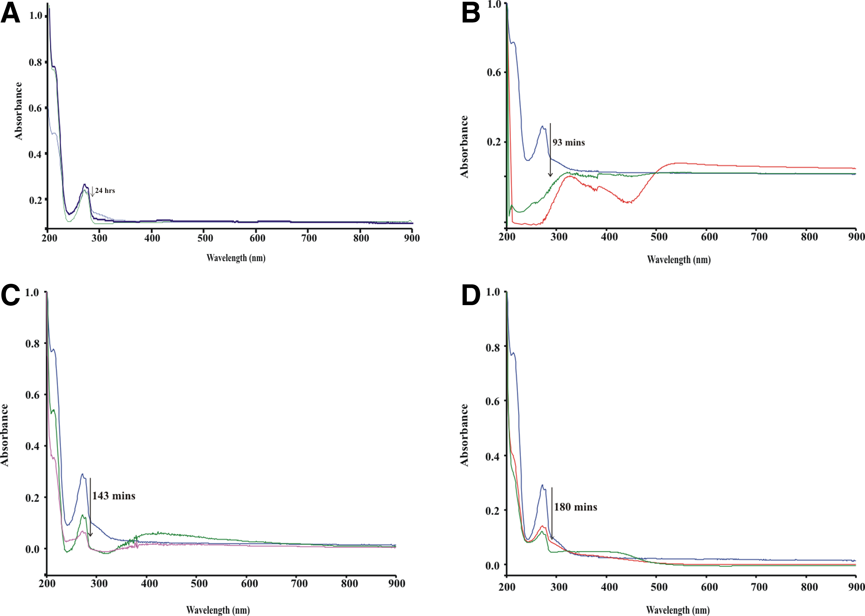
UV-Vis spectra of photocatalytic degradation of m-cresol

Kinetic plot of ln A versus time for the degradation of m-cresol.
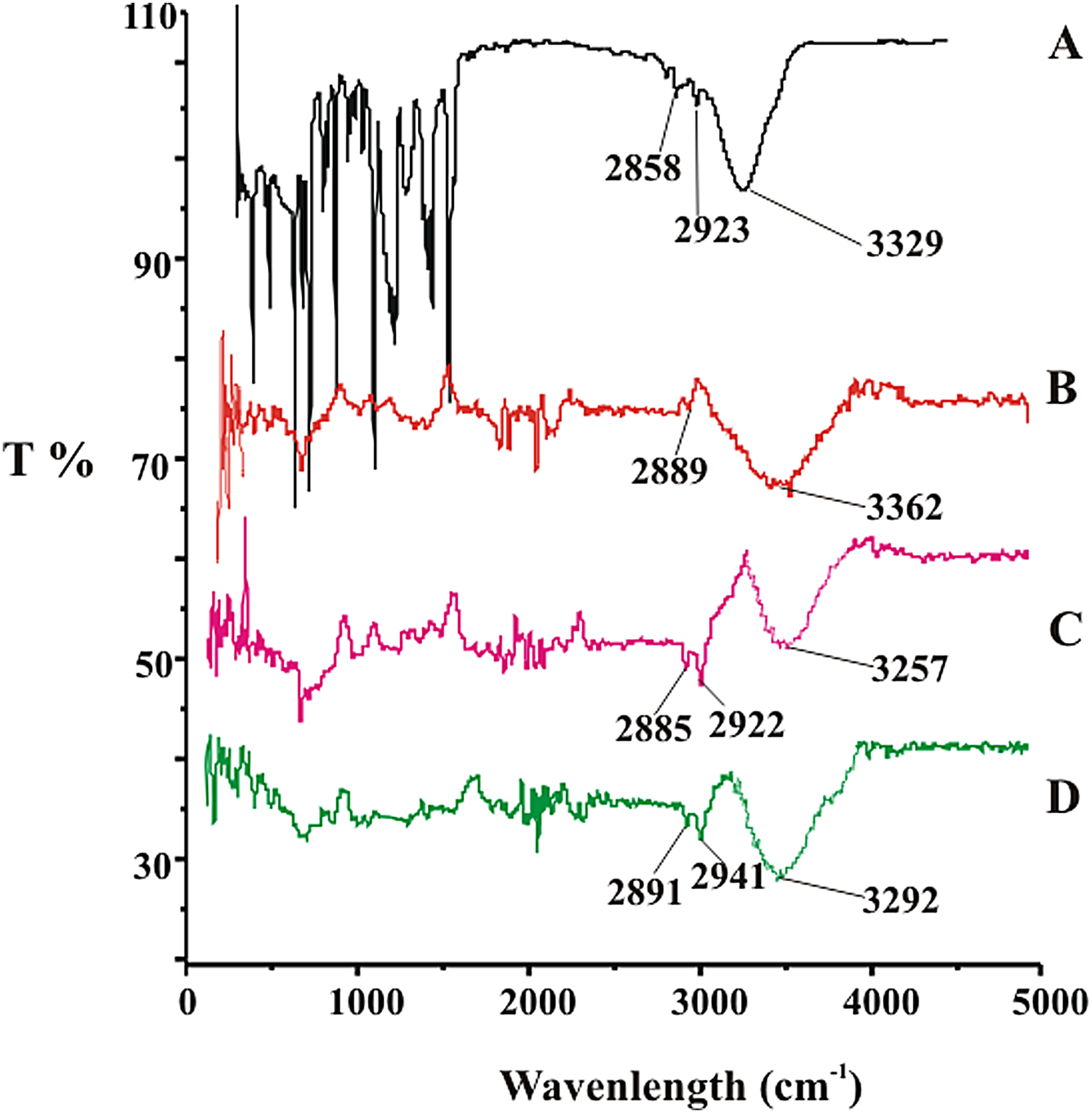
IR spectra of
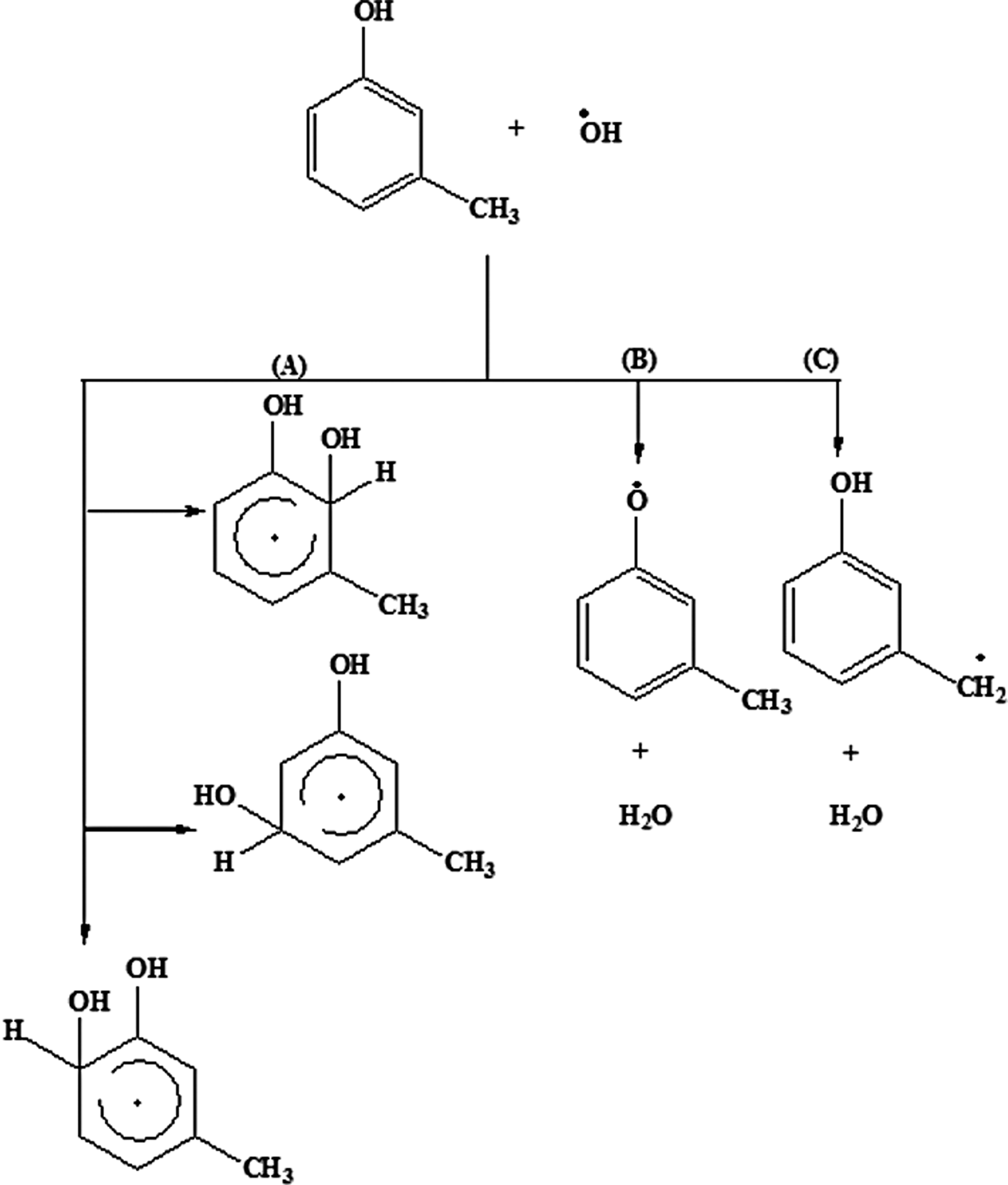
Possible pathways for the photocatalytic degradation of m-cresol.
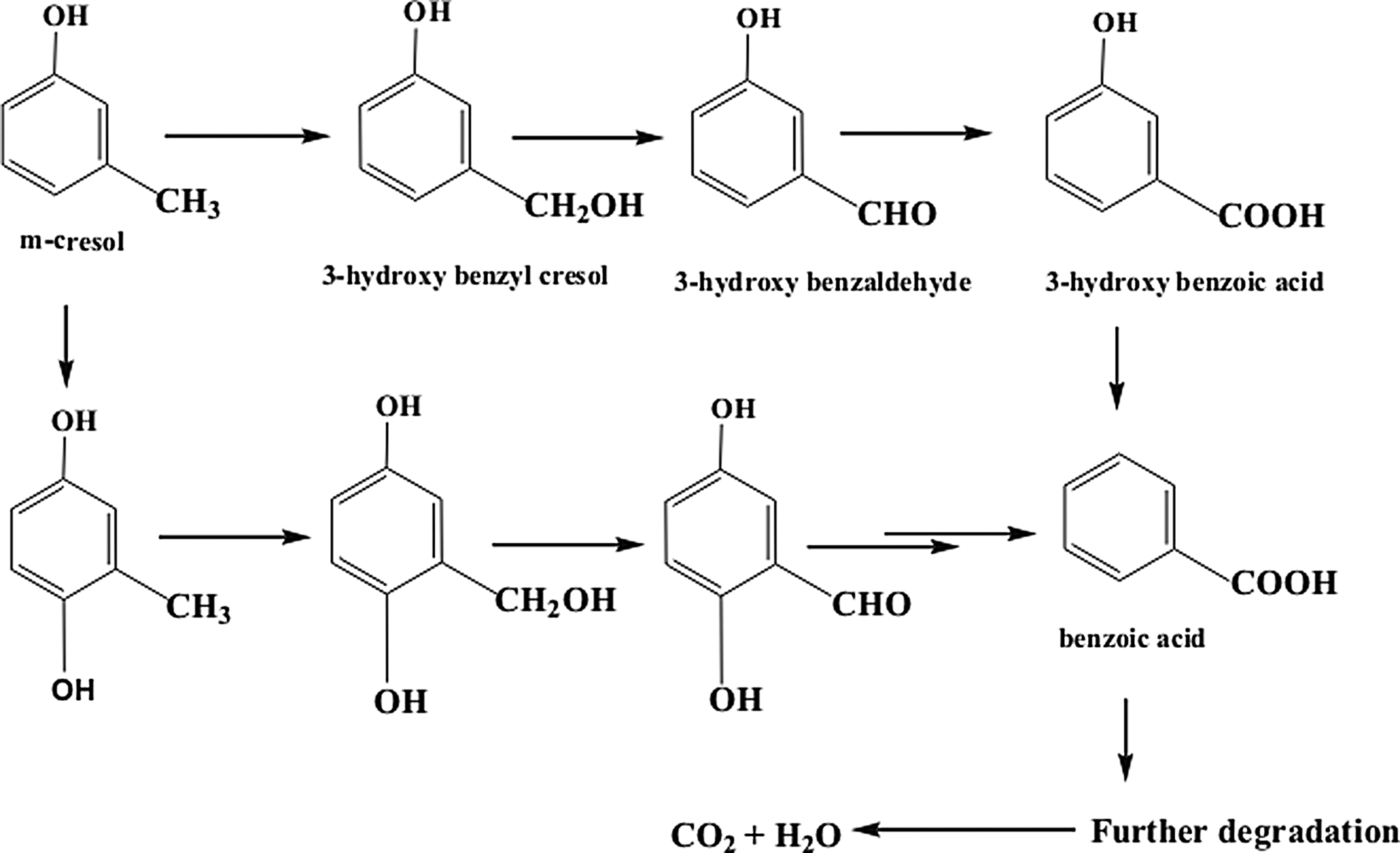
Degradation mechanism of m-cresol.
NPs have excellent catalytic properties in chemical reactions comparing with bulk catalysts in many cases (Beatriz, 2010). Their catalytic behavior varies with sizes, shapes, structures, and solvents (Xie et al., 2013). Interaction of metal atoms and solvent on the surface of NPs changes the electronic properties and geometric structures of NPs and indirectly affects their catalytic properties (Hou et al., 2015). The catalytic performance of NPs largely depend on the exposed facets; the catalytic activity can be enhanced with high-index facets and moreover shaped NPs usually have greater catalytic potential than flat NPs (Li et al., 2002; Narayanan and El-Sayed, 2004; Tian et al., 2008; Li and Liu, 2011; Xie et al., 2013; Zhang et al., 2013). From finding obtained from XRD result, it is confirmed that in AgW NPs, (110) plane of FCC is exposed. In case of AgG and AgE NPs, the most stable plane of FCC (111) is more pronounced. The (110) plane is more reactive for the catalytic properties than (111) plane because of its high surface energy (Kazumi et al., 2014). Surface energy (
surface energy of (111) and (110) plane calculated using above equation (i) are
Where ΔHS = molar enthalpy of sublimation, NA = number of atoms in one mole crystal and a0 = edge length
It can be concluded that surface energy of (110) plane is higher that (111) plane. Nanocrystals of high surface usually possess a high density of atomic steps, edges, and kinks, which can serve as catalytically active sites. Because of high surface energy, the work functions of the FCC planes is in the following order Φ(111) > Φ(100) > Φ(211) > Φ(123) > Φ(310) > Φ(110) (Jian and Shao-Qing, 2014). Work function of (111) plane is high so its catalytic potential is low compared with (110) plane with low work function value. Because of this reason catalytic activity of AgW NPs with exposed (110) plane has higher catalytic property than AgG and AgE NPs with stable (111) plane.
Conclusion
Silver particles of different shape and size particles were successfully fabricated using leaf extract of NA under ultrasonic radiation in different solvents. Ultrasonic radiation maintained the size of Ag particles in nanoscale thereby preventing growth of particles. Variation in shape and size of the NPs may be due to the different complexing ability of Ag ions with leaf extract in different solvents. NPs of silver possess good catalytic activity toward degradation of m-cresol. AgW NPs completely degrade m-cresol in 93 min, while AgG and AgE NPs consume more time and have low catalytic efficacy. Degradation of m-cresol by Ag NPs followed first order kinetic and calculated rate constant value following the order—kAg water > kAg ethylene glycol > kAg ethyl acetate. Dominating catalytic activity of AgG NPs can be attributed to its facet structure, the high atom density of facet NPs, and high surface energy. This protocol for the degradation of m-cresol is an environmentally benign method and there is no use of harmful reducing agents, but easy, safe and cost-effective.
Footnotes
Acknowledgments
The authors are thankful to SAIF, NEHU for recording TEM images and Department of Physics, Manipur University for allowing them to take SEM images and XRD.
Author Disclosure Statement
No competing financial interests exist.
References
Supplementary Material
Please find the following supplemental material available below.
For Open Access articles published under a Creative Commons License, all supplemental material carries the same license as the article it is associated with.
For non-Open Access articles published, all supplemental material carries a non-exclusive license, and permission requests for re-use of supplemental material or any part of supplemental material shall be sent directly to the copyright owner as specified in the copyright notice associated with the article.