
Abstract
Abstract
Layered double hydroxide (LDH)-based nanocomposites, fabricated by interacting LDHs with organic anions, are an emerging and active area in healthcare, environmental remediation, catalysis, and storage. Ciprofloxacin, sulfanilamide, and oxazolidinone, which exhibit antibacterial activity, are successfully intercalated in Zn-Al LDHs through anion exchange method. Synthesis of the precursor was confirmed by X-ray diffraction (XRD), transmission electron microscopy (TEM). Intercalation was successfully evaluated from XRD data. It has been found that both the nature and position of aromatic ring substituents effect the value of the basal distance and the host–guest hydrogen bond network. Evidence on interactions of intercalated anions with the inorganic layer has been confirmed from Fourier transform infrared absorption spectroscopy. Morphology of composites was studied by field emission scanning electron microscopy, and thermal stability was studied by thermogravimetry and differential thermal analysis. The novel nanocomposites demonstrated antibacterial activity against both Gram-positive (Staphylococcus aureus) and Gram-negative (Escherichia coli) bacteria. The designed organic–inorganic fusions may offer a promising antimicrobial nanomaterial for varied applications.
Introduction
I
Layered double hydroxides (LDHs) can act as a base material because they are smart nanocontainer materials that serve as reservoirs and delivery carriers of functional molecules (Li et al., 2006; Hoyo, 2007; Sokolova et al., 2008). LDHs, which are also called as anionic (anion exchanging) clays, are potential candidates to form inorganic/organic composite materials based on the incorporation of the organic species into the interlayer space. The stacked sheets are held together by hydrogen bonding, and the exchangeable anions are accompanied by water molecules, which lead to incorporation of a large range of organic guest (Lakraimi et al., 2006). Depending upon the nature and character of the intercalated species the properties and the applications of the LDH composites differ accordingly. A wide range of applications for immobilization (Aisawa et al., 2007; Costantino et al., 2008, 2009; Bugatti et al., 2011), sensors (Mousty et al., 2007; Chen et al., 2008; Vial et al., 2008; Vreysen et al., 2008; Lopez et al., 2010), photoluminescence (Sun et al., 2007, 2010; Chen et al., 2010; Tanaka et al., 2010), and flame resistance (Zammarano et al., 2005; Costa et al., 2007; Zou et al., 2007; Ding et al., 2009; Zhang et al., 2008; Zhang et al., 2010) are the current area of research. LDHs are capable of protecting the accommodated species from light, oxygen, and water vapor effects (Silion et al., 2008) and, thus, are immensely reliable for application in pharmaceutical formulations and the sustained releases of drug delivery. Drugs such as antitumor drug podophyllotoxin (Qin et al., 2010), anti-inflammatory drug fenbufen (Li et al., 2004), and antibacterial drug cefazolin (Ryu et al., 2010) are intercalated so far for controlled release formulation. This anion exchange property can also be extended to agro-industries for intercalation of pesticides so as to remove the excess of pesticides from the soil and ground water, for instance 2,4-dinitrophenol and 2-methyl-4,6-dinitrophenol (Chaara et al., 2010), 2, 4-dichlorophenoxyacetate (Lakraimi et al., 1999), humic and fulvic acid (Vreysen et al., 2008), and atrazine (Hoggan et al., 2009). However, only a few studies on the application of antibacterial LDH nanocontainer have been reported (Costantino et al., 2009; Ryu et al., 2010).
In the present work antibacterial LDH composites were prepared by intercalating some synthetic antibacterial reagents like ciprofloxacin, sulfanilamide, and oxazolidinone into the layers of Zn-Al LDH. A study of the intercalation chemistry and the corresponding antibacterial activities is presented in this study.
Experimental
Materials
All the chemicals used were of analytical reagent grade without any further purification. Zinc sulfate (ZnSO4.7H2O), aluminum sulfate [Al2(SO4)3.16H2O], and sodium hydroxide (NaOH) were purchased from Merck. Organic reagents used for intercalation are ciprofloxacin, sulfanilamide, and oxazolidinone, which were purchased from Spectrochem chemicals, India. The bacterial strains used in this work are Escherichia coli (MTCC 739, Gram negative) and Staphylococcus aureus (MTCC 96, Gram positive).
Methods
Preparation of Zn-Al LDH precursor
LDH having Zn/Al molar ratio 2 (denoted as SO4-LDH) was prepared by coprecipitation method, ∼14.37 g of ZnSO4.7H2O and 7.88 g of Al2(SO4)3.16H2O was dissolved in 50 mL of distilled water. 1 M of NaOH solution was added to the above solution under vigorous stirring to maintain a constant pH of 10.0. The whole precipitation was carried out under N2 atmosphere to avoid carbonate intercalation. The white precipitate which was formed was allowed to age for 4 h at 150°C. Then it was washed extensively with deionized water and dried at 40°C under vacuum.
Preparation of organic-LDH matrices
The intercalation compounds were obtained through anion exchange between sulfate ion of precursor LDH and anionic organic compound in a mixture solution of water: organic solvent. Approximately 0.5 g of finely ground LDH precursor was dispersed in 100 mL of water: ethanol mixture solution containing 0.7 g of organic compound under vigorous stirring both at room temperature and at 60°C to check the effect of temperature on intercalation. Both the experiments were carried out under N2 atmosphere to avoid carbonate intercalation. The pH of the exchange medium was adjusted by slow titration of 1 M NaOH until pH 10.0 was achieved. The mixture was left under stirring for 24 h. The resulting product was collected by washing the precipitates thoroughly with deionized water and ethanol and was dried at 40°C for 24 h. Finally the antibacterial activity was studied with all the samples by spot inoculation method.
Characterization
Powder X-ray diffraction (XRD) patterns of the as-prepared precursor and the composite samples were investigated by X-ray diffractometer with model no. Rigaku Ultima IV in the 2 theta range of 5° to 80° with a scan rate of 3°/min. In this study, the voltage applied is of 30 kV, and Cu Kα is used as X-ray source. The high magnification morphological analysis was carried out by transmission electron microscope (FEI, Tecnai G2; Philips) operating at 200 kV. Thermal analysis (both thermogravimetry [TG] and differential thermal analysis [DTA]) measurements were carried out by Mettler-Toledo TGA/SDTA851e thermo analyzer in the temperature range of 25–1,000°C with a heating rate of 10°C/min in air flow (50 mL/min). The surface morphology was studied through field emission scanning electron microscopy (FESEM) with model no. SUPRA 55 ZEISS. Fourier transform infrared (FTIR) spectra were recorded in the range of 400–4,000 cm−1 wave number on a FTIR spectrophotometer supplied by Perkin Elmer India Pvt Ltd., and KBr was used as a reference. The drug loading capacity was estimated using VARIAN Cary 50 Bio UV-Vis spectrophotometer.
Antimicrobial activity
Antimicrobial activity of the samples was evaluated against two test organisms, namely E. coli and S. aureus, both qualitatively and quantitatively. The qualitative test was done by spot inoculation method to evaluate the potency of each sample, whereas the quantitative evaluation of antibacterial property of the samples was done by the colony count method.
Spot inoculation method
The bacterial inoculum was prepared with pure cultures of the test organisms cultured in 5 mL nutrient broth (HiMedia) for 6 h, and the cell density was measured as OD at 600 nm. Then, 107 cells of the test organisms were inoculated in 25 mL of nutrient agar (HiMedia) at 45°C, mixed well, and poured in sterile petri plates. One milligram of each sample was then spot inoculated at well-marked places and incubated at 37°C up to 96 h. The antibacterial effects of the composites were observed in the form of inhibition zones around the spots at regular intervals after 24 h of incubation at 37°C.
Colony count method
The samples were taken in a beaker at a concentration of 1 mg/mL in 25 mL of sterile tap water with 0.85% NaCl (pH = 6.6) (to maintain the osmotic balance of bacterial cells during the experiment), and 107 cells/mL of E. coli or S. aureus was added to it. The sample beaker was covered with sterile aluminum foil and placed on a magnetic stirrer under normal room temperature and light conditions. One milliliter of sample was drawn at various intervals of time and serially diluted up to 104 times. This was followed by plating 0.1 mL of the sample on NA plates by adopting spread plate method and incubation at 37°C without illumination for 18–24 h. Another set of control was prepared without the sample in the same way as above. After incubation the numbers of colonies were counted in all the plates, and the colony forming unit per mL was calculated for each sample at different time intervals using the following formula:
CFU/mL = no. of colonies × dilution factor/volume inoculated,
where dilution factor is the reciprocal of the dilution in which the plate count was taken, and volume inoculated is 0.1 mL.
Results and Discussion
Analyses of precursor
XRD pattern of precursor and effect of aging temperature
The XRD pattern of the aged and unaged Zn-Al LDH precursor is given in Fig. 1. The patterns of the unaged and aged sample are similar to each other (Fig. 1a), which match perfectly with the standard XRD patterns of zinc aluminum hydroxide sulfate (JCPDS-44-0600).
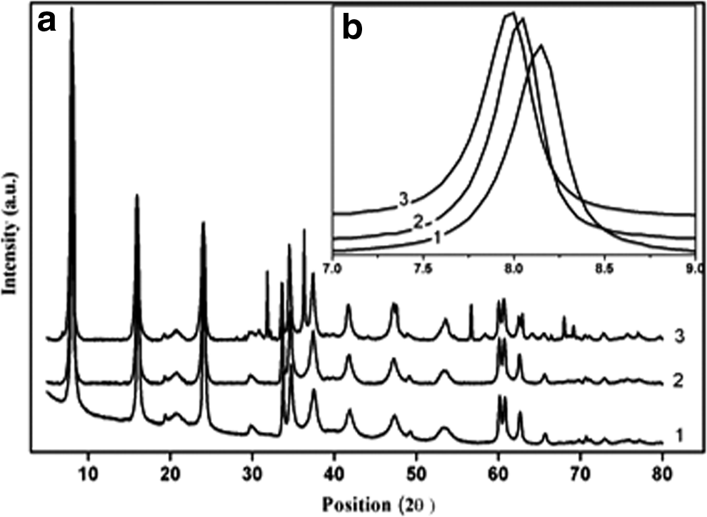
Aging increases the crystallinity along with agglomeration by eliminating the excess water from the crystal. At the closer view (Fig. 1b) the 2θ position decreases from 8.12° to 7.95° at the same time the d-values increases from 10.87Å to 11.09Å from unaged to 150°C aged sample, which is close to the standard position of 7.94° and d-value of 11.12Å.
Oswald ripening results in a wide particle size distribution (Forano et al., 2006a). The particle size distribution depicts the Oswald trend; as with aging the narrow particle size distribution peak becomes broader (Fig. 2). The crystallite size of the unaged sample as calculated from Scherrer's formula is 26.32 nm, which increases to 28.9 and 72.4 nm with aging at 80°C and 150°C, respectively (Table 1). The increase in the crystallite diameter with increase in the aging temperature supports the phenomenon that aging increases crystallinity.
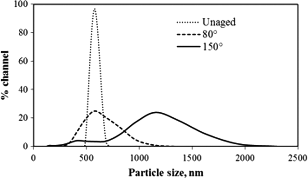
Particle size of Zn-Al LDH unaged and aged.
MCD, mean crystallite diameter.
Analysis of the micromorphology/TEM
The micromorphology and structure of the Zn/Al-LDH precursor were characterized by transmission electron microscopy (TEM) and selected-area electron diffraction (SAED). The TEM image of the Zn/Al-LDH precursor (Fig. 3a) shows typical hexagonal platelets with particle size of average 0.15 μm determined after five measurements. The SAED pattern (Fig. 3d) indicates that the Zn/Al-LDH nanoplatelets are highly crystalline; the spots can be indexed to a hexagonal structure and showing [110] and [111] planes.

TEM images of Zn-Al LDH.
FESEM study
The FESEM image shows that the LDH precursor has sheet-like morphology as represented in Fig. 4. The images demonstrate that the crystals are very thin, flat, and some are broken, and they have flake-like structures. Some of the layered crystals can be seen transversely, allowing assessment of the thickness, which is very less.
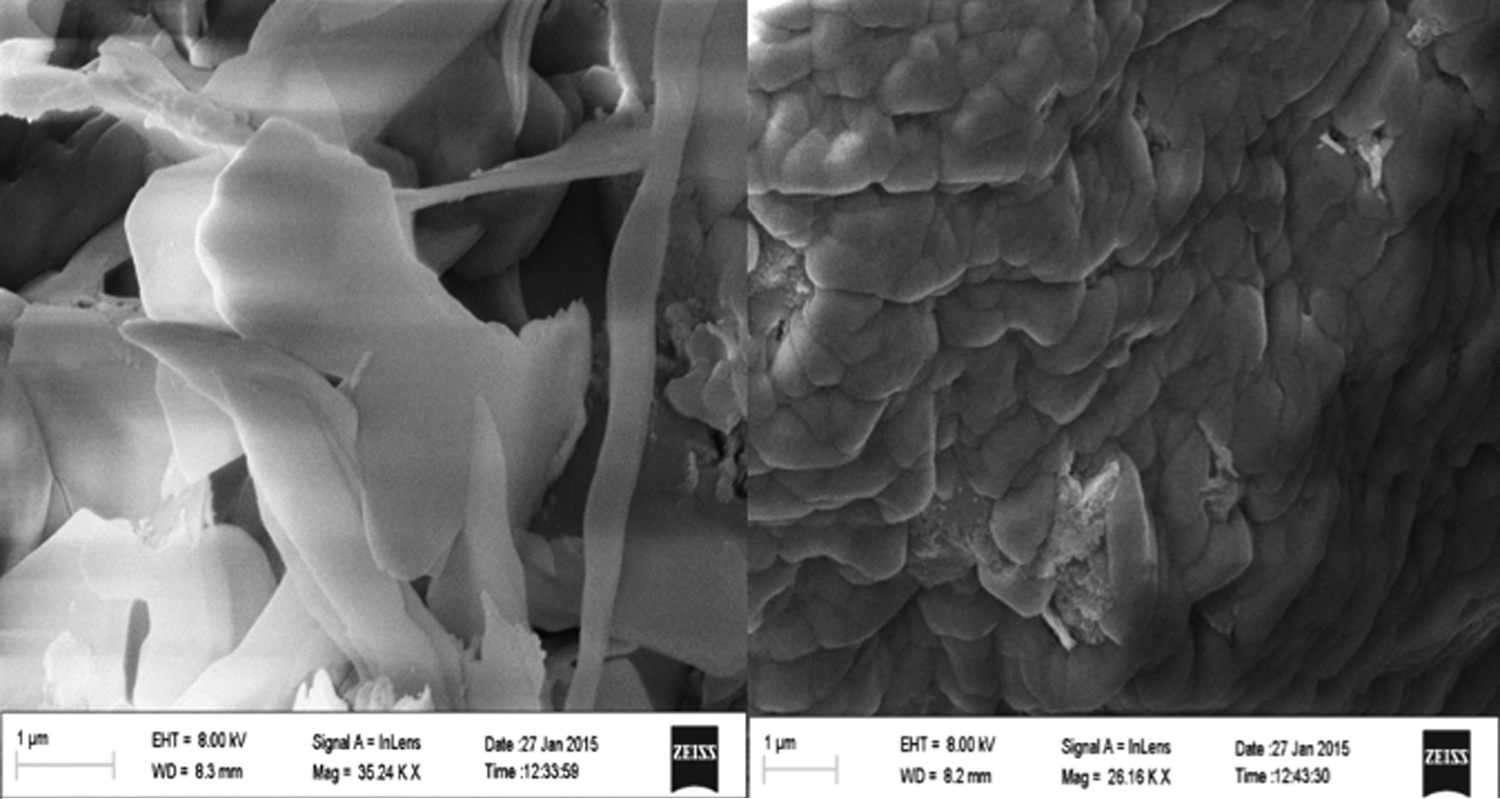
Field emission scanning electron microscopy images of Zn-Al LDH precursor.
Analyses of the composite
X-ray diffraction
The XRD patterns of the hybrid composites are presented in the Fig. 5. The basal plane spacing of the pristine LDH is 11.09Å, while the same for the organic LDH composites is 20.7 with the composites prepared at 60°C through ion exchange (Fig. 5A). With a successful intercalation there is an increase in the basal plane. Herein, the basal reflections of LDH are shifted to lower 2θ values; as anion exchange is completed, it shows the formation of organic-LDH intercalated compound. The interlayer anions are exchanged by organic molecules and after intercalation the products maintain the characteristic features of LDHs. The (110) reflection (2θ = 59°) showed no obvious shift compared with Zn/Al LDH precursor, indicating that no significant change occurred in the LDH host layers after intercalation.
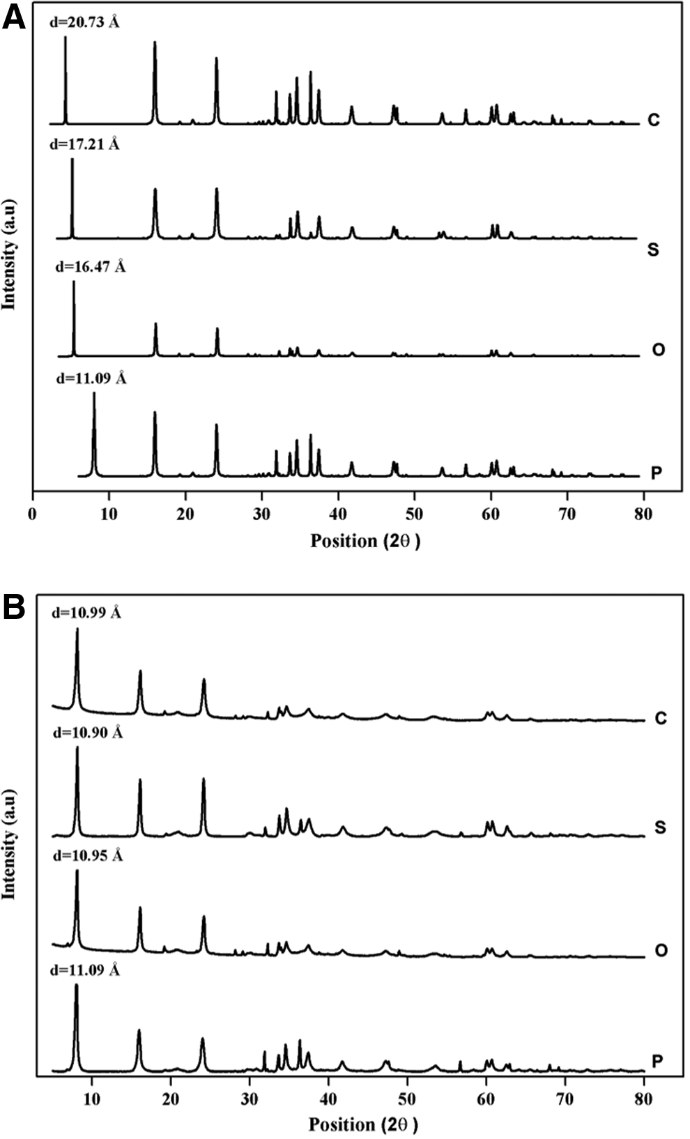
XRD images of organic composites
However, when the composites are prepared at room temperature, there is no increase in basal spacing (Fig. 5B), that is, 10.99Å, 10.90Å, and 10.95Å for ciprofloxacin, sulfanilamide, and oxazolidinone, respectively. Sometimes due to intercalation the gallery height decreases as the decrease in the d-values is generally attributed to the enhanced electrostatic attraction between the hydroxide layers and the interlayer organic species (Forano et al., 2006c). However, the values 10.90Å, 10.95Å, and 10.99Å cannot be assigned as contraction due to intercalation because the general basal plane spacing is 11Å, which is supposed to be with intercalated sulfate and alkali groups. When the alkali cations are removed by washing, the value decreases to 10.9Å (Roy et al., 2001). Sometimes contraction of the basal spacing occurs when anions are grafted (Prevot et al., 1998). Oxoanions strongly interact with the hydroxyl surface groups and a dense packing is achieved because of the symmetry compatibility between the oxoanion polyhedral and the octahedral layers (Forano et al., 2006b). From the d-values of the organic-LDH composites, the intercalation status cannot be predicted as it offers three possibilities as follows:
1. The species are not intercalated at all. 2. The species are not intercalated in the interlayers, but are adsorbed on the external surfaces. 3. The species are intercalated but lying flat or grafted.
In reference with the other characterization the correct status of intercalation can be predicted as per the predicted possibilities.
The thumb rule of intercalation is the replacement of the interlayer anions with the new entity. The amount of sulfate anion present in the interlayer is calculated to be 67.2% (0.336 g/0.5 g sample). For anion exchange the amount of intercalating substance should be taken as around double of the existing anion (Kooli et al., 1996; Costatino et al., 2009; Wang et al., 2011). Hence, the amount of ciprofloxacin, sulfanilamide, and oxazolidinone is taken as 0.7 g/0.5 sample. Ultimately the loading capacity was estimated through UV-Vis spectra and presented in Table 2. It was found that in all the three cases and in both the temperatures the loading was nearly same as the amount of sulfate. This also gives an evidence for the intercalation in room temperature category.
LDH, layered double hydroxide; RT, room temperature.
Thermal treatment
Sequences of thermal treatments are release of adsorbed water up to 100°C followed by release of interlayer water at around 250°C and dehydroxylation of the cationic bilayers and decomposition of the interlayer anions (Costantino et al., 2009; Bugatti et al., 2011; Barahuie et al., 2015). The thermal analyses are same for both the conditions, that is, at room temperature, as well as at 60°C. Results are presented in Fig. 6. The first endothermic peaks (E1, E4, and E7) appearing at around 100°C corresponds to the removal of surface water (adsorbed water) (Silion et al., 2008) and are not detected in the pristine material. The simultaneous mass loss is also reported in the thermogravimetric plot as 8.64%, 2.10%, and 7.51% (T1, T4, and T7). The second endothermic peaks at around 200°C in all the DTA curves (E2, E5, E8, and E10) represent the loss of interlayer water, which is also clear from the mass loss of the thermogravimetric plot 13.77%, 12.38%, 8.65%, and 13.47% with regions (T2, T5, T8, and T10). In DTA of the organic composites, the endothermic peaks at around 350–400°C (E3, E6, and E9) are for the decomposition of the organic species (which is actually absent in the DTA curve of the pristine material). The XRD of all the three composites at 400°C heat treatment is presented in Fig. 7. The figure exhibits the fact that the layers collapse at this temperature because of the absence of the basal peaks, as well as generation of new peaks showing partial conversion to spinel form. After the layers crumble the organics get exposed and degrades. The corresponding weight losses are 8.56%, 10.11%, and 8.69%, respectively, for T3, T6, and T9. The facts strengthen the presence of the organics inside the layers, which degrades only after the layers collapse.
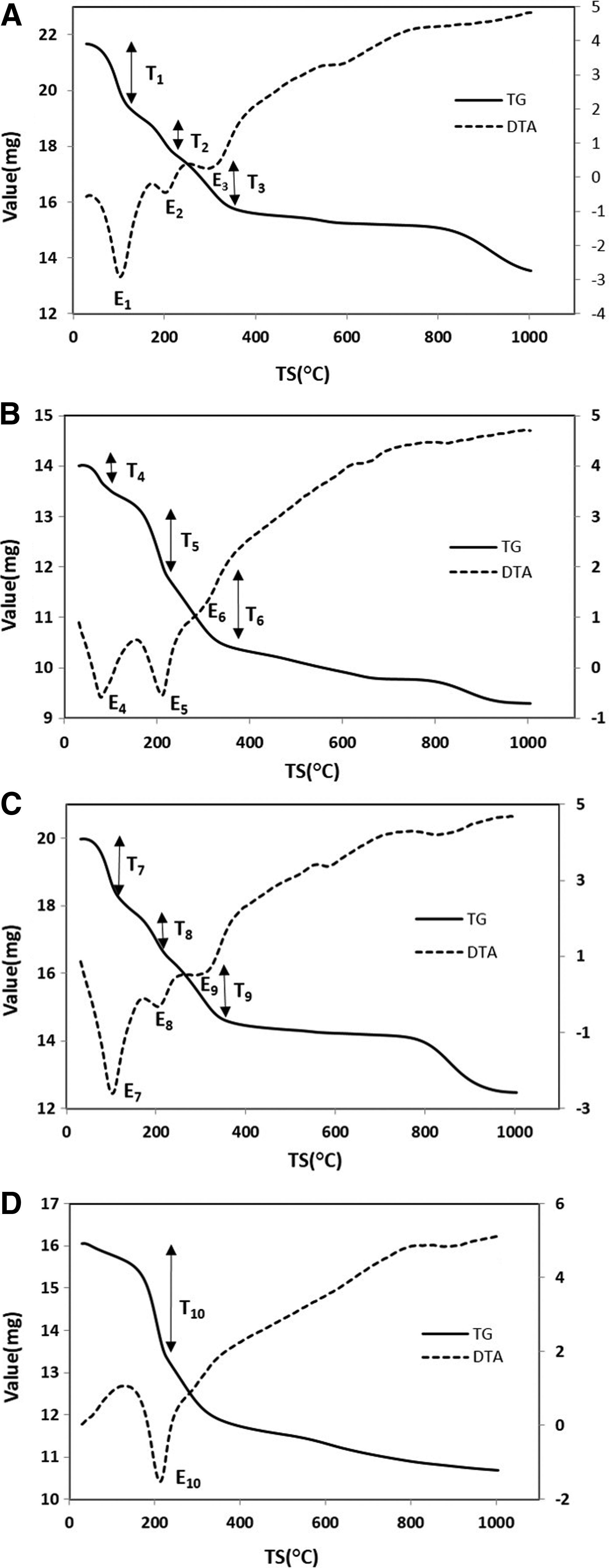
Thermogravimetry and differential thermal analysis curve of
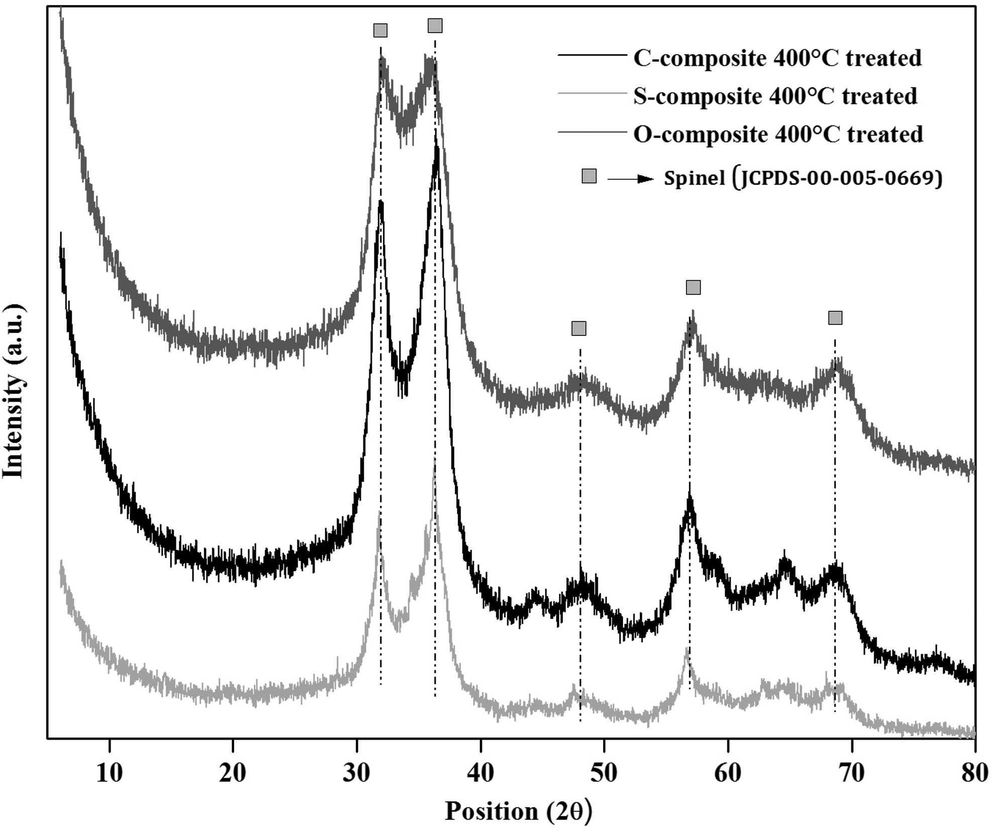
XRD of the 400°C heat treated composites.
Fourier transform infrared spectroscopy
Bands of Fourier transform infrared spectroscopy (FITR) are presented in Fig. 8. The detail description and significance of the FTIR bands of precursor, as well as all the composites, are presented in Table 3. The band at 2,437 cm−1 represents the adsorbed water (Wang et al., 2012), which is present in precursor and all the three composites, but certainly absent in the entire 100°C treated sample showing the removal of all the surface water. The band at 2,147 cm−1 of all the composites represents the interlayer water (Wang et al., 2012), which is still present after 100°C treatment and boost up the fact of intercalation.

Fourier transform infrared spectra of Pristine LDH and organic-LDH composites.
Study of release mechanism of organic anions by spectrophotometer
The release of organic anions from the composites was conducted in mild acidic solution (pH 4.0) and neutral medium (pH 7.0) taking phosphate-buffered solution (PBS) and physiological saline water as the release medium (Wang et al., 2012). Approximately 2.5 g of hybrid material was immersed in 25 mL of the release medium under stirring at room temperature (30°C) solution. Samples (5 mL) were withdrawn at predetermined intervals up to 36 h and replaced with an equivalent amount of fresh medium. The accumulated release of organic anions to the medium was measured at different wavelengths (λmax = 275 nm for ciprofloxacin, 260 nm for sulfanilamide, and 260 nm for oxazolidinone, respectively), and the release profiles are given in Fig. 9.

Release profile of organic anions from LDH matrices, acidic medium:
Figure 9 shows a faster release rate at the beginning followed by a slower rate until the equilibrium was attained (Wang et al., 2012). The release of the anion from the inorganic lamella was independent of the arrangement of the anion in between the interlayer space, which can be observed from both Fig. 9A and B. In both the cases the release rate was more or less same. The exceptionally slow sustained release of the organic anions in both the cases was ascribed due to the coordination bond between the positive bilayer units with the intercalated anions. Organic anions were strongly held to the extent that their diffusion from the interlayer into the exchange medium solution was retarded, owing to the fact that total release of the anions was not achieved (Wang et al., 2011). Based on the release profiles, the equilibrium percentage of released anions was only around 10–15%. Release mechanism was produced by anion exchange reaction, which can be described by the following reaction.
Host-guest, as well as guest, interactions are the major characteristics that affected the diffusion processes.
The slow and sustained release process may be interpreted on the basis of ion-exchange process between the guest organic anions pillared in between the host inorganic lamella, which are more stable in neutral medium rather than acidic medium.
Based on the above analyses of the two media, it can be predicted that the release mechanism of the three composites involves only ion-exchange process between the intercalated organic anions with that of phosphate, as well as chloride ions from the media, and the diffusion through the particle is the rate limiting step (Wang et al., 2011).
Structural modeling
Since the ratio of guest species: LDH precursor is similar in all the cases, we have to look into the orientations of different guests by comparing the length of each of the guest anion with the interlayer spacing. The molecular dimensions of ciprofloxacin, sulfanilamide, and oxazolidinone compound, calculated using the chemical bond lengths and atomic angles, are 7.173Å, 3.224Å, and 4.134Å along the y-axis and 12.24Å, 7.339Å, and 6.498Å along the x-axis, respectively. All the three molecules showing coordinate axes are presented in Fig. 10.
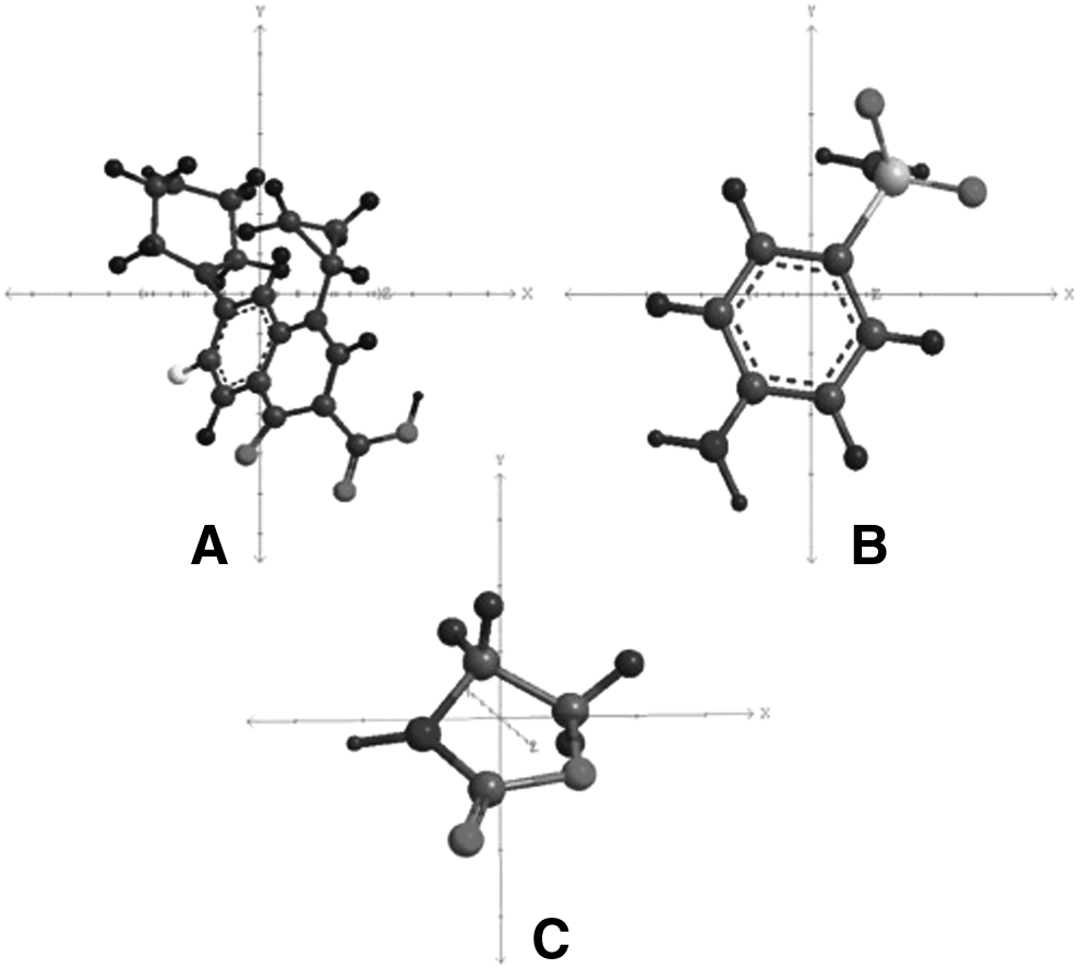
Molecules showing coordinate axes,
Considering the synthesis at 60°C temperature, according to the XRD data the intercalation of the ciprofloxacin molecule leads to an increase of the interlayer distance of the LDH sheet by 15.93Å. The obtained interlayer distance is larger than the longitudinal length of the ciprofloxacin molecule (12.24Å). Therefore unilayer arrangement of ciprofloxacin in between the inorganic lamella is not the case in this study. So bilayer arrangement can be predicted (Fig. 11A). In this study, bilayer means one molecule along x-axis and one molecule along y-axis intertwined together to form a bilayer. In contrast, the coupled organic anions as a quasi-guest ion-pair through the p–p interaction of phenyl groups are more likely to link to the LDH layer through COOH groups at the top and bottom layers of the same interlayer by electrostatic attraction and hydrogen bonds (Wang et al., 2011). In this way, the benzene planes are vertically bilayer-positioned as a quasi-guest ion-pair form in the gallery space (Park et al., 2010). In this study, the gallery height (15.93Å) is 3.483Å smaller than the bilayer arrangement (19.413Å). This may be due to the strong hydrogen bonding between highly electronegative –COOH groups of the guest anion and –OH groups of the host inorganic sheets, which attracts the positive bilayer closer or nearer to each other resulting in decrease of interlayer distance (Costantino et al., 2009).

Structural modeling of intercalation at 60°C temperature
Similarly, sulfanilamide leads to an increase of gallery height of the inorganic lamella by 12.41Å, which is larger than the longitudinal length of sulfanilamide molecule (7.339Å). Therefore the organic anion was more likely to adopt a bilayer arrangement within the interlayer space (Zou et al., 2007). The proposed orientation of the organic anion in the interlayer space was presented in Fig. 11B. The model takes into account the length of the hydrogen bonds established between the anions and the LDH hydroxyl groups.
The interlayer distance in the case of oxazolidinone composite was 11.67Å, which was larger than the length of oxazolidinone molecule along its long axis. If the guest molecule will accommodate itself in a bilayer mode in between the inorganic lamella, then the interlayer distance (11.67Å) was nearly equal to length of intertwined molecule (10.362Å). Therefore, all the three organic anions are more likely to adopt a bilayer arrangement within the interlayer space.
Considering all the characterizations as evidences in favor of intercalation at room temperature (Fig. 12), we can say that the organic anions were present in the interlayer region. From the XRD data the interlayer distance in case of ciprofloxacin composite is 6.19Å, for sulfanilamide it is 6.10Å, and for oxazolidinone composite it is 6.15Å, respectively.
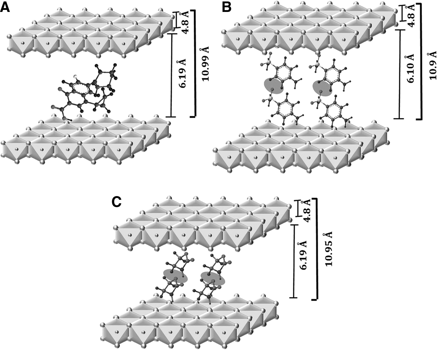
Structural modeling of intercalation at room temperature
In case of ciprofloxacin, the obtained gallery height is smaller than the horizontal length (7.173Å). Similarly for sulfanilamide and oxazolidinone composite, the interlayer distance is larger for unilayer arrangement (3.224Å for sulfanilamide and 4.134Å for oxazolidinone) and smaller for bilayer arrangement (6.448Å for sulfanilamide and 8.268Å for oxazolidinone).
Therefore calculating the dihedral angle, θ, and assuming that COO− was attached to the brucite-like layer of LDH, the length of the organic anions in the LDH gallery was 6Å (at 33.23°), 5.9Å (at 23.8°), and 5.9Å (at 16.4°), respectively, as derived from the optimized geometries. Therefore, we believe that the ciprofloxacin molecule orientation in the LDH gallery was a unilayer model (Fig. 12A) and that sulfanilamide and oxazolidinone molecule orientation in the LDH gallery were bilayer models without overlapping as shown in Fig. 11B and C, respectively.
Antibacterial activity
Ciprofloxacin is a second-generation synthetic antibacterial agent of fluoroquinolone group. The role of ciprofloxacin group is to stop the synthesis of bacterial DNA and protein. Sulfanilamide is the sulfonamide group attached to the aniline. Its role is to completely inhibit the bacterial enzymatic reactions. Oxazolidone is a heterocyclic organic compound containing both nitrogen and oxygen in a five-membered ring. The antibacterial effect of oxazolidinone includes inhibition of bacterial protein synthesis.
The antibacterial activities of the composites were carried out by spot inoculation method with two bacteria E. coli and S. aureus. In the disc diffusion test the antibacterial species containing composites were diffused in the agar medium, and the corresponding zone was obtained. The inhibition zones can be seen in Fig. 13. The antibacterial activity test results were observed for 48 h, and the corresponding decrease in zone size is documented in Table 4.
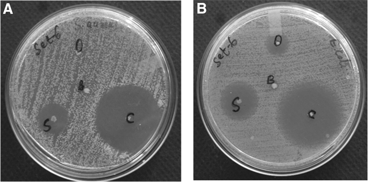
Inhibition zones of
Zone size (in mm) includes the 3 mm spot size.
For the sake of comparison the antibacterial activity of pure antibiotics was studied. The amount of sample for spot inoculations is same for all, that is, 1 mg each (Table 5). It was found that the zone sizes of the pure compounds are comparable for ciprofloxacin but lesser for sulfanilamide and oxazolidinone from the hybrid composites in Table 4. That means the 1 mg of composite contains less than 1 mg of organics, which is compared against 1 mg of pure compound, and still the zone sizes of the composites are comparable or bigger than the pure ones. Therefore the activity of the antibiotics was enhanced after intercalation.
Zone size (in mm) includes the 3 mm spot size.
The ciprofloxacin-LDH composite showed the highest inhibition activity against both E. coli and S. aureus, followed by sulfanilamide-LDH composite. However the inhibition zones of oxazolidinone-LDH composite are comparatively small in case of E. coli and faint in case of S. aureus. Although the size of ciprofloxacin-LDH composite is largest compared to the other two antimicrobial agents, its activity is very good, whereas the size of oxazolidinone-LDH composite is smallest and it showed lesser zone sizes against both the pathogens. This may be attributed to two reasons—first, the release of oxazolidinone species from the composite is very less and second the test organisms may be resistant toward the antibacterial agent.
Quantitative test results showed that ciprofloxacin incorporated LDH material reduced S. aureus counts from 107 to 0 in 5 h, whereas the same material reduced E. coli counts from 107 to 102 in 6 h. The sulfanilamide and oxazolidinone incorporated LDH had least effect on both the test organisms as shown in the Figs. 14 and 15 below.
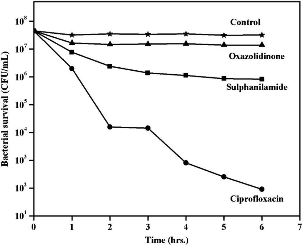
Quantitative test of antibacterial activity of LDH composite samples against E. coli.
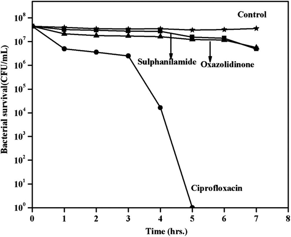
Quantitative test of antibacterial activity of LDH composite samples against S. aureus.
Conclusions
The XRD, FTIR, and TG-DTA studies show the successful intercalation of organic antibacterial reagents, that is, ciprofloxacin, sulfanilamide, and oxazolidinone in the inorganic interlayer space through anion exchange mechanism. The presence of antibacterial material makes the composite exhibit antibacterial property. The antibacterial activity was evaluated with respect to the inhibition zone size in mm. The amount of material inoculated was varied, as well as the amount of organic species added, and the antibacterial activities were found to be increasing with increase in the inoculation amount and addition amount for oxazolidinone-LDH composite and for all the three organic species, respectively. Due to strong coordination bond between drug–host lattices, the intercalated anions were slowly released, following diffusion–anion exchange mechanisms, in which diffusion from the interlayer anions was slow.
Footnotes
Acknowledgments
The authors are very much thankful to Prof. B.K. Mishra, Director, CSIR-IMMT, Bhubaneswar for his encouragement and for providing all the research facilities. The authors are also thankful to MoES, India for the financial support.
Author Disclosure Statement
No competing financial interests exist.