
Abstract
Abstract
F-N co-doped TiO2/C/ZnFe2O4 (ZCT-FN) catalyst with excellent UV-Vis photocatalytic activity and recyclable performance was successfully synthesized by the sol–gel method. X-ray power diffraction results showed that ZCT-FN was anatase titanium dioxide with good crystallinity. X-ray photoelectron spectroscopy and UV-Vis revealed that F and N were co-doped into the lattice of TiO2, and a red shift of the absorption edge was brought out owing to F and N co-doping. Comparing to undoped TiO2/C/ZnFe2O4 and TiO2/ZnFe2O4, ZCT-FN exhibited an excellent photocatalytic activity for degradation of methyl orange both under UV- and Vis-light irradiation. Visible light photocatalytic degradation activity of ZCT-FN is nearly 4.2 times higher compared with P25. The high activity of ZCT-FN can be attributed to the synergetic effects of strong absorption and narrow band gap induced by F, N co-doping and narrow band gap semiconductor ZnFe2O4. The carbon layer between outer layer TiO2 and ZnFe2O4 core can effectively suppress the photodissolution behavior of catalyst. Moreover, ZCT-FN can be easily separated after the photocatalytic reaction and remain as stable photocatalytic activity after five cycles. This work not only offers a controllable method for the fabrication of F-N co-doped TiO2/C/ZnFe2O4 core-shell structure hybrids but also provides an effective and conveniently recyclable photocatalyst for the practical application in the purification of wastewater.
Introduction
U
To overcome the reuse of TiO2 nanoparticles (NPs), a way to integrate TiO2 with magnetic NPs was proposed, which can recycle and remove the magnetic particles by external magnetic field (Yu et al., 2013). Magnetite (Fe3O4) and maghemite (γ-Fe2O3) were commonly used as magnetic cores in previous works. However, it has been demonstrated that the heat treatment has important impact on its Fe3O4 properties, especially on Fe3O4, because it may transform into the antiferromagnetic Fe2O3 phase (Belessi et al., 2009). Furthermore, Fe2O3 and Fe3O4 are unstable in acidic environment. Because of superparamagnetic behavior, large surface area to volume ratio, and high saturation magnetization (MS), MFe2O4 (M = Ni, Zn, Co) with a spinel structure have been applied in several application fields such as catalysis and environmental remediation. Among them, spinel ZnFe2O4 has been widely investigated due to its good photochemical stability, sensitivity to visible light with a relatively narrow band gap of 1.9 eV, and low cost (Fu and Wang, 2011). It was demonstrated that the ZnFe2O4/TiO2 composite nanotube arrays can sensitize TiO2 to visible light region and have an enhanced photocatalytic activity (Wang et al., 2013).
However, the direct contact of TiO2 layer with magnetic cores will reduce the photocatalytic activity because the magnetic cores may act as recombination centers for electrons and positive holes (Beydoun et al., 2000, 2002; Belessi et al., 2009), and on the other hand, the magnetic cores are sensitive and unstable in the acidic conditions. A suitable way is to insert a barrier layer between the photocatalysts and the magnetic iron oxide (Shi et al., 2012). It has been (Yu et al., 2011) reported that the SiO2 intermediate layer in the γ-Fe2O3/SiO2/TiO2 sandwich-like structure enhanced the enrichment of pollutant molecules. Recently, some researchers found that the carbon interlayer can suppress the occurrence of photodissolution (Zhang et al., 2015).
To improve the photocatalytic activity under Vis-light, much efforts have been made, including doping TiO2 with metals (Hirakawa and Kamat, 2005; Enache et al., 2006; Chen et al., 2009), nonmetals (Ohno et al., 2004; Li et al., 2005, 2014; Cong et al., 2007), and co-doping with double nonmetals such as N-S (Parida et al., 2010), N-B (Feng et al., 2011), and N-F (Huang et al., 2006). Habibi-Yangjeh and Akhundi (2016) fabricated a series of novel magnetically separable visible light-driven photocatalysts such as g-C3N4/Fe3O4/Ag2CrO4, ZnO/Ag/Ag2WO4, Fe3O4/ZnO/CoWO4, and so on. (Pirhashemi and Habibi-Yangjeh, 2017; Shekofteh-Gohari and Habibi-Yangjeh, 2017). According to our previous report (Wei et al., 2008), N-S co-doped TiO2 showed a high photocatalytic activity for degradation of methyl orange (MO) comparing to S doped TiO2 and undoped TiO2 due to the synergetic effects induced by N and S co-doping. Recently, our group prepared the N doped TiO2/ZnFe2O4 hybrid, which exhibited good degradation performance only under UV-light irradiation (Yao et al., 2015). Therefore, there are still challenges to prepare the TiO2 composites with both enhanced UV-Vis photocatalytic activity and easy recyclable performance.
In this study, we provide a facile and feasible way to synthesize a core-shell nanostructured F-N co-doped TiO2/C/ZnFe2O4 (ZCT-FN) composites (Fig. 1), which possess excellent UV-Vis light photocatalytic activity and superior magnetic responsibility. The resulting products were characterized by X-ray power diffraction (XRD), transmission electron microscope (TEM), vibrating sample magnetometer, X-ray photoelectron spectroscopy (XPS), diffuse reflectance spectra (DRS), and photoluminescence (PL) spectra. In addition, the catalytic activities of ZCT-FN for degradation of MO under UV- and Vis-light irradiation were compared with undoped TiO2/C/ZnFe2O4 (ZCT) and TiO2/ZnFe2O4 (ZT), as well as the recoverable performance was investigated. This work may be helpful for preparing novel UV- and Vis-light photocatalysts with the good photocatalytic activity and recoverable ability.
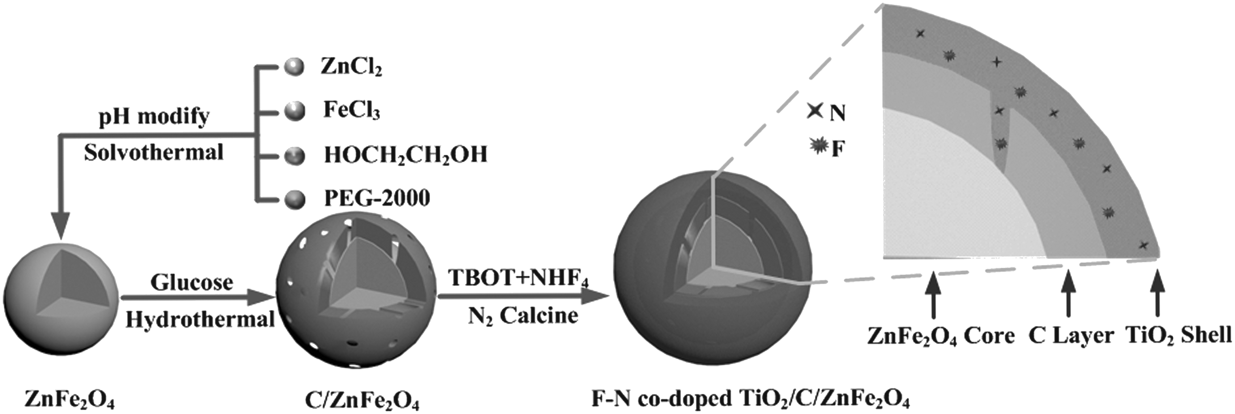
Schematic illustration of the preparation method of ZCT-FN. ZCT-FN, F-N co-doped TiO2/C/ZnFe2O4.
Experimental
Reagent and materials
Zinc chloride, ferric chloride, polyethylene glycol-2000, sodium acetate, sodium hydroxide, ethylene glycol, tetrabutyl titanate (TBOT), ammonium fluoride, ethyl alcohol, glucose anhydrous, and MO were purchased from Sinopharm Chemical Reagent Company. All of these regents were of analytical reagent grade and used without further purification.
Simple synthesis
ZnFe2O4 NPs were synthesized according to the literature (Xue et al., 2014). Core-shell structured C/ZnFe2O4 NPs were prepared through a hydrothermal reaction (Li et al., 2008; Kong et al., 2011) with minor modification. Typically, 120 mg of obtained ZnFe2O4 NPs was added to 60 mL 0.3 M glucose solution and then the flask was placed in an ultrasonic bath with stirring for 24 h. After that, the mixture was transferred and sealed into a Teflon-lined stainless steel autoclave (100 mL in capacity). The autoclave was heated at 180°C for 6 h and then cooled to room temperature. The obtained C/ZnFe2O4 was washed with ethanol and distilled water.
The synthesis of ZCT-FN photocatalysts with core-shell structure was carried out typically through the sol–gel method using TBOT as Ti source and NH4F as F, N source. First, 200 mg as-prepared C/ZnFe2O4 and NH4F (0.1088 g) were added into 70 mL ethanol with stirring. Then, the flask was placed in ultrasonic bath to completely disperse the powder. After 2 h, 10 mL TBOT was transferred to the above suspension and then 5 mL distilled water and 35 mL ethanol were added into the mix solution drop by drop. After reaction, the precursor was repeatedly washed and dried at 80°C in a vacuum oven for 12 h. The obtained product was calcined in nitrogen protection at 350°C, labeled as ZCT-FN. Undoped ZCT and ZT and pure TiO2 were synthesized by the same process only without NH4F or glucose or ZnFe2O4NPs as precursor.
Characterization
XRD was used to characterize the phase structures and particle sizes of all as-prepared samples, using a Rigaku D/max 2500 X-ray diffractometer with Cu Kα radiation (λ = 1.54156 Å) at a scan rate of 0.02°s−1. DRS of samples were performed using a Varian Cary 5000 spectrometer equipped with a Cary Lab sphere DRA-CA-301 DR accessory. The morphology and structure of catalysts were examined by high-resolution transmission electron microscope (HRTEM; JEOL JEM-2100F) and TEM (HITACHI H-800). Energy dispersive X-ray spectroscopy analysis was also performed during the HRTEM measurements. XPS was conducted using an ESCALAB 250 spectrometer. Bonding energies were standardized by assigning the C 1s peak to 284.6 eV. Magnetic properties (M-H curve) were measured at room temperature (300 K) on a superconducting quantum interference device magnetometer (MPMS XL-7) made by Quantum Design Corp. The PL spectra were measured at room temperature using Fluorolog-3-TAU fluorescence spectrophotometer with an excitation wavelength of 420 nm. Photocurrent density was measured using a CHI electrochemical analyzer (CH instruments 660e) in a standard three-electrode configuration, with the working electrode (an effective area of 2 cm2), a platinum foil as the counter electrode, and a saturated calomel electrode as the reference electrode. Na2SO4 (0.01 M) was used as the electrolyte. A 300 W high-pressure xenon lamp (CEL-HXF300) with a filter to remove light of wavelength below 370 nm was used as the visible light source.
Photocatalytic performance measurement
The performances of the as-prepared catalysts were evaluated in photodegradation of MO aqueous solution. The experiments were implemented in a Pyrex cylindrical photoreactor, which included mainly five parts: 25 W mercury lamp (UV source) or 300 W xenon lamp (Vis source); the reactor with two-layer Pyrex glass bottles, the space between the two layers filling with the circulated water to control the solution temperature; magnetic rotor, and air-lift agitator. Before the experiment under UV- or Vis-light irradiation, the desired MO solution in the presence of the catalyst was first stirred in dark at 25°C for 30 min to reach the adsorption–desorption equilibrium. Afterward, the photodegradation was started by illuminating the reactor with magnetic stirring and air bubbling. Subsequently, aliquots (4.0 mL) were drawn at varying irradiation time, and the concentration was examined by a 722P spectrophotometer at 464 nm. To evaluate photocatalyst stability, ZCT-FN was magnetically separated, washed, dried, and reused for the next cycle under the same conditions. The photocatalytic reaction rate constants (k) used the pseudo-first-order model were as expressed as Eq. (1):
where C and C0 were the actual and initial concentration of MO solution, respectively; k is the apparent reaction rate constant.
Results and Discussion
X-ray power diffraction
Crystallinity and structure of as-prepared samples were confirmed by XRD. Figure 2 shows the XRD patterns of ZnFe2O4, ZC (ZnFe2O4/C), ZCT, and ZCT-FN. It can be seen that the ZnFe2O4 sample obtained has highly crystalline cubic spinel structure, agreeing well with the standard XRD pattern of ZnFe2O4 (JCPDS#22-1012). By comparison with ZnFe2O4, there are no new diffraction peaks in ZC after coated with a carbon layer, which suggests that the carbon layer is likely in an amorphous state (Jiang et al., 2016), and ZnFe2O4 NPs are still highly crystalline after the carbon coating. After coated with TiO2 layer and calcination, ZCT and ZCT-FN samples have the strong and sharp characteristic peaks of TiO2, agreeing well with the standard anatase TiO2 XRD pattern (JCPDS#21-1272). However, there are no diffraction peaks of ZnFe2O4, which suggests that most of ZnFe2O4 NPs are coated with TiO2 layer (Li et al., 2015). Moreover, no additional phases are evident from the XRD patterns.
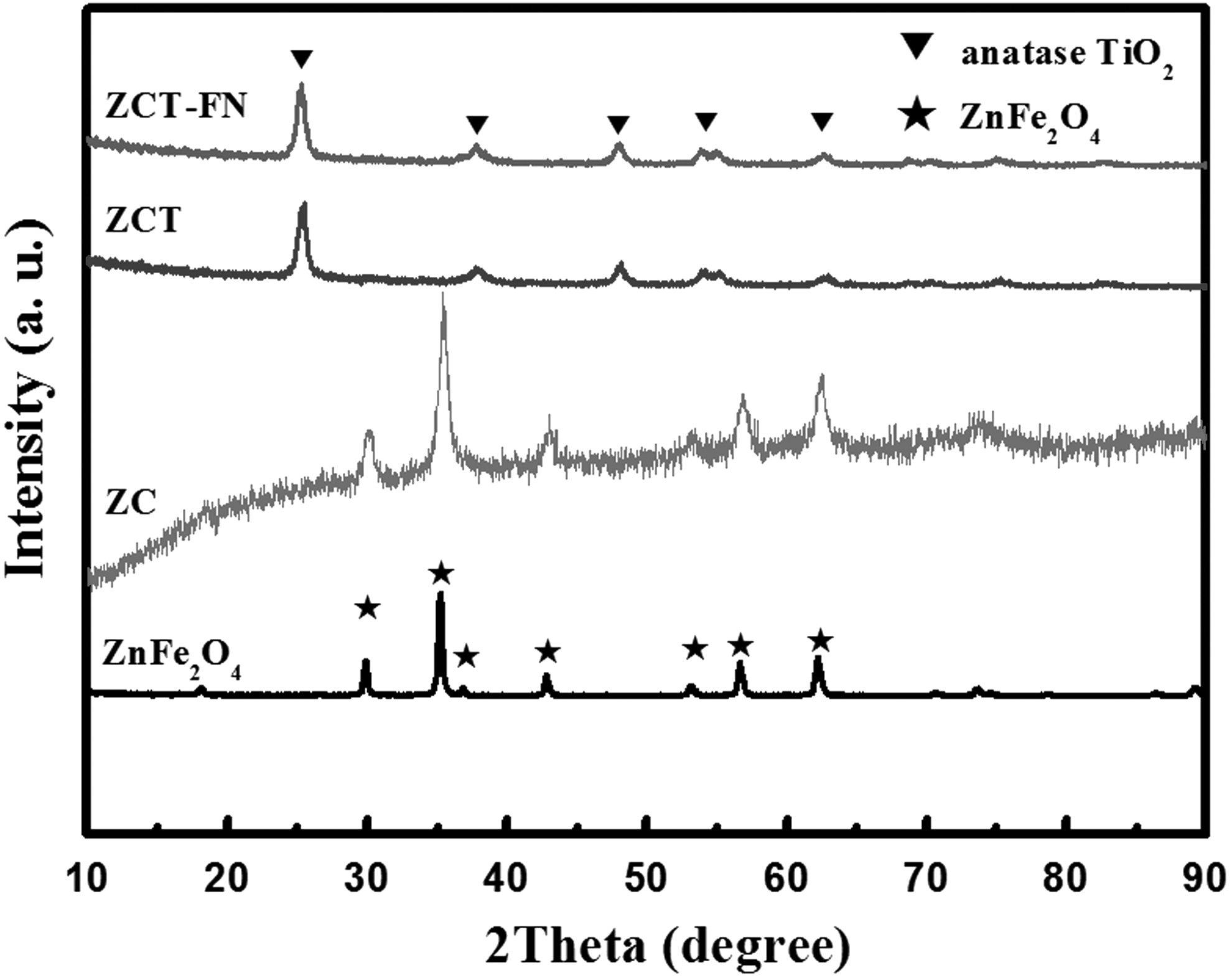
X-ray power diffraction patterns of samples. ZC, ZnFe2O4/C; ZCT, TiO2/C/ZnFe2O4.
Transmission electron microscope
Figure 3a–c shows TEM images of samples. In Fig. 3a, the ZnFe2O4 NPs have spherical shape with a diameter of about 120–150 nm. As shown in Fig. 3b, it is obvious that a uniform carbonaceous layer is coated on ZnFe2O4 microspheres, and the upper right insert image of Fig. 3b indicates that the thickness of the carbon layer is about 5–10 nm. After the sol–gel process and calcination, the average diameter of (ZCT-FN) is about 320–450 nm, and the average thickness of the TiO2 layer is about 100–150 nm. The HRTEM of ZCT-FN (Fig. 3d) shows that two different lattices are observed with d spaces of 2.50 Å and 3.50 Å, corresponding to the (311) plane of ZnFe2O4 and the (101) plane of anatase TiO2, respectively, which are quite similar to the literature values (Li et al., 2012).

Transmission electron microscope images of
Vibrating sample magnetometer
Magnetic properties of the samples were studied by MPMS XL-7 magnetometer measured at 300 K as shown in Fig. 4. The MS values of ZnFe2O4, ZC, and ZCT-FN are 77.5, 33.9, and 12.8 emu/g, respectively. It indicates that all samples have superparamagnetic behavior. The magnetization curve and demagnetization curve are coincidence, no hysteresis phenomenon is found, and remnant magnetization and coercivity are equal to zero. But the hysteresis loops of ZC and ZCT-FN are weaker compared with ZnFe2O4, and it reveals that the coated layers affected the magnetic properties of ZnFe2O4 microspheres. Fortunately, the maximum MS of ZCT-FN (12.8 emu/g) is enough strong to be separated from solution with the help of an external magnetic force.
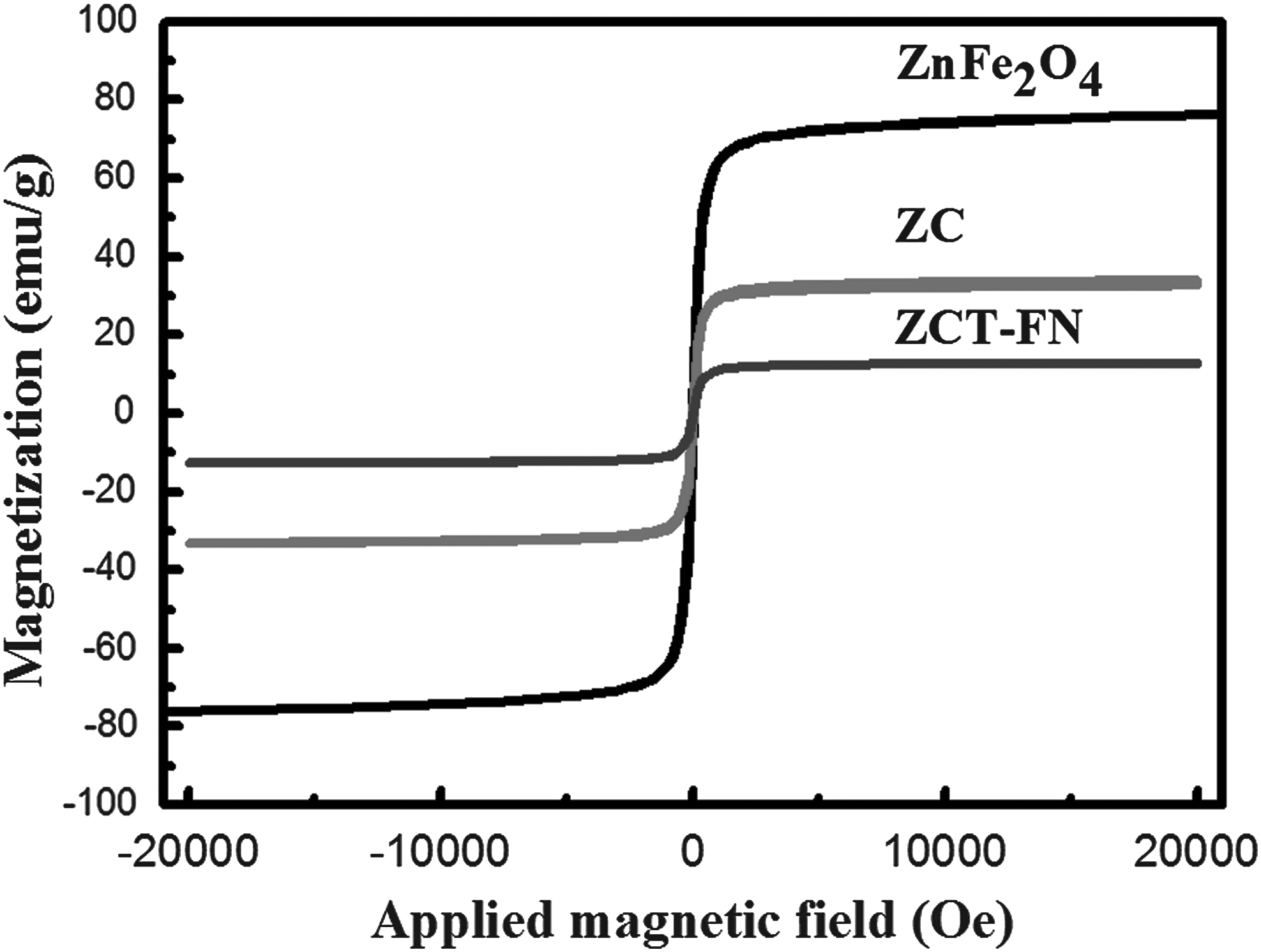
Vibrating sample magnetometer hysteresis loops of samples.
X-ray photoelectron spectroscopy
Surface composition of ZCT-FN samples was also analyzed using XPS. Figure 5a gives the whole XPS survey spectrum, which reveals the coexistence of elements C, N, Ti, O, F, Fe, and Zn. For the high-resolution XPS spectra for C 1s (Fig. 5b), four different peaks centered at 284.5, 285.6, 286.6, and 288.4 eV are assigned to C C/C–C in aromatic rings, C–O, C–OH (epoxy and alkoxy), and CO groups, respectively (Min et al., 2011). For the N 1s XPS spectrum in Fig. 5c, two peaks at 398.3 and 399.8 eV can be assigned to O-Ti-N linkages in the TiO2 lattices and NO bond, respectively (Min et al., 2013; Asahi et al., 2001; Pelaez et al., 2009; Camillo et al., 2012). From Fig. 5d, the F 1s XPS spectrum appears two peaks at 684.5 and 688.5 eV. The stronger peak located at 684.5 eV corresponds to F− ions physically adsorbed on TiO2 (Hattori et al., 1999), and the weak peak located at 688.5 eV is assigned to F− ions in Ti-F bonds (Lin et al., 2005; Yang et al., 2013). It is indicated that small amount of F− could be doped into the lattice or on the surface of TiO2 because of the NH4F decomposition after calcination (Li et al., 2010). Briefly, the characterization analysis proves that ZCT-FN hybrids are successfully synthesized.
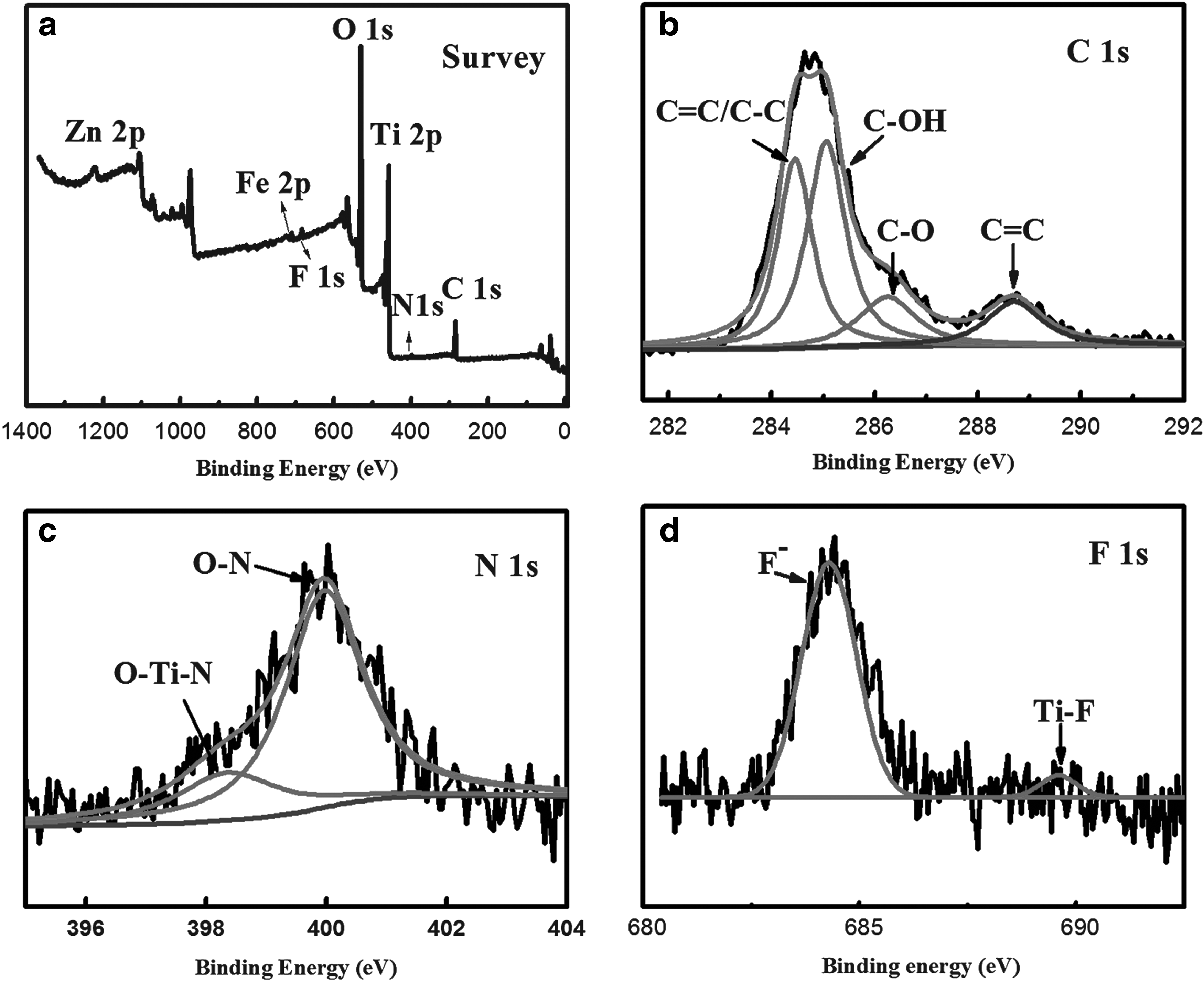
X-ray photoelectron spectroscopy spectra of ZCT-FN hybrids.
Diffuse reflectance spectra
To study the optical properties, the samples were also analyzed by UV-Vis DRS. As shown in Fig. 6, P25 mainly responds to the ultraviolet light, and its energy band gap (Eg) value is about 3.39 eV roughly estimated by Kubelka–Munk function. By coupling with TiO2, ZT shows the higher absorption in a wide region from 300 to 800 nm due to the formation of new level between the valence band (VB) and conduction band (CB) of ZnFe2O4, which promotes the carrier's mobility and further enhances the absorption (Zhu et al., 2014). The Eg value of ZT is about 2.67 eV. With the carbon middle layer between ZnFe2O4 and TiO2, ZCT shows a red shift to the absorption threshold and better absorption intensity than ZT with Eg of 2.03 eV. It is because doping TiO2 with carbon may achieve absorption of visible, as well as UV, light (Shim et al., 2015). Compared with ZCT and ZT, a significant absorption in Vis-light region for ZCT-FN is observed, and its Eg value is about 1.82 eV. As previously reported, the fundamental absorption edge of TiO2 does not cause a red-shift when it is doped only with F atoms (Yang et al., 2013). Therefore, it can be inferred that the enhanced absorption in Vis-light region of ZCT-FN arises from the results of co-doping N and F atoms. As known (Lettmann et al., 2001), there is a positive correlation between the optical absorption performance and the photocatalytic activity; thus it provides a strategy to obtain high photocatalytic activity by improving its photoabsorption.

Photoluminescence
PL emission spectra have been widely used to reveal the efficiency of charge carrier transfer, capture, and recombination in the semiconductor material. In general, a lower PL intensity may indicate a lower recombination rate of photo-induced electron-hole pairs (Li et al., 2005). Figure 7 shows the PL spectra of P25, ZT, ZCT, and ZCT-FN samples under the excitation wavelength of 420 nm. Peak at 570 nm is assigned to the oxygen vacancy (Vo) with one trapped electron (Li et al., 2011; Pirhashemi and Habibi-Yangjeh, 2017; Shekofteh-Gohari and Habibi-Yangjeh, 2017). Among them, P25 has the highest peak intensity, implying the fastest recombination rate of the electrons and holes. Compared with that of ZT, the weaker PL intensity of ZCT represents the fact that the middle carbon layer can prevent the injection of charges from TiO2 particles to magnetic particles. Meanwhile, the weakest PL intensity of ZCT-FN indicates that the charge separation efficiency may have been improved, leading to enhanced photocatalytic activity due to F, N co-doping.
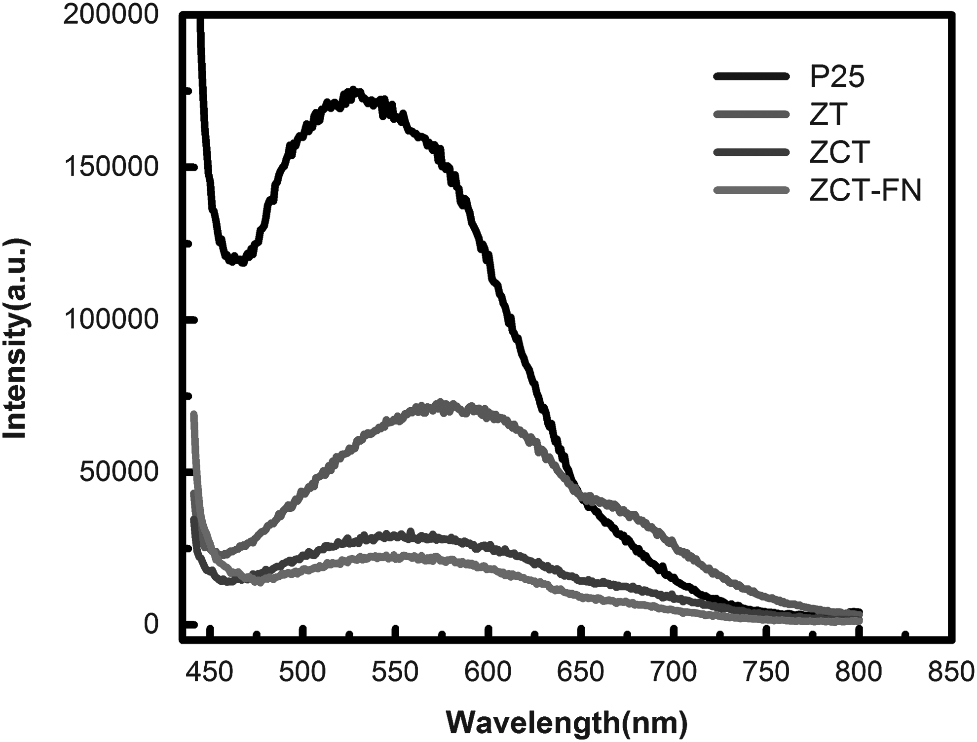
Photoluminescence spectra of P25, ZT, ZCT, and ZCT-FN.
Photocurrent density
Figure 8 shows the photocurrent density variations of P25, ZT, ZCT, and ZCT-FN electrodes with measured potential in 0.01 M Na2SO4 solution. The dark current densities in all cases can be negligible. Upon irradiation, a significant increase in the photocurrent density is observed throughout the potential window at ZCT-FN electrode. The saturated photocurrent density of ZCT-FN electrode is about 1.54 μA cm−2, higher than those over ZCT (0.67 μA cm−2), P25 (0.52 μA cm−2), and ZT (0.43 μA cm−2) electrodes by a factor of 2.3, 3.0, and 3.6, respectively. As a result, the N, F dopants incorporated in the TiO2 network could capture photoelectrons and, thus, also inhibit the recombination (Li et al., 2011). This result is consistent with the charge transport properties of the PL spectra.
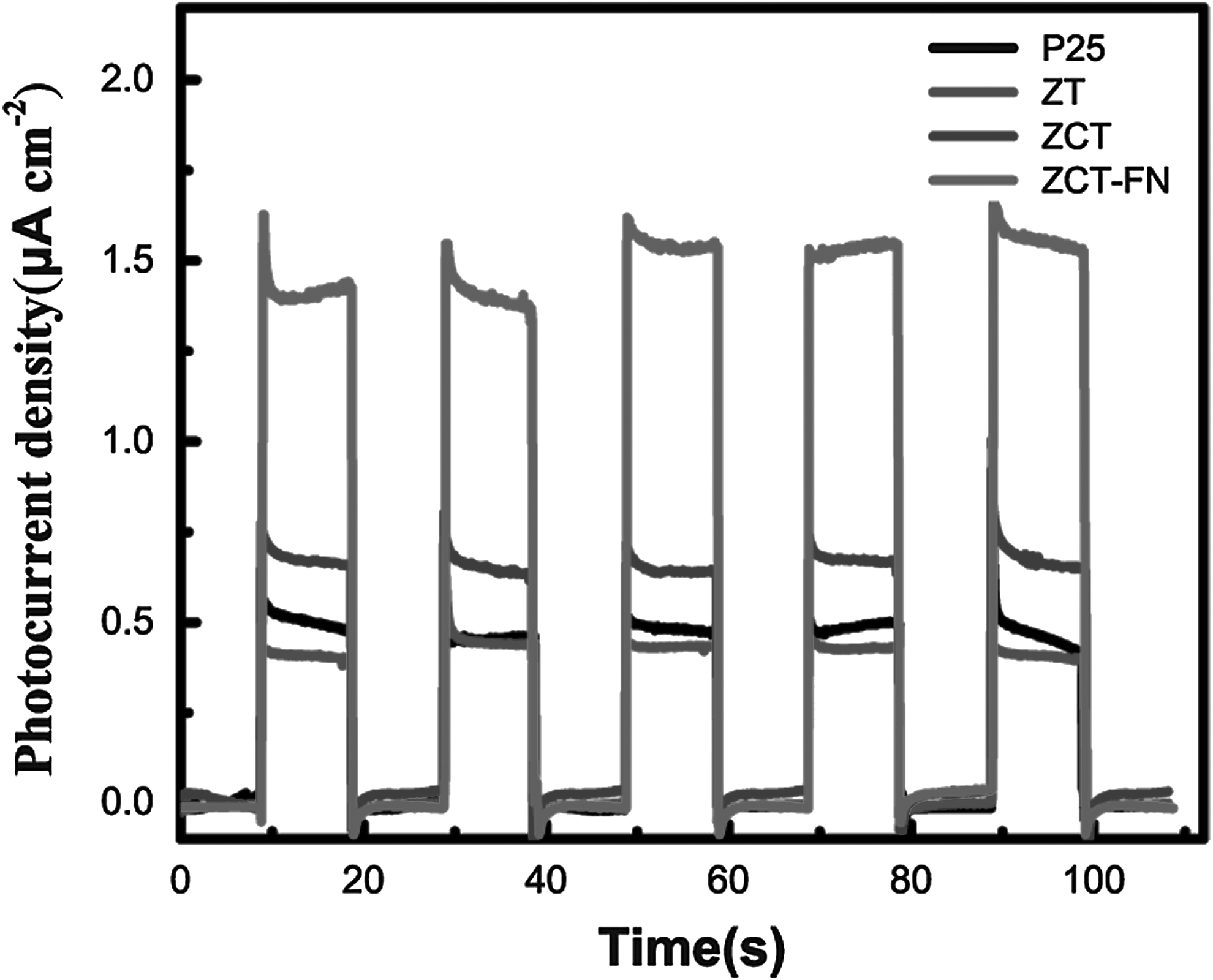
Photocurrent density of P25, ZT, ZCT, and ZCT-FN.
Photocatalytic activity
Figure 9 shows the photocatalytic activity of ZCT-FN, ZCT, ZT, and P25 samples under UV- and Vis-light irradiation. As shown in Fig. 9a and b, the degradation rate of MO on ZCT with the first-order degradation rate constant k about 0.0127 min−1 is obviously higher than that on ZT (0.00359 min−1) under UV-light irradiation; this is because the carbon layer between the TiO2 shell and ZnFe2O4 core can effectively decrease the interaction and then prevent the injection of charges from TiO2 particles to magnetic particles (Shi et al., 2012). For F, N co-doped ZCT-FN, its rate constant k (0.01617 min−1) is higher compared with undoped ZCT, but is lower compared with P25 (0.05409 min−1) due to the lower content of TiO2 in ZCT-FN compared with P25 at the same dosage of catalysts. From the Fig. 9c, d, it is obvious that ZCT-FN possesses the highest Vis-light photocatalytic performance, and its rate constant k is about 4.2 times greater compared with commercial P25. ZCT-FN displays a high photocatalytic activity under both UV- and Vis-light irradiation. It may be arisen from the synergetic effects of F, N co-doping, a middle carbon layer, and narrow semiconductor ZnFe2O4. First, the carbon layer between outer layer TiO2 and ZnFe2O4 core can effectively suppress the photodissolution behavior of catalyst. Second, the presence of N species can generate N2p midgap above the TiO2 VB and absorb visible light (Asahi et al., 2001; Lin et al., 2005). And TiO2 oxygen sites substituted by nitrogen and fluorine atom can also form isolated impurity energy levels above the VB and induce the formation of reduced Ti3+ which forms an isolated defect energy level below the bottom of the CB of TiO2 (Ihara et al., 2003; Yang et al., 2013). Third, the Vo produced due to F doping may lead to another energy level, which is also under CB of TiO2 (Li et al., 2005). A synergetic result of the existed defect energy state belt related to Ti3+ and Vo and the N2p midgap state could narrow the band gap of F-N co-doped TiO2 catalysts, which not only enhances the visible light absorption but also serves to capture the photogenerated electron, resulting in the improvement of the charge separation efficiency (Li et al., 2014).
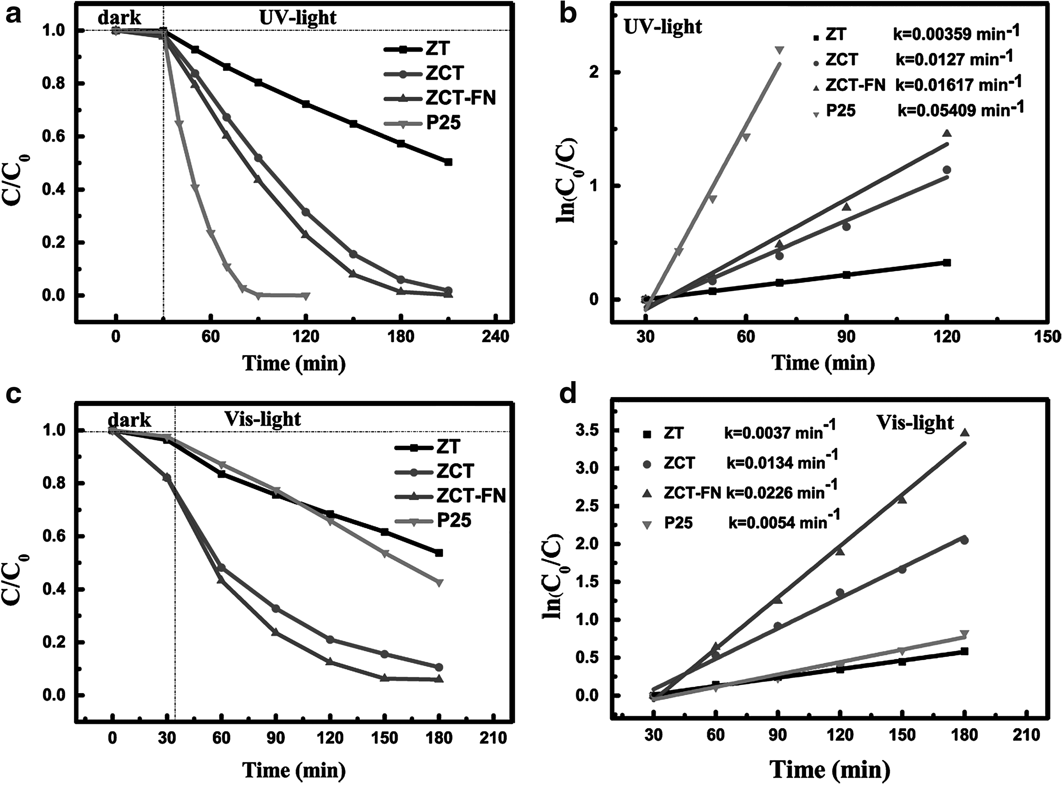
Photodegradation of MO on ZCT-FN, ZCT, ZT, and P25 under
Besides photocatalytic activity, high stability and facile separation of catalysts are also important for the photocatalytic nanomaterials in water remediation. Renewable photocatalytic activities were investigated with ZCT-FN for five times under UV- and Vis-light irradiation, and the results are shown in Fig. 10. After five recycling tests, the degradation efficiency under UV- and Vis-light irradiation for 6 and 8 h was still achieved at 90% and 95%, respectively. It indicates that the as-prepared catalysts have an excellent chemical stability and can have a great potential in organic dyestuff wastewater purification.
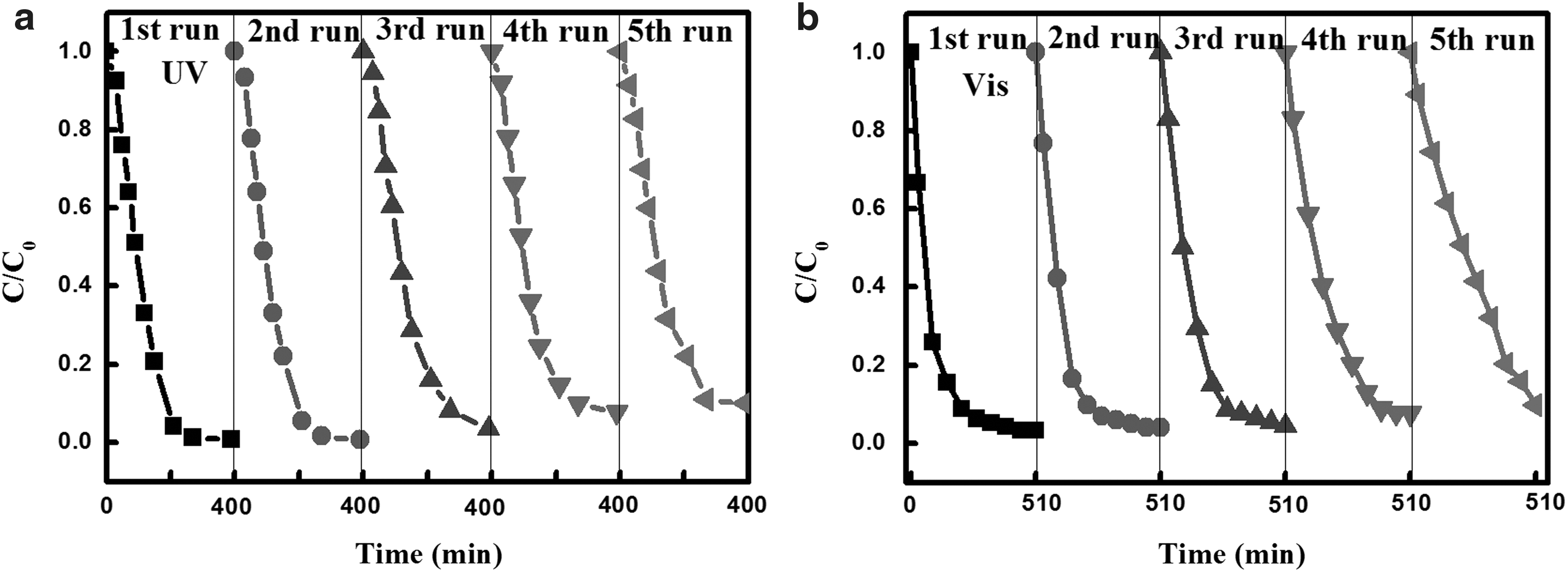
Cycling photocatalytic degradation of MO over ZCT-FN under
Above that, it can be confirmed that the catalyst ZCT-FN obtained not only displays the enhanced UV- and Vis-light photocatalytic performance but also possesses the reuse capability.
Conclusion
In conclusion, we have presented a simple and controllable way to synthesize magnetically separable ZCT-FN core-shell photocatalysts. The results show that ZCT-FN produces an excellent photocatalytic activity under visible-light irradiation, as well as under UV-light irradiation. Moreover, the hybrid can be easily separated from the reaction solution because of the ZnFe2O4 inside, which retains its photocatalytic efficiency through five cycles of operation. As indicated, we provide an effective and recyclable photocatalyst for eliminating organic pollutants from wastewater.
Footnotes
Acknowledgment
The authors acknowledge the financial support from the National Natural Science Foundation of China (Grant No. 51372062).
Author Disclosure Statement
No competing financial interests exist.