
Abstract
Abstract
Use of in situ fluorescence sensors is gaining popularity in water and wastewater treatment and water reuse industries. Tryptophan-like (TRP-like) fluorescence is particularly well suited to tracking fluorescent compounds derived from bacteria as well as a wide range of chemicals of concern for drinking water. Despite the merits of fluorescence sensors, they are affected by environmental factors such as temperature, which can influence photophysical properties of fluorescence. Although temperature correction factors have been well established for humic-like fluorescent dissolved organic matter (fDOM) sensors, there is a need to assess the suitability of existing temperature compensation models in diverse water types and derive new corrections that apply specifically to TRP-like fluorescence. Temperature experiments were conducted using a submersible fluorometer and a range of water types, including creek water, water from different treatment stages at a water reuse facility, synthetic wastewater, and prepared samples containing TRP and fulvic acids. Results from this study revealed that at low fluorescence intensities, no temperature corrections were needed for TRP-like and fDOM fluorescence sensors. For most water types, a published temperature compensation constant for fDOM fluorescence produced a fairly good fit to the reference temperature, but had limited applicability for TRP-like fluorescence. The best fit to the reference temperature for TRP-like fluorescence was achieved using a method to minimize the root mean square error (RMSE) between modeled and measured TRP-like fluorescence. Application of temperature corrections resulted in a difference of as much as 2500 RFU in water types with high DOC concentrations at high (>30°C) and low (<10°C) temperatures, which has important implications for use of fluorescence sensors for source water protection and other monitoring applications in these conditions. For TRP-like fluorescence, empirical relationships and temperature compensation constants were also derived and recommended for samples with varying tryptophan and organic carbon concentrations.
Introduction
F
Despite the growing application of in situ optical sensors, their sensitivity is affected by environmental conditions such as pH, metal ions, photodegradation, and temperature (Vodacek and Philpot, 1987; Hudson et al., 2007; Downing et al., 2012). Depending on climatic conditions, the natural range of temperatures in source waters, water and wastewater treatment plants, and water reuse facilities can vary widely. An inverse relationship has been observed between temperature and the intensities of different fluorophores (Watras et al., 2011; Khamis et al., 2015). This inverse relationship has been shown to be, in part, the result of increased collisional quenching of fluorescent molecules (Vodacek and Philpot, 1987). An increased temperature causes the molecules to undergo collisions with other molecules, which results in the loss of excitation energy as heat instead of emitted light. According to Ruhala and Zarnetske (2016), increased temperature will cause electrons to return to the original ground state through radiationless decay, thereby reducing the fluorescence signal. Decreased temperature, on the other hand, increases viscosity, which may influence collisional quenching, but which may also result in solvent relaxation and subsequent shift of the emission spectra (Lakowicz, 2006). Temperature also influences the fluorescence efficiency of fluorophores through mechanisms that are nonradiative and do not require bimolecular collisions. For example, in the case of tryptophan (TRP) and other indoles, the temperature dependence of fluorescence quantum yields and lifetimes results from solvent quenching, following an Arrhenius-type expression (Kirby and Steiner, 1970; Chen and Barkley, 1998). Similarly, for isoquinoline, the increase in quantum yield at low temperature was shown to be due to internal conversion processes, in which molecules in the S1 state were depopulated by the higher singlet state at lower temperatures (Huber et al., 1976).
In addition to temperature effects on intermolecular and general photophysical behaviors of fluorescent molecules, it is also important to recognize the effects of complex formation on static quenching. The effects of complex formation, which can result in both increased and decreased fluorescence intensities (Coble et al., 2014), may be difficult to disentangle from those of temperature. Similarly, fluorescence quenching by inner filter effects, which commonly occur in samples with high absorbance (Hudson et al., 2007), will also influence fluorescence intensities, reducing the intensity of light available to excite a fluorophore or reducing the light emitted by the fluorescing molecule by other light absorbing molecules (Coble et al., 2014).
Several studies have focused on understanding the effect of temperature on in situ fluorescence measurements, and these have generated useful correction models and factors (Watras et al., 2011; Ryder et al., 2012; Khamis et al., 2015). For example, Watras et al. (2011) developed a temperature compensation model for fluorometers equipped with fluorescence DOM (fDOM) sensors as follows:
where T is temperature (°C), ρ is the temperature-specific coefficient of fluorescence (°C–1), and subscripts r and m represent the reference and measured values. Using two in situ fluorometers, a Sea Point fluorometer and a Turner C3 submersible fluorometer in six temperature experiments with a reference temperature of 20°C, Watras et al. (2011) determined that a ρ coefficient of −0.0155 and −0.009 was optimal for fDOM fluorescence temperature compensation for Sea Point and C3 submersible fluorometers, respectively. The same fDOM temperature compensation model was evaluated by Ryder et al. (2012), who also showed that the ρ coefficient was better approximated by a ratio of the slope of linear regression line of fluorescence intensity plotted against temperature from the same curve.
In the case of TRP-like fluorescence, appropriate correction factors have not yet been developed. The sensitivity of fluorescence to temperature depends on the composition of fluorophores at a molecular level. Therefore, it would be expected that fluorophores with different compositions, for example those that fluoresce in the humic-like region compared to the TRP-like region, are not likely to have the same correction factors. Nevertheless, in the absence of correction factors identified directly for TRP-like fluorescence, other studies have applied the fDOM temperature compensation model for TRP-like fluorescence, and found that it produced only marginal improvement (Khamis et al., 2015; Shutova et al., 2016). The effect of environmental temperature changes appears to be more pronounced for TRP-like fluorescence than for fDOM or humic-like fluorescence (Baker, 2005; Carstea et al., 2014).
Given the increasing application of TRP-like fluorescence to monitor water quality in source waters before treatment (Ahmad and Reynolds, 1999; Carstea, et al., 2018; Goffin et al., 2018; Sorensen et al., 2018) and in the final produced water of water reuse facilities, there is a need to assess the suitability of existing temperature compensation models in such water types, specifically for TRP-like fluorescence, and suitable correction factors are lacking.
In this study, temperature quenching experiments were performed using different water types, including treated water from a water reuse facility, creek water, and water prepared with a range of DOM sources prepared in the laboratory. This enabled us to evaluate temperature compensation models, including the algorithm and constants developed by Watras et al. (2011), referred to as Method 1 in this study, and slope to intercept ratio method as recommended by Ryder et al. (2012) and Watras et al. (2011), referred to as Method 2 in this study. A range of DOM concentrations was used, including both dilute and concentrated solutions, and a range of water types and applied recommended constants for the correction of fDOM and TRP-like sensor data. For comparison, ρ coefficients for the Watras et al. (2011) algorithm were also generated using a root mean square error (RMSE) model (Method 3 in this study) with TRP-like fluorescence data. Based on the analysis of Method 3, we made recommendations for ρ coefficients at different TRP-like fluorescence intensity ranges.
Methods
Experimental setup
In situ fluorescence was measured using a Turner C3 submersible fluorometer equipped with three sensors for TRP-like, fDOM, and chlorophyll fluorescence. The excitation and emission wavelength for the three sensors are shown in Table 1. Detailed physical and electrical properties of the fluorometer are shown in Supplementary Table S1.
Source: Turner Designs (2015).
Excitation wavelength of the TRP-like sensor same as the LED wavelength of 275 nm.
TRP like, tryptophan like; fDOM, fluorescent dissolved organic matter.
Temperature experiments were carried out using 17 different water samples, including river water, samples from the Pure Water San Diego Advanced Water Purification Facility (AWPF), and prepared solutions of ultrapure water and TRP and fulvic acids. River water was collected as grab samples from Alvarado Creek (32o46’42.3”N, 117o03’48.97”W) on 15 January 2017, a small tributary of the San Diego River that is located on the campus of San Diego State University. Water from the DPWF was collected from three different points along the treatment train: (1) at the inlet of the facility (pre-ozone water), (2) after the ozonation of the influent (post-ozone water), and (3) after the ultraviolet-advanced oxidation process (UV-AOP water) between 15 December 2016 to 24 January 2017. Detailed information about the sampling locations (a, b, and c) and the treatment train of the facility are shown in Supplementary Fig. S1. Prepared solutions were also made by dissolving sphagnum moss, peat moss, International Humic Substances Society (IHSS) Suwannee River Fulvic Acid (SRFA), and laboratory-grade TRP in ultrapure water. Mixing ratios of the different constituents are described in the Supplementary Methods. The DOC concentrations of 13 of the 17 samples used are given in Supplementary Table S2 and their respective fluorescence intensities are provided in Supplementary Tables S3 and S4. DOM fluorescence intensities of all samples are presented in relative fluorescence units (RFU).
All samples were placed in buckets (4 L) and first cooled to approximately 4°C in a dark refrigerator. The C3 fluorometer connected to a laptop was then inserted into an opaque bucket of cold water samples, which was then placed into an incubator and gently warmed from 4°C to 30°C with constant mixing for 7–8 h for each sample. All experiments were carried out in a dark room with no lights, and the incubator was also closed for the span of each experiment to maintain dark conditions in case of any light interruptions. Fluorescence intensity data and corresponding temperature were continuously logged onto a computer at an interval of 1 s. The first 7 to 15 values (before readings stabilizing) were discarded.
Development of correction factors
A temperature compensation algorithm with temperature correction coefficient, ρ, of −0.0155 developed by Watras et al. (2011) for fDOM [Eq. (1)] was initially used to correct all the sensor data (Method 1). A value of −0.0155 was chosen as constant for comparison from Watras et al. (2011) because this constant produced a better fit to the reference temperature than the constant that Watras et al. recommended for a C3 submersible fluorometer (−0.009), as explained in the results section (Supplementary Fig. S2). Sensor data were then corrected using ρ values calculated from the slope to intercept ratio as recommended by Ryder et al. (2012) (Method 2). Using the RMSE model (Method 3), ρ values were also determined for different water types. In this model, the Goal-seek function in Microsoft Excel was utilized to optimize the ρ value so that the sum of the error squared between “correct” intensity values at a temperature of 20°C and the calculated temperature from Equation (1) was minimized. The Goal-seek program conducts up to 100 runs until it arrives at the optimal ρ value (Hossain et al., 2013). The procedure for using the solver program is presented in the Supplementary Methods.
Statistical analyses
Two sample t-tests were used to assess the statistical difference between constants produced by Method 3 and Method 2 with a 95% significance level using MINITAB v.17. Similarly, a one sample t-test was used to assess the statistical difference between the constants calculated from the Method 3 and the constant, Method 1.
Results and Discussion
Consistent with previous studies, the controlled temperature experiments for all samples showed that the increase in temperature was inversely proportional to fDOM and TRP-like fluorescence intensities (Watras et al., 2011; Ryder et al., 2012; Carstea et al., 2014; Khamis et al., 2015). Furthermore, the slopes of each regression line increased with increasing DOM concentration in each of the water sources (Fig. 1). This indicates that temperature effects are more pronounced for waters with high DOM fluorescence intensities (Fig. 1 and Supplementary Tables S3 and S4) and colder temperatures, as has been previously observed by Watras et al. (2011). Consistent with previous findings (Carstea et al., 2014; Khamis et al., 2015), it was observed that temperature effects were more pronounced for TRP-like fluorescence compared to fDOM or other humic-like fluorescence, as reflected in the steeper slopes of the relationships between TRP-like fluorescence intensity and temperature (Fig. 1). In addition, there was a nonlinear response of TRP-like fluorescence intensity to temperature change in the highest concentration sample, especially at very low temperatures (<9°C). Khamis et al., (2015) observed similar trends for TRP-like fluorescence in high concentration samples in experiments using laboratory-prepared standard TRP solutions. This nonlinearity is exacerbated by the high concentrations, which are likely to produce inner filter effects that are not compensated for by the portable instruments. Indeed, these effects were only present in samples S15–S17 (Supplementary Table S2), which had TRP concentrations that exceeded the recommended range for the Turner C3 fluorometer (3–5,000 ppb). These results make a case for future development of inner filter correction modes in online monitoring, potentially with the use of multiple sensors (fluorescence and absorbance) in tandem. Using a 3D benchtop fluorometer, Miller et al. (2010) noted that such highly concentrated samples must be diluted before analysis.
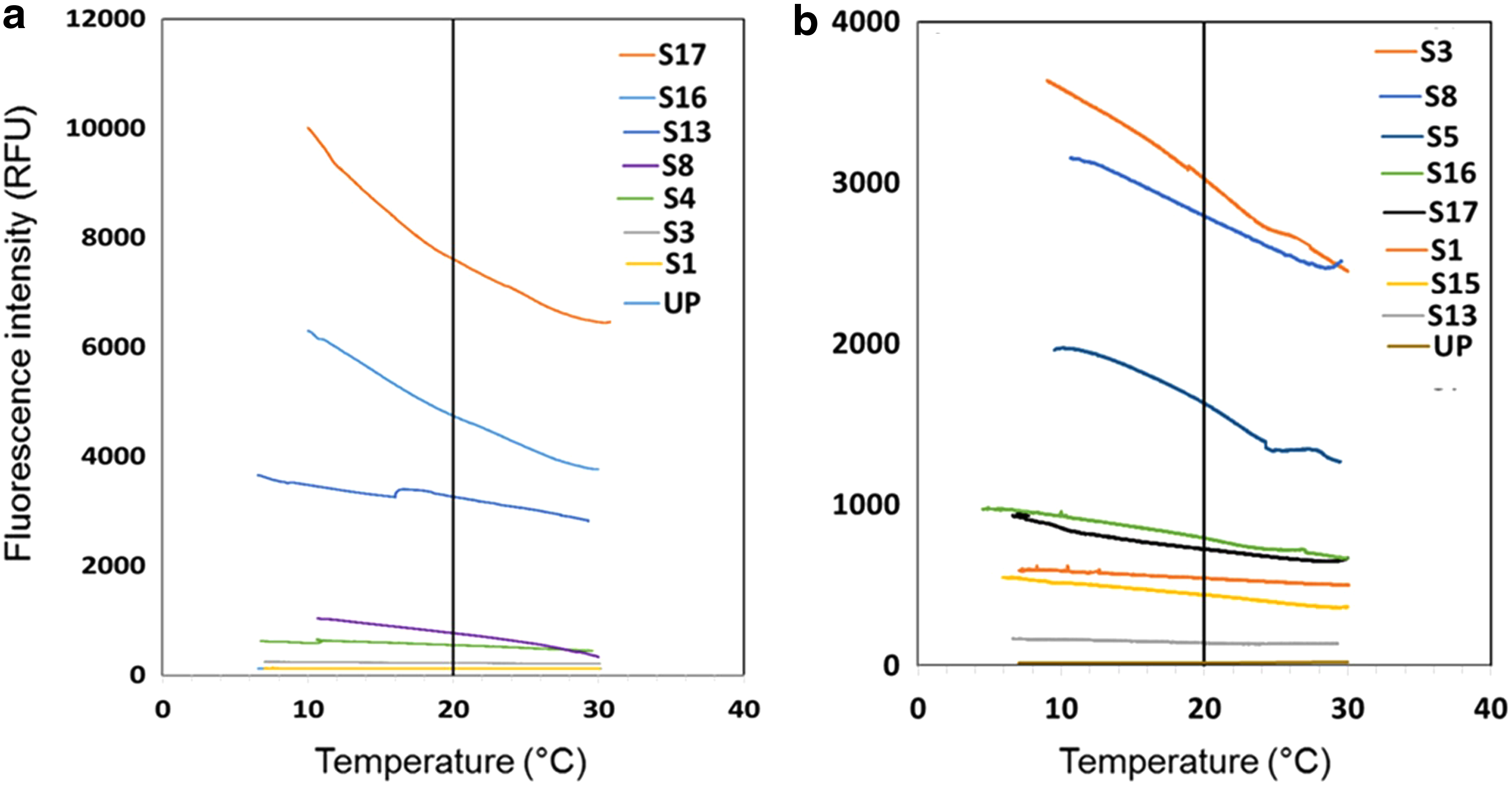
Effect of temperature changes on
Figure 1 provides guidance that may be useful for determining the need for temperature correction. For example, at low reference fluorescence intensities (<200 RFU for TRP-like), results from this study revealed that the effects of temperature were negligible. For fDOM fluorescence at intensities <150 RFU, this study was consistent with those of Watras et al. (2011), who also observed no temperature effects in their analysis of diluted water samples. These results indicate that at low fluorescence intensities, as described above, there is no need to apply temperature corrections.
The closeness of fit of fluorescence intensity to the reference fluorescence intensity at 20°C was evaluated using the temperature compensation Equation (1) with three different models for the correction constant, ρ: (1) the recommended empirical constants of Watras et al. (2011) of −0.009 for the Turner C3 fluorometer and −0.0155 for the SeaPoint fluorometer, which is referred to as Method 1, (2) constants calculated using Method 2, and (3) constants calculated using Method 3. In Fig. 2, the results for different water types (post-ozone treated water and synthetic wastewater made from mixing milk powder and yeast extract and creek water) are presented representing low and high intensity fluorescence, respectively. Based on preliminary observations from Supplementary Fig. S2, we compared the fluorescence intensities calculated using two constants of Watras et al. (2011) (−0.009 and −0.0155) to fluorescence intensities at the reference temperature. In the case of TRP-like fluorescence, the average percentage deviation between fluorescence intensities at the reference temperature and fluorescence intensities calculated using the −0.009 constant was 332%, while in the case of the −0.0155 constant, the average percentage deviation was only 32%. In the case of fDOM sensor fluorescence, the average percent deviation between fluorescence intensities at the reference temperature and fluorescence intensities calculated using the −0.009 constant was 90%, while in the case of the −0.0155 constant, the average percent deviation was 10.3%. This indicates that −0.0155 was more appropriate for correction of both TRP-like and fDOM sensor fluorescence than −0.009. When Method 1 (using the −0.0155 constant) was applied for temperature compensation on all samples, fDOM sensor intensities deviated from the reference intensity at 20°C within ±57%. However, neither the −0.0155 constant nor the −0.009 constant were able to adequately correct TRP-like fluorescence, producing fluorescence intensities deviating by as much as 114% from the reference fluorescence intensity. Temperature compensation using Method 2 produced intensity values that matched the reference intensity within ±11% for the fDOM sensor and ±17% for the TRP sensor. Constants calculated using Method 3 produced corrected fluorescence intensities that most closely matched the reference intensity (within ±0.47% for fDOM and ±1.79% for TRP like).

Response of different temperature constants to changes in temperature for representative low and high TRP-like and fDOM fluorescence intensities for TRP sensor (left panels) and fDOM sensor (right panels). The water samples used were as follows: post-ozone water
Application of the temperature compensation factors in water with low fluorescence intensities (Fig. 2) resulted in either an overcorrection of TRP-like fluorescence intensities (e.g., when the −0.0155 factor was applied) or no effect (for Method 1 and 2 corrections). In very high fluorescence intensity samples (>2500 RFU for fDOM and >4000 RFU for TRP like), corrections using both Methods 1 and 2 constants could not remove the temperature effects. Only constants calculated using the Method 3 were able to remove the temperature effects.
To evaluate the goodness of fit between corrected and reference fluorescence intensities, the y-intercept values produced by temperature compensation using the three methods under study versus the reference fluorescence intensities at 20°C for 17 samples were plotted. Theoretically, the reference values at 20°C and y-intercept fluorescence intensities values should be equivalent and there should be a 1:1 slope if there is no deviation. For TRP-like fluorescence, the application of Method 3 constants produced the line with a slope closest to 1.0 (1.035) compared to Methods 1 and 2, which produced slopes of 1.23 and 1.193, respectively (Fig. 3). Similarly, Method 3 constants for the correction of fDOM values also produced the slope closest to 1.0 (Fig. 3). These results suggest that Method 3 achieves temperature corrections that produce corrected values for both TRP-like and humic-like fluorescence, which are closest to the reference intensity values at 20°C for all sample types. The temperature compensation constants obtained using Method 3 were consistently higher (mean = −0.0209, SD = 0.0109) than the constants calculated from slope to intercept ratio (mean = −0.0139, SD = 0.00603). Paired sample t-tests indicated these differences were significant (p < 0.05).
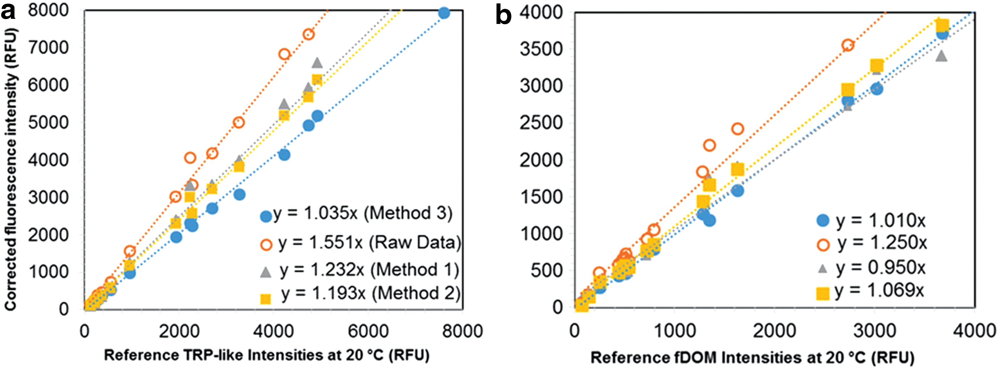
Scatterplots showing corrected fluorescence intensities (y-intercept values for each sample after applying different correction constants) plotted against reference fluorescence intensities using different temperature correction constants for
Not only is the difference in the calculated constants caused by the type of method used for calculating the constants but also by the composition of the solutions. As explained in the introduction, fluorescence quenching can also result from intermolecular interactions between different components of DOM. For example, Wang et al. (2015) observed that fluorescence of the protein-like components was greatly quenched by the humic-like components, while fluorescence of the humic-like components was not impacted by the protein-like components. All samples used in this study had a mixture of both humic-like and protein-like DOM. Three-dimensional excitation emission matrix spectra show the co-occurrence of humic-like and protein-like DOM in each water type (Supplementary Fig. S3). Because there were no pure solutions (solutions with single component) used in this study, a clear understanding of the extent of TRP-like DOM fluorescence quenching by humic-like DOM with temperature was not possible. However, a linear regression analysis between TRP-like reference fluorescence intensities and TRP concentration for the synthetic samples in this study gave an R2 value of 0.87 and p < 0.0001. These results not only reflect a significant linear correlation between TRP concentrations and fluorescence intensities but also highlight that there is additional variability that may be due to formation of molecular assemblies between TRP-like and humic-like components, as described by Wang et al. (2015).
Practical Considerations
Although Method 3 did produce corrected values for TRP-like fluorescence that matched the reference intensity at 20°C for a range of water types, Method 3 requires the collection of data at different temperatures (including the reference temperature) and operation of a solver routine for error analysis of modeled and measured intensities. Method 2, which was recommended by Ryder et al. (2012) and Watras et al. (2011), also generates the constant using a range of fluorescence data collected at different temperatures. For practical applications in which fluorescence intensities collected at a range of temperatures may not be available, empirically derived constants, such as that used in Method 1 for fDOM fluorescence (Watras et al., 2011), are needed. Empirical constants also are advantageous for faster or real-time correction of data (e.g., for use in alert or warning systems of contamination).
Using a set of 17 samples, including six different water types at different concentrations, (Supplementary Table S2) constants calculated from the Method 3 were plotted against TRP-like intensities measured at 20°C (Fig. 4). The best fit line through these points generated a logarithmic Equation (Fig. 4):

Scatterplot of temperature correction constants and TRP-like intensities at 20°C for diverse water types (samples S1–S17 from Supplementary Table S2). The equation of the relationship of the logarithmic fit is shown (R2 = 0.57, p < 0.05). Relationships from plots of TRP-like fluorescence and tryptophan concentration yield a conversion of 1 RFU = 0.00169 mg tryptophan/L. The gray lines above and below the fit represent upper and lower 95% prediction confidence intervals, respectively.
where y is the temperature compensation constant (from Method 3 analysis) and x is the TRP-like fluorescence intensity at 20°C. In samples for which the preferred Method 3 cannot be applied, as long as the TRP-like fluorescence intensity at 20°C is known, the temperature compensation constant, y, may be calculated using Equation (2). Therefore, Equation (2) should be applied with caution and only as a guide for temperature compensation constants for diverse water types.
For most intensity values between 500 RFU and 6000 RFU, it was observed that applying a temperature compensation constant of −0.021, derived in Supplementary Tables S5 and S6, produced corrected values that were very similar (no statistically significant difference) to the correction coefficients calculated using Method 3. In addition, for circumstances in which reference intensities are not available at 20°C, it would also be possible to select a different reference temperature and use Method 3 to determine an appropriate constant for temperature compensation using Equation (1).
This study recommends that temperature compensation constants for TRP-like fluorescence are applicable to studies using a Turner C3 submersible fluorometer, but may also be a useful starting point for studies using other in situ fluorescence sensors. Watras et al. (2011) evaluated their temperature compensation model on two different fluorometers, at Turner C3 fluorometer and a SeaPoint UV fluorometer, both of which use LED light for fDOM excitation, and found that the ρ constant was slightly different (−0.009 for the C3 and −0.0155 for the SeaPoint) depending on the sensor type. Therefore, empirical constants for TRP-like fluorescence may need to be reevaluated for other sensor models. In addition, there are other environmental factors known to affect fluorescence or cause quenching, such as turbidity, which were not the focus of this study, but for which correction models exist and should be applied. In any case, the empirically derived equations tested in this study and in other studies (Watras et al.; Khamis et al.) are a useful starting point for fluorescence data correction. Future work should revisit the fundamental relationships between temperature and both radiative and nonradiative effects to also develop temperature compensation models based on theoretical principles governing fluorescent behavior of molecules.
Conclusions
The aim of this study was to assess suitability of available temperature compensation models from the literature for correcting the temperature effects on TRP-like fluorescence and developing appropriate temperature compensation constants. A C3 submersible fluorometer was used to study the effects of temperature on fDOM sensor and TRP sensor fluorescence in the laboratory. Results support the use of the −0.0155 constant suggested by Watras et al. (2011) to correct temperature effects for fDOM sensor intensities as long as intensities are <3500 RFU. It was observed that application of the fDOM constant to TRP-like fluorescence correction underestimated the true TRP-like fluorescence values. Therefore, using Method 3, a relationship was developed between TRP-like fluorescence intensities and temperature compensation constants, and an empirical constant was proposed. For all water types, application of Method 3 produced the best fit to the reference temperature for all three fluorescence sensors. The empirically derived constants for TRP-like fluorescence temperature compensation may be of use in situations in which in situ fluorescence measurements are made in a narrow temperature range or without available fluorescence intensities at the reference temperature. The basis for the development of Equation (1) was the constant slope: intercept ratio regardless of CDOM concentration. However, in this study, we have observed that it may not be true, especially at very high concentrations. In addition, there was a significant difference between constants calculated using the slope: intercept ratio and the RMSE method for the TRP-like fluorescence. We recommend that there is also a need to reconcile the model with theories of DOM photophysics.
Footnotes
Acknowledgments
We acknowledge support from the San Diego State University Presidential Leadership Fund for the Water Innovation and Reuse Laboratory. Funding was also provided in part from the National Science Foundation through water reuse project NSF CBET 170591. We thank Anita Sanchez and Chelsi Pascua for assistance with experimental work.
Author Disclosure Statement
No competing financial interests exist.
References
Supplementary Material
Please find the following supplemental material available below.
For Open Access articles published under a Creative Commons License, all supplemental material carries the same license as the article it is associated with.
For non-Open Access articles published, all supplemental material carries a non-exclusive license, and permission requests for re-use of supplemental material or any part of supplemental material shall be sent directly to the copyright owner as specified in the copyright notice associated with the article.