
Abstract
Abstract
This study investigates removal of selenite [Se(IV)] by reductive precipitation during treatment with an Advanced Reduction Process (ARP) that uses dithionite activated by ultraviolet (UV) irradiation. Our screening experiments evaluated a number of ARP and found that the dithionite/UV ARP was most effective in removing soluble selenite. Furthermore, this work considers effects of operating conditions such as dithionite dose, solution pH, initial selenite concentration, and light intensity on reduction of Se(IV). Selenite [Se(IV)] was completely removed in 120 min when initial Se(IV) concentration was 0.023 mM, dithionite dose was 1 mM, and the initial pH was ∼4.5. Higher dithionite doses, lower pH, and higher incident UV irradiance increased soluble Se(IV) removal. Selenium in the solids was effectively removed from solution by conversion to solids that were removed by filtration. Scanning emission microscopy/energy dispersive X-ray spectroscopy (EDS), X-ray diffraction, and X-ray Photoelectron Spectroscopy results showed that Se(IV) was reduced by the dithionite/UV ARP to form solids identified as elemental Se or as a compound composed of both Se and S (e.g., SemSn). Elemental Se was the primary solid, especially at higher initial Se(IV) concentrations. In the dithionite/UV ARP, rapid removal of soluble Se(IV) at low pH is attributed to photolysis of dithionite or a dithionite decomposition product (e.g., bisulfite, metabisulfite, thiosulfate, and trithionate) that is initially present in the dithionite solution.
Introduction
A
Selenium is an essential trace element to humans; however, it can be toxic at high concentrations (de Albuquerque et al., 2012). The maximum contaminant level (MCL) for selenium set by the USEPA for drinking water is 50 μg/L and levels above the MCL may cause adverse health effects to people, including hair or fingernail loss, numbness, and kidney and liver damage (US EPA, 2007; Stivanin de Almeida et al., 2009). Removal of selenium can be achieved using various physical, chemical, and biological methods that include membrane separation, ion exchange, adsorption, chemical reduction, electrocoagulation, and bacterial reduction (Rovira et al., 2008; Zhang et al., 2009; Santos et al., 2015). The disadvantages of a biological process are that it requires more time for the reduction and can result in incomplete reactions that can result in formation of more toxic compounds (Hartmanis and Stadtmant, 1982; Murphy, 1988). Adsorption and ion exchange are commonly applied physical methods to remove selenium from wastewater (Balistrieri and Chao, 1990; Peak, 2006; Rovira et al., 2008; Su et al., 2008; Han et al., 2012; Kim et al., 2012). The performance of an adsorbent is dependent on the conditions such as pH, concentration range, ionic strength, and temperature, and this can make it difficult to evaluate the adsorption capacity (Soda et al., 2011). Zerovalent iron (ZVI) has been widely used as reductant to reduce Se(VI) to Se(IV) or elemental Se. The advantage of chemical reduction using ZVI is that ZVI is easily available and relatively low cost. However, the disadvantages of this method have been demonstrated to include pH and temperature dependence, the need for long hydraulic residence time, and the formation of iron corrosion products (CH2MHILL, 2010; Santos et al., 2015). Sodium dithionite is also a strong reductant, and it is able to reduce Se(IV) rapidly in acidic environments (Geoffroy and Demopoulos, 2009).
In this study, we apply an ARP using dithionite as a reagent and UV irradiation as an activation method to treat solutions containing selenium. The dithionite/UV ARP has been previously applied to a variety of contaminants, including perfluorooctanoic acid, perchlorate, dichlorophenol, 1,2-dichloroethane, and trichloroethylene (Vellanki et al., 2013; Liu et al., 2014; Jung et al., 2015). The dithionite/UV ARP was also found to be effective in degrading nitrate (Bensalah et al., 2014), but when applied to chlorate, it was found that much of the chlorate was removed immediately after mixing with reagents and before UV irradiation began (Jung et al., 2017b). This unexpected pattern of removal was attributed to reaction of chlorate with products of dithionite decomposition that existed in the original dithionite solution.
Our recent work found that both As(III) and Se(IV) are removed by dithionite activated by UV irradiation (Duan et al., 2017a, 2017b). Se(IV) concentrations at pH 7 were observed to rapidly decrease and then increase after dithionite was consumed and irradiation continued (Duan et al., 2017b). It was found that As resolubilization could be avoided by frequent addition of dithionite that would maintain a nonzero concentration (Jung et al., 2017a). Additional experiments demonstrated that resolubilization only occurs in the absence of dithionite and the presence of UV irradiation (Duan et al., 2017b; Jung et al., 2017a). This indicates that resolubilization occurs when products of dithionite degradation are photolyzed to produce radicals that react with arsenic or selenium to form soluble products. Experiments at higher initial Se concentrations (0.025 mM) than used by Duan et al. (0.005 mM) showed that resolubilization of selenium did not occur. Previous work with ARPs focused on evaluating the effectiveness of dithionite/UV ARP toward a variety of contaminants. Dithionite is highly soluble in water, but it decomposes in solution to produce various products. Few studies have addressed the question of what compound in dithionite solutions, particularly those at low pH, is responsible for reacting directly with selenium or indirectly through products of photolysis. Products of dithionite decomposition include sulfites, thiosulfate, polythionates, sulfides, and elemental sulfur (Burlamacchi et al., 1968; Čermák and Smutek, 1975). Decomposition pathways varied depending on whether or not the solution is irradiated by UV. More detailed discussion of dithionite decomposition reactions and identification of potential reactive species for Se(IV) reduction was presented in a later section.
The novelty of this study is the investigation of potential reactive species for Se(IV) that could be formed in irradiated and nonirradiated dithionite solutions. This investigation considered experimental results and published data, as well as evaluating the effects of operating conditions such as dithionite dose, solution pH, initial selenite concentration, and light intensity on removal of Se(IV) from solution. Also, this study identified the type of solids formed from Se(IV) solution during the treatment by the dithionite/UV ARP.
In summary, the objectives of this study were to (1) evaluate the effectiveness of the dithionite/UV ARP to treat water containing Se(IV), (2) study the effects of the system variables [pH, reducing agent dose, pH, initial Se(IV) concentration, light intensity] on Se(IV) reduction, (3) examine the surface morphology, element composition, and surface chemistry of precipitates formed in Se(IV)-dithionite solution using scanning emission microscopy (SEM)/energy dispersive X-ray spectroscopy (EDS), X-ray diffraction (XRD), and X-ray Photoelectron Spectroscopy (XPS) analysis, and (4) study the compounds produced by dithionite decomposition products to determine which ones can be photolyzed to produce reducing radicals that can convert soluble selenium to solid forms.
Materials and Methods
Experimental procedures
The combination of dithionite and UV-L irradiation was chosen as an effective combination for Se(IV) removal based on results of screening tests. All reagent solutions were prepared using reagent grade chemicals and deoxygenated deionized water within an anaerobic chamber (Coy Laboratory Products, Inc., Grass Lake, MI). The chamber was filled by N2 and equipped with an oxygen and hydrogen analyzer (Coy Laboratory Products, Inc.). Deoxygenated deionized water (18.2 MΩ) was acquired by a Barnstead Nanopure filter system and purged with N2 gas (purity >99.99%) for at least 2 h and then stored in an anaerobic chamber. And the sources of Se(IV) and Se(VI) were Na2SeO3 (sodium selenite, 99%; Sigma) and Na2SeO4 (sodium selenate, ≥95%; Sigma), respectively. Dithionite stock solution was prepared at high concentration (25.3 mM) by adding sodium dithionite (Na2S2O4, sodium hydrosulfite, >82%; Sigma) to deoxygenated deionized water, and it was vigorously shaken by hand. An appropriate volume of this solution was immediately added to a reactor and diluted before irradiation was initiated to start an experiment. The time from dissolving sodium dithionite to beginning the experiment did not exceed about 3 min. NaHSO3 [sodium bisulfite, ≥58.5% (SO2), Acros Organics] and Na2S2O5 (sodium metabisulfite, 99.1%; Fisher Chemical) were used to prepare solutions that would contain different amounts of sulfite ion (SO32−), bisulfite ion (HSO3−), and metabisulfite ion (S2O52−). Sodium thiosulfate (Na2S2O3·anhydrous, reagent grade) was purchased from Amresco, Inc. If needed to adjust the pH, 1 N HCl (hydrochloric acid; 37%; ACS Reagent) or 1 N NaOH (sodium hydroxide, 97%; ACS Reagent) was added. The solution pH was always monitored during the experiment using a Thermo Triode pH meter after calibrating with 3 pH buffers (4, 7, and 10). The standard initial concentrations of selenium and dithionite in a reactor were 0.025 and 1.08 mM, and the solution pH was ∼4.5, unless otherwise specified. Experimental reactors were quartz cells of 17 mL capacity and 1-cm thickness, which were placed below low-pressure mercury UV lamps installed in a UV box (14.5 [H] × 33 [D] × 26 [W] cm; BioLink BLX). Five T-8C UV lamps (Viber Lourmat) of 8 W capacity were used and produced incident irradiance of 4,600 ± 300 μW/cm2 at a distance of 12 cm. The UV lamps were warmed up for 10 min before the experiments began and light intensity during experiments was measured using a UV light meter (ST-512, UVC, 220–275 nm, calibration point at 254 nm). At regular time intervals, an individual reactor was sacrificed by removing about 10 mL and filtering it using a 0.2 μm membrane filter paper (Suport®-200; PALL Life Sciences) with a disposable syringe. Total soluble Se concentration was determined in the filtrate. The filter paper, including precipitates, was separated from the filter holder and dried inside the anaerobic chamber before surface characterization.
Analytical procedures
Total soluble selenium concentration in the sample was measured using an inductively coupled plasma-optical emission spectrometry (ICP-OES, Thermo Scientific iCAP 6000 Series). Method detection limit and method quantification limit for selenium using ICP-OES were 0.022 and 0.071 μM, respectively. Species of selenite [Se(IV)] and selenate [Se(VI)] were analyzed by an ion chromatograph (Dionex ICS-5000), which was equipped with a dual gradient pump, AS autosampler, and eluent generation module. The eluent used was 4.5 mM Na2CO3/0.8 mM NaHCO3 with a flow rate of 0.25 mL/min. The injection volume was 1,200 μL, and ions were detected with suppressed conductivity using an anion self-regenerating suppressor 300 (ASRS 300, 2 mm). Four selenite solution samples were prepared at 0.127 mM using Na2SeO3, and the average and standard deviation of measured concentrations were 0.130 mM and 0.15, respectively. Dithionite concentration was determined by UV absorbance at 315 nm using a Perkin Elmer UV-visible spectrophotometer (Lambda 25) and a molar extinction coefficient of 8,043 (M−1cm−1) (McKenna et al., 1991). Surface morphology and elemental composition of precipitates were characterized using FEI Quanta 400 SEM/ EDS. Phase changes on the surfaces of the solids were characterized by XPS (Kratos Axis Ultra DLD). The spectra peak of C 1s at 284.5
Results
Selenium removal
Figure 1 shows total soluble Se concentrations in reagent control, light control, and experimental reactor with dithionite/UV. In dithionite solution activated by UV light, total soluble Se concentration rapidly dropped in the first 5 min and then slowly decreased. The solids formed had a yellow–orange color (Supplementary Fig. S1). In the reagent control, the light control, and the experimental reactor, the initial pH values were 8.4, 5.7, and 4.6, respectively. At every reaction time, solution pH was measured (Supplementary Fig. S2 and Supplementary Table S1). In Fig. 1, the concentration of total soluble Se approaches a low, but nonzero value. This could be due to consumption of dithionite or by the presence of some selenate [Se(VI)] in the initial solution that resists removal. To evaluate the possibility that selenate was present in solutions prepared from sodium selenite, solutions were prepared from sodium selenite and sodium selenate and the concentrations of Se(IV) and Se(VI) were measured in them by ion chromatography (Supplementary Fig. S3 and Supplementary Table S2). Solutions prepared from sodium selenate showed only one peak, which was identified as that for Se(VI). However, solutions prepared from sodium selenite showed two peaks, which were identified as being for Se(IV) and Se(VI). The concentration of Se(VI) measured in the sodium selenite solution was consistently about 12–14% of initial total soluble Se concentration. This indicates that approximately 12–14% of initial selenium in the reagent sodium selenite was present as selenate. The same result was observed in our pervious study (Jung et al., 2016a). Therefore, the approach to a constant soluble Se concentration is probably due to a combination of negligible dithionite and the presence of selenate, which resists removal. The rapid initial removal of soluble Se shown in Fig. 1 is probably the result of removal of selenite in the presence of dithionite during irradiation and the slower removal may be due to low concentrations of dithionite.

Concentrations of total soluble Se in controls and experimental reactor. Conditions: [Total Se]0 = 0.027 mM, [S2O42−]0 = 1.02 mM, UV-L, the solution pH was not buffered (measured pH values are shown in Supplementary Fig. S2). UV, ultraviolet.
Effect of dithionite dose
Figure 2 shows the effect of dithionite dose (0.052–2.6 mM) on soluble selenite [Se(IV)] concentration in solutions treated by the dithionite/UV ARP. Soluble Se(IV) concentrations were calculated by assuming that 12% of the initial total Se concentration was due to presence of selenate in the reagent sodium selenite. In the absence of dithionite, soluble Se(IV) concentrations were almost constant with time. After 60 min, soluble Se(IV) concentrations were reduced by 11%, 65%, 83%, and 92% at dithionite doses of 0.26, 0.52, 1.04, and 2.6 mM, respectively. When dithionite dose was 0.052 mM, soluble Se(IV) removal percentage was 6.1% during 240 min, which was similar to a reagent control (6.5%). It is likely that the cessation of Se(IV) removal observed at lower dithionite doses is due to consumption of dithionite. When dithionite is present, the initial pH ranged between 4 and 5.5 and the final pH decreased to about pH 3.7 (Supplementary Fig. S4). This pH decrease may be explained by the protons produced by the dithionite decomposition reactions (Lindholm, 1999). Dithionite undergoes various reduction reactions producing bisulfite, dithionate, bisulfate, and protons (Lindholm, 1999; Jung et al., 2017b). Geoffroy and Demopoulos (2009) observed that selenite was reduced to elemental Se or Se-S solids even in the absence of irradiation, but this occurred at a pH (pH 1.3) much lower than used in our experiments. Figure 1 shows negligible loss of Se(IV) by reaction with dithionite at pH between 5 and 6 in the absence of UV. In the presence of irradiation, our hypothesis is that Se(IV) is removed by a reaction with a product of dithionite photolysis, which will be discussed in more detail in the following section. As shown in Fig. 2, higher dithionite doses will produce more active species from dithionite photolysis, which will result in higher Se(IV) removal. When dithionite dose was 2.6 mM, applied UV-L achieved a complete soluble Se(IV) removal of an initial concentration at 0.023 mM in 120 min, and UV fluence was calculated to be 33.8 J/cm2.

Effect of initial dithionite dose on soluble Se(IV) concentration during treatment by dithionite/UV ARP. Conditions: [Se(IV)]0 = 0.023 mM, light intensity (T-8C) ∼4,700 μW/cm2, and the initial pH = ∼pH 4.5 (not buffered). ARP, advanced reduction process.
Effect of pH
Figure 3 shows the effect of pH on soluble Se(IV) concentrations during treatment by the dithionite/UV ARP. The solution pH was monitored at every reaction time (Supplementary Fig. S5). These results show substantially improved removal of Se(IV) at lower pH, both in terms of removal kinetics and removal extent. This is similar to the results reported by Geoffroy and Demopoulos (2009) at much lower pH (pH 1.3) and in the absence of irradiation. Higher soluble Se(IV) removal at acidic pH (pH ∼4.4) could be explained by a mechanism, in which products resulting from nonphotolytic decomposition of dithionite are involved in Se(IV) removal. This is based on the expectation that little dithionite would remain at low pH, as it decomposes rapidly there. The lack of Se removal in the absence of irradiation (Fig. 1) indicates that these decomposition products are photolyzed into more reactive species that are responsible for the removal of soluble Se.
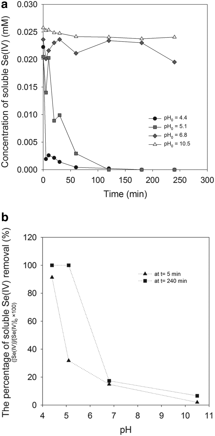
Effect of pH on soluble Se(IV) during treatment by the dithionite/UV ARP. Concentration of soluble Se(IV)
Figure 4 shows absorption spectra of solutions of dithionite at different pH and solutions of dithionite and selenite after different periods of UV irradiation. Dithionite solutions at pH above 5 show the expected peak at 315 nm, but that peak is not observed for the sample at pH 2.51, due to rapid decomposition of dithionite at low pH. The peak at 315 nm also disappears after irradiation for 5 min in the solution of selenite and dithionite. The fact that dithionite decomposes more rapidly at lower pH and that greater removal of Se(IV) occurs at lower pH, supports the hypothesis that the removal mechanism depends on products of dithionite decomposition.
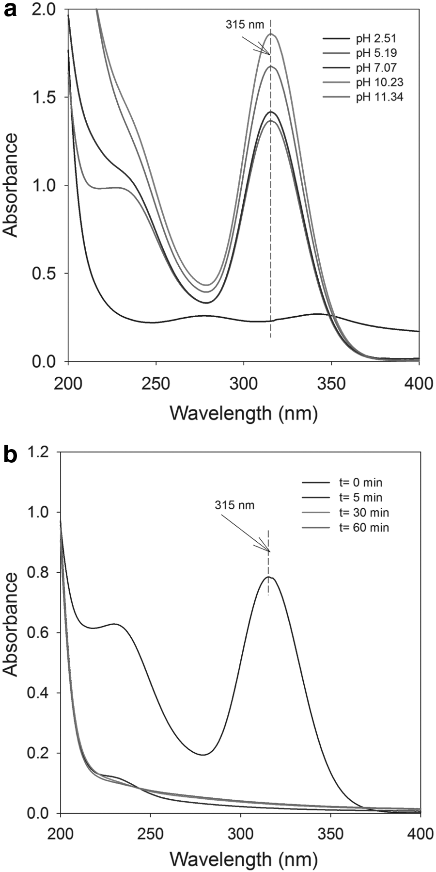
Absorption spectra of solutions prepared with dithionite alone
Effect of initial Se concentration
Figure 5 shows the effect of initial Se(IV) concentration on its removal by the dithionite/UV ARP and solution pH was also measured during treatment (Supplementary Fig. S6). In all of these experiments, the molar ratio of dithionite dose to initial Se(IV) concentration was constant at 40. The data plotted in Fig. 5 are relative concentrations (C/C0), so the negative slopes of the relative concentrations (−d[C/C0]/dt) are the rates of removal (−dC/dt) divided by the initial concentration. In such a plot, if the slopes for the different sets of data were all the same, that would indicate that the rates were proportional to the initial concentrations. Since the slopes in Fig. 5 tend to increase with increasing initial Se(IV) concentration, that means that the rates increase more strongly than linearly. This stronger response may be due to the presence of higher concentrations of dithionite when higher initial concentrations of Se(IV) were used. Figure 5 also shows that Se(IV) is removed to greater extents when initial Se(IV) concentrations increase. This is probably due to the higher concentrations of dithionite that result in it being present for a longer period of time. The rate of dithionite photolysis is controlled by the incident light intensity, so when a solution starts with more dithionite, it will take longer for it to completely photolyze. Additional experiments were conducted to evaluate the effect of initial Se(IV) concentration, but they were conducted at similar dithionite dose (Supplementary Fig. S7). At similar dithionite dose, the slopes of relative Se(IV) concentration were roughly the same, indicating that the rates of soluble Se removal increased with initial Se(IV) concentration.
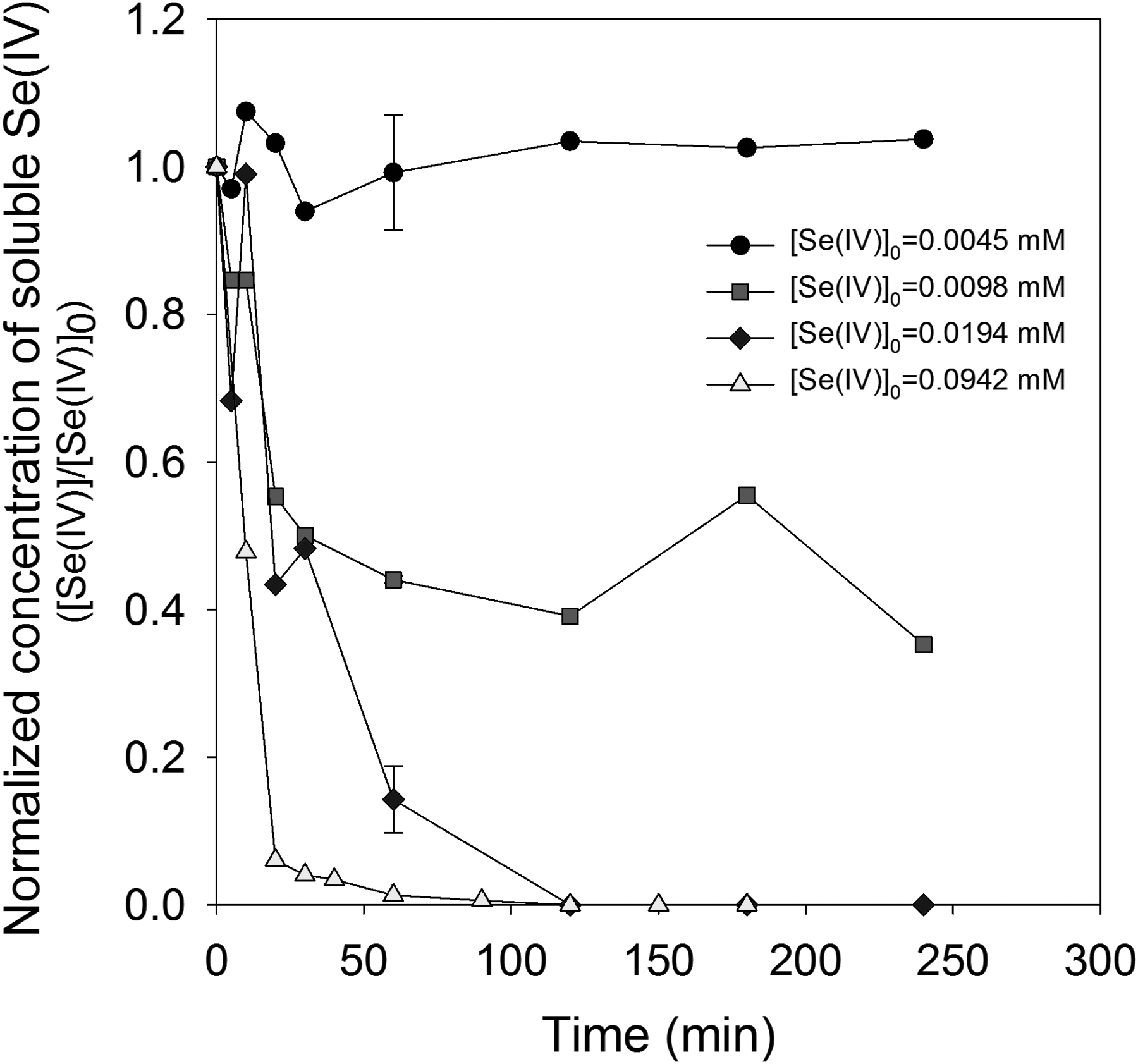
Effect of initial selenite concentration on soluble Se(IV) concentration during treatment by dithionite/UV ARP. Conditions: initial molar ratio of dithionite to Se(IV) = 40. UV-L light intensity ∼4,700 μW/cm2, and the initial pH ranged between 4.3 and 5.4.
Resolubilization was not observed in these experiments with Se(IV), although it has been observed in our concurrent study on As(III) removal by the dithionite/UV ARP (Jung et al., 2017a). The lack of resolubilization could be due to the selenium solid phases not being susceptible to reaction with photolysis products being produced in the system as were arsenic solid phases.
Effect of light intensity
Figure 6 shows the effect of incident light intensity on soluble Se(IV) concentrations during treatment by the dithionite/UV ARP. Higher light intensities resulted in more rapid Se(IV) removal, but eventually complete removal was observed since sufficient dithionite was provided. More rapid removal is probably due to the higher rates of photolysis caused by higher light intensities.
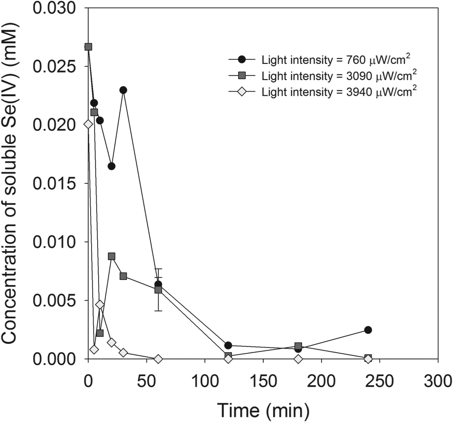
Effect of light intensity on soluble Se(IV) concentrations in dithionite/UV ARP. Conditions: [Se(IV)]0 = 0.023 mM, [S2O42−]0 = 1 mM, and initial pH = ∼pH 4.5 (not buffered).
Solid characterization
Orange–yellow-colored solids were formed during treatment by the dithionite/UV ARP, as shown in Supplementary Fig. S1. Geoffroy and Demopoulos (2009) found that red amorphous elemental selenium was formed by reactions of selenious acid with sodium dithionite at low pH. Our previous results showed that selenite in sodium sulfide solution was reduced to elemental Se or Se-S precipitates (Jung et al., 2016a). XRD patterns showed that gray elemental Se solid was a major solid in experiments in the absence of UV, whereas Se-S precipitates (Se3S5) with an orange color were found in those with UV irradiation. Duan et al. (2017b) have found that solids formed in Se(IV) solutions being treated by the dithionite/UV ARP were unstable due to resolubilization of Se(IV), regardless of UV irradiation. They proposed that the solids were elemental Se based on published data, but this was not confirmed with experimental evidence.
In this study, solids were formed during treatment by the dithionite/UV-L ARP, and they were characterized using SEM/EDS, XRD, and XPS analysis (Figs. 7–9 and Supplementary Figs. S8 and S9). In the experiment where the concentrations of initial Se(IV) and dithionite were 0.127 and 5 mM, respectively, the SEM image of the solids showed layered shapes of long rectangles (Fig. 7a) as well as irregularly shaped precipitates (Fig. 7b). The red circle in Fig. 7a identifies an area that EDS analysis showed to have a higher elemental intensity of Se (49.8%) than S (22.6%). The red circle in Fig. 7b identifies an area with higher elemental intensity of S (43.0%) than Se (9.5%). Fu et al. (2012) have described synthesized sulfur as forming particle shapes that appear melted and similar irregularly shaped particles are shown in Fig. 7b. Higher initial Se(IV) concentration (2.54 mM) was used to produce sufficient solids for XRD analysis. These solids were dark red or dark gray, unlike the orange-colored solids produced at lower initial Se(IV) concentrations (Supplementary Fig. S1). The SEM image of these solids shows them as aggregates of small irregular particles (Fig. 7c). XRD analysis showed an XRD pattern consistent with that of elemental Se (03-065-1876) (Fig. 8). However, some elemental S and mixtures of Se and S would be expected to be present. In our previous study on Se(IV) removal by sulfide/UV ARP (Jung et al., 2016a), orange-colored solids were formed, and they were identified by XRD analysis as having the formula Se3.02S4.98. Figure 9 shows the high-resolution Se 3d spectra of solids formed at low initial Se(IV) concentration (0.127 mM). These spectra were described by five peaks placed at binding energies of 56.3, 55.9, 55.6, 55.2, and 55 eV. According to the NIST XPS database, the binding energies of elemental Se range between 54.6 and 57. 5 eV, so all peaks shown in Fig. 9 were attributed to elemental Se (Supplementary Table S3).

SEM images for solids produced during treatment by dithionite/UV ARP. Conditions:
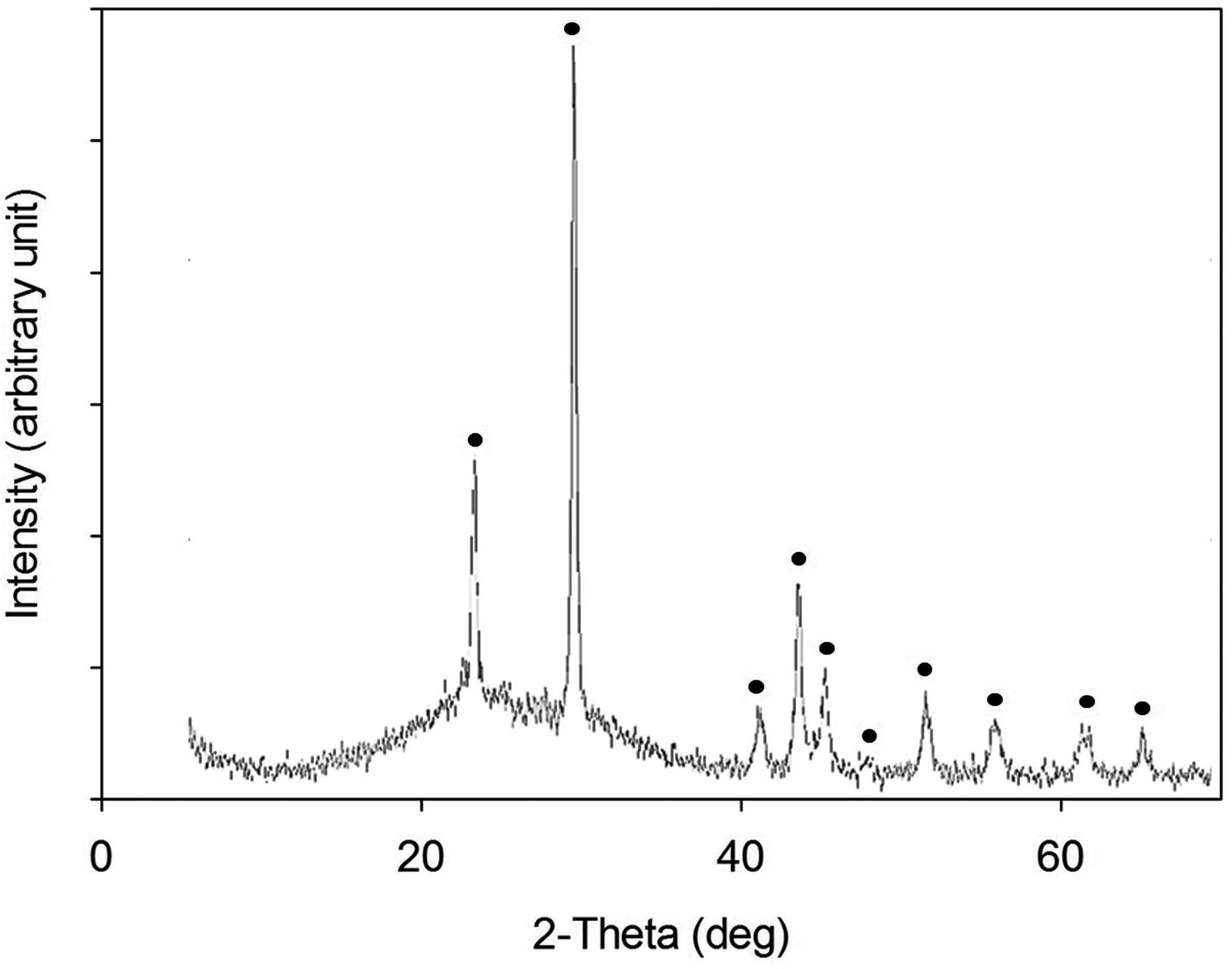
XRD pattern of solid formed during treatment by the dithionite/UV ARP. Conditions: [Total Se]0 = 2.54 mM, [S2O42−]0 = 7.75 mM, light intensity ∼5,300 μW/cm2, and pH 4.1 (not buffered). The red dots are XRD pattern of elemental selenium (Se0) (03-065-1876). XRD, X-ray diffraction.
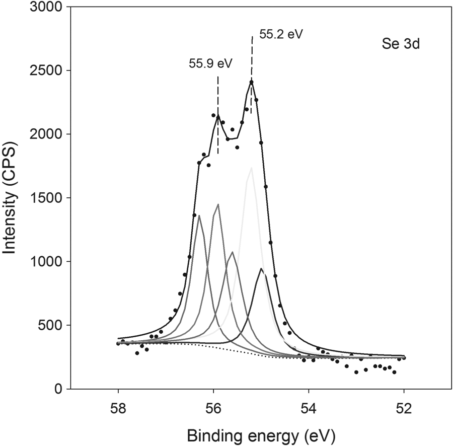
High resolution of Se 3d of solids produced during treatment by the dithionite/UV ARP. Conditions: [Total Se]0 = 0.127 mM, [S2O42−]0 = 12.7 mM, pH = 5.6 (not buffered), and UV-L light intensity ∼4,700 μW/cm2.
Effect of dithionite decomposition products
In the absence of irradiation, dithionite produces various decomposition products, such as bisulfite/sulfite/metabisulfite (HSO3

Comparison of soluble Se(IV) concentrations with different reducing agents. Conditions: [Se(IV)]0 = 0.022 mM, [S2O42−]0 = [HSO3−]0 = [S2O52−]0 = [S2O32−]0 = [S2O62−]0 ∼1 mM, and the initial pH and light intensities were pH 4.5 and 3,940 μW/cm2, pH 3.7 and 4,990 μW/cm2, pH 3.9 and 5,000 μW/cm2, pH 3.9 and 4,730 μW/cm2, and pH 3.8 and 4,730 μW/cm2 for experiments including dithionite, bisulfite, metabisulfite, thiosulfate, and trithionate, respectively.
Discussion
Our results are consistent with a mechanism, in which Se(IV) is removed from solution by formation of solid phases produced by reaction with photolysis products of dithionite or photolysis of compounds formed from nonphotolytic decomposition of dithionite. Dithionite can react to form two sulfur dioxide radicals in the absence of irradiation (S2O42
Trithionate is known to absorb UV light (Meulenberg et al., 1992), but no information could be found in the literature on its photolytic reactions. Tetrathionate (S4O62
In summary, there is not a single product of dithionite decomposition that can be clearly identified as the active agent of Se(IV) removal in solutions prepared from dithionite at low pH. As shown in Fig. 10, thiosulfate and trithionate showed rapid removal, but with diphasic behavior, and solutions with sulfite species showed rapid removal, but after an extended lag period. It appears that all of these species may play a role in Se(IV) removal and the relative importance of each is difficult to discern in complex mixtures formed in solutions of dithionite at low pH. Although more studies are required, it is clear that irradiated solutions prepared with dithionite at low pH led to the most effective removal of soluble Se considering both the kinetics and extent of Se(IV) removal.
Conclusion
Soluble Se(IV) was effectively removed in dithionite solutions at low pH with UV irradiation. Generally, reducing reagents (e.g., sulfite, ferrous iron) without activation were not able to reduce Se(IV). Various dithionite decomposition products formed by nonphotolysis reactions can undergo photolysis and be involved in soluble Se(IV) reduction. HSO3
Footnotes
Acknowledgments
This study was made possible by grants from the Qatar National Research Fund under its National Priorities Research Program award number NPRP 6-729-2-301 and NPRP 8-1406-2-605. The article's contents are solely the responsibility of the authors and do not necessarily represent the official views of the Qatar National Research Fund.
Author Disclosure Statement
The authors have declared no potential conflict of interest.
References
Supplementary Material
Please find the following supplemental material available below.
For Open Access articles published under a Creative Commons License, all supplemental material carries the same license as the article it is associated with.
For non-Open Access articles published, all supplemental material carries a non-exclusive license, and permission requests for re-use of supplemental material or any part of supplemental material shall be sent directly to the copyright owner as specified in the copyright notice associated with the article.