
Abstract
Layered double hydroxide (LDH) and LDH-graphene (LDH-G) composites were successfully fabricated using a facile one-step coprecipitation route. Scanning electron microscopy and energy dispersive spectroscopy, Brunauer–Emmett–Teller analysis, X-ray photoelectron spectroscopy, and X-ray powder diffraction patterns were used to characterize the composites before and after reacting with Fe(II). The LDH-G composites exhibited a noticeable enhancement in Fe(II) removal compared to the LDH. The maximum adsorption capacity was 654.66 mg/g for LDH-G. The better performance for Fe(II) removal was due to the unique structural characteristics of the LDH-G. The coexistence of oxidation and adsorption was the key factor for the improvement of Fe(II) removal in the presence of oxygen. The characterizations of the samples after the reaction reveal that the removal of Fe(II) was a complex process, including multiple surface adsorption and oxidation steps. The effective and stable Fe(II) removal capability of the LDH-G particles indicated great potential as an effective and environmentally friendly agent, which can replace existing technology for Fe(II) removal from groundwater.
Introduction
Exploring hierarchical composites by combining various building blocks together into a well-designed structure is a hot topic for material science (Zhao et al., 2012). The combination of various building blocks to form a novel composite can inherit advantages of materials and even lead to the multifunctional material with unexpected properties (Cao et al., 2016). Recently, the combination of graphene and layered double hydroxide (LDH) has been intensively investigated. LDH and graphene are both novel materials. The combination of both materials together to form a novel composite can take full advantage of each kind of material, which is an effective way to prepare multifunctional materials.
Graphene and LDH nanosheets usually suffer from irreversible aggregation owing to van der Waals interactions, which hinders their practical applications (Miao et al., 2015). Hybridizing graphene with LDH can effectively ameliorate the aggregation of the nanosheets and endow the composites with enhanced synergistic properties over the individual counterparts by combining the respective advantages of each component (Huang et al., 2012). Designing novel LDH-G hybrids for the removal of heavy metals has recently attracted enormous interest. Metal cations can be removed by LDH through precipitation, surface adsorption, isomorphic substitution, and chelation (Liang et al., 2013) (Tran et al., 2018). Graphene oxide (GO) contains oxygen functional groups such as epoxy (C-O-C), hydroxyl (-OH), and carboxyl groups (-COOH) on its basal planes and edges, which can provide bonding interactions to synthesize hybrids with other materials and enhance the adsorption of heavy metal ions (Komarala et al., 2016).
Gbb et al. (2017) reported the synthesis of three-dimensional (3D) porous Mg-Al LDH/pRGO and its adsorption of Pb(II) from aqueous solution with a maximum adsorption capacity of 116.2 mg/g. The high adsorption efficiency was due to a unique 3D porous network and the presence of oxygen-containing functional groups on graphene. Tan et al. (2015) also used rGO/LDH to remove U(VI) ions from aqueous solution, and the maximum adsorption capacity calculated from the Langmuir isotherm was 277.8 mg/g at 298 K. The GO-supported LDH can effectively prevent the agglomeration of LDH nanosheets by electrostatic repulsion or steric hindrance. This can increase the reactive active sites, thus improving the interaction activity with various heavy metal ions. Consequently, it is reasonable to speculate that novel LDH-G composites endowed with additional performance will be excellent candidates for realizing convenient heavy metal removal from contaminated water.
Iron is an essential nutrient for human beings and animals, which is widely spread in groundwater (Cairo et al., 2006; Doggaz et al., 2018). It usually exists in water as soluble forms of ferrous iron [Fe2+ or Fe(OH)+] or complexed forms of ferric iron [precipitate Fe(OH)3 or bacterial form] (Ghosh et al., 2008). Groundwater contains iron mainly due to the process of rain filtering through soil, rocks, and minerals. An industrial origin, such as mining, the iron and steel industry, and metal corrosion, can also discharge iron into the water (Chaturvedi and Dave 2012). The presence of iron in drinking water is not directly harmful to human health, but it is undesirable for its unpleasant taste, discoloration, and odor (Doggaz et al., 2018). Therefore, a permissible limit of Fe(II) in water is set as 0.3 mg/L for secondary drinking water according to the U.S. Environmental Protection Agency (EPA) (El kady et al., 2016).
Iron can be in divalent and trivalent forms with different physicochemical properties, so various physicochemical phenomena occurred for Fe(II) removal (Doggaz et al., 2018). Contact oxidation filtration is a common technique for Fe(II) removal from groundwater. Natural manganese sand is currently used as a filter with a main mechanism of catalytic oxidation (He et al., 2010). Fe(II) ions are adsorbed on the active filter membrane and then rapidly oxidized into Fe(III) under the catalytic action of the active filter membrane. Fe(OH)3·2H2O is then considered the main chemical composition of the active filter membrane (Yuhui et al., 2003). However, the Fe(OH)3 colloid is unstable, which may influence the effluent quality and cause coloration problems.
Previous studies have shown that LDH-G composites have superior adsorption capacities for various metal ions, and Fe(II) ions are likely to be adsorbed on the surface of LDH-G composites and oxidized in the presence of oxygen. To the best of our knowledge, no attempt has been made to remove Fe(II) by LDH-G composites through adsorption-oxidation, especially for the precise characterization of the detailed mechanism.
In the current work, LDH-G composites were fabricated using a facile coprecipitation method for the removal of Fe(II). The main objectives of this study were to evaluate the performance of the synthesized LDH and LDH-G composites for the removal of Fe(II) from aqueous solution. Emphasis is placed on the exploration of the mechanisms of Fe(II) removal on LDH-G composites compared with LDH composites.
Experimental
Materials
Purified natural graphite powder was purchased from Sinopharm Chemical Reagent Co. Ltd. Other reagents were analytical grade and purchased from Sigma-Aldrich (Shanghai) Co. Ltd. without further purification. Solutions were made anoxic (<0.02 mg/L) by boiling and cooling under a stream of N2 gas to remove dissolved oxygen. Glassware was soaked in dilute hydrochloric acid for 24 h and then rinsed thrice with distilled water before use.
Fabrication of LDH-G and LDH composites
GO was synthesized by Hummers methods, as described elsewhere (Hummers and Offeman, 1958). The LDH-G composites were synthesized using the coprecipitation method (Xie et al., 2014). In a typical procedure, 0.1 g of GO powder in 100 mL water was ultrasonicated for 1 h and then stirred vigorously with a magnetic stirrer. A 100 mL mixed metal solution of MgCl2·6H2O and AlCl3·9H2O was prepared in deionized water with a total metal ion concentration of 0.4 mol/L and a Mg2+/Al3+ molar ratio of 3:1. Another 100 mL alkaline solution of 0.6 mol/L NaOH and 0.2 mol/L Na2CO3 was also prepared. Both the mixed metal solution and alkaline solution were simultaneously dropwise added to the GO suspension under vigorous stirring at room temperature. The pH was maintained at 10 ± 0.5 during the process. The suspension products were stirred for another 4 h at room temperature and then aged in a water bath at 65°C for 4 h. The precipitate was centrifuged and thoroughly washed with distilled water until the washings were neutral. The precipitate was then dried at 65°C overnight. The resulting powder was designated LDH-G. For comparisons, pure LDH was prepared in a similar process but without the addition of GO.
Characterization
The images of the synthesized samples before and after adsorbing Fe(II) were captured by a scanning electron microscope (SEM) (JSM-7500F, Japan). The specific surface areas and pore structures of the samples were detected by nitrogen adsorption based on Brunauer–Emmett–Teller (BET) and Barrett–Joiner–Halenda (BJH) methods using N2 adsorption-desorption at 77 K on a surface area analyzer (ASAP2020). The X-ray diffraction (XRD) patterns were carried out by a powder diffractometer using Cu Kα radiation at a scanning speed of 2° min−1 (PANalytical B.V., Holland). X-ray photoelectron spectroscopy (XPS) tests were performed on an AXIS Ultra DLD (Shimadzu, Japan) using a monochromatic Al Kα X-ray source.
Experimental procedures
The experiments were carried out in a 250 mL glass conical flask by adding 0.2 g of adsorbents into 200 mL of Fe(II)-containing solution with continuous stirring over a magnetic stirrer at 500 rpm. If not otherwise specified, the solution pH and temperature in the Fe(II)-containing solutions were 6 and 30°C ± 2°C, respectively. All experiments were conducted in triplicate. For a predetermined time interval, supernatant was immediately filtered through a 0.45-μm membrane for complete particle removal. The supernatant was used to determine the concentrations of Fe(II) and total Fe [including Fe(II) and Fe(III)] measured using the 1,10-phenanthroline method (Tamura et al., 1974). The removal capacity qe (mg/g) and removal rate R (%) are calculated with Equations (1) and (2):
where C0 is the initial concentration of Fe(II) (mg/L), Ce is the equilibrium or residual Fe(II) concentration (mg/L), V is the volume of the solution (L), and W is the mass of adsorbent (g).
Results and Discussion
Characterization
The X-ray diffraction (XRD) patterns of the original GO, LDH, and LDH-G are presented in Fig. 1. In the XRD pattern of GO (Fig. 1a), a single broad peak of GO was observed at 12.3°, which corresponded to an average interlayer spacing of ∼0.72 nm and indicated the presence of oxygen-containing functional groups attached on the surface of the graphite sheets (Shen et al., 2011). For the pure LDH (Fig. 1b), the intense diffraction peaks indexed to the (003), (006), (009), (015), (018), (110), and (113) planes represented a typical hydrotalcite-like structure (Chao et al., 2018). The basal spacing (d003) of the pure LDH was ∼0.78 nm, which was consistent with the report in the literature (Yue et al., 2017).
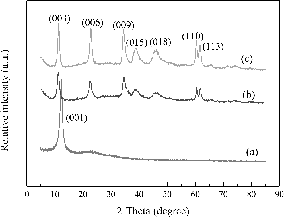
The XRD patterns of
In the case of LDH-G (Fig. 1c), the XRD pattern exhibited no peaks for GO, which was similar to the LDH and suggested that the LDH platelets were deposited on both sides of GO (Wen et al., 2013). The deposition weakened the surface energy of the graphene sheets and inhibited the restacking of the exfoliated GO (Miao et al., 2015). Furthermore, the diffraction pattern of the LDH-G composites exhibited sharp and intense peaks compared to that of the LDH phase. The GO phase gave a weak and broad peak, which was overshadowed by the strong LDH peaks. Hence, the identification of GO peaks in the XRD spectrum of LDH-G was difficult. It had also been observed that the XRD patterns of LDH-rGO composites do not show any reflections of rGO (Komarala et al., 2016).
The morphologies of the original samples were investigated by SEM observation. The SEM image of the pure LDH (Supplementary Fig. S1a) shows that the product was composed of a number of inhomogeneous flakes. The flakes were smooth and flat with most of the flakes stacked together to form agglomerates. For the LDH-G composites (Supplementary Fig. S1c), the flakes were loose and not compactly packed, and some of the staggered flakes enclosed an irregular 3D space (Zhou et al., 2019).
A favorable specific surface area was generally desired because it can provide many surface active sites to bind pollutants. The pore volume and BET specific surface area of the LDH-G composites were 0.52 cm3/g and 72.71 m2/g, respectively, which were larger than those of the pure LDH (0.32 cm3/g and 55.09 m2/g, respectively). In addition, the average pore sizes of the LDH and LDH-G were 22.58 and 28.36 nm, respectively, which suggested the presence of mesopores (2–50 nm) (Yu et al., 2018). It had been proven that the addition of GO can decrease the aggregation of LDH, resulting in exposed LDH sheets and an enlarged specific surface area (Fang et al., 2012). The porous and loosened 3D structures supported fast ionic conducting channels and provided potential adsorption sites for LDH-G application.
Fe(II) removal
The addition of LDH and LDH-G dose significantly influenced the Fe(II) adsorption. The variation of the adsorbent dose on Fe(II) removal is shown in Supplementary Fig. S2. As expected, the removal efficiencies of Fe(II) increased significantly with an increased adsorbent dose. In fact, the increase in adsorption of Fe(II) with the adsorbent dose was attributed to the enhancement of the adsorptive surface area and the availability of a large number of active surface sites (Abdi et al., 2017).
The pH dependence on the adsorption process of Fe(II) is shown in Supplementary Fig. S3b. The adsorption process was strongly dependent on the solution pH. The removal efficiencies of Fe(II) with LDH and LDH-G increased with increasing pH. Supplementary Figure S3a shows the zeta potential values versus pH for LDH and LDH-G; a decrease in zeta potential is noticed as the basicity of the solution was increased for both LDH and LDH-G. In the case of LDH, the surface charge was positive within the pH range 1–12, and the electrostatic repulsion between the positively charged surfaces and Fe(II) could be the reason for the low removal efficiencies that arose (Tran et al., 2018). For LDH-G, the surface charges became more negative with an increasing pH.
The surface of LDH and LDH-G were positively charged at a lower pH, implying that Fe(II) adsorption was favored at high pH. At lower pH, the concentration of H+ ions was high, which caused competition for adsorbent sites between the H+ ion and Fe(II) (Panneerselvam et al., 2011). Moreover, the dissolution effect of solid particles was also reduced as the pH increased (Zou et al., 2017). The enhanced Fe(II) adsorption for LDH-G at high pH (pH >4) can be explained by the strong electrostatic interactions between the negatively charged surfaces and positively charged Fe(II).
The kinetic behaviors of Fe(II) on LDH and LDH-G in the presence of oxygen at different initial Fe(II) concentrations are illustrated in Fig. 2. At an initial Fe(II) concentration of 100 mg/L, nearly all of the Fe(II) in the solution was removed by both the LDH and LDH-G within 60 min, but a faster velocity was obtained for the LDH-G. At an initial Fe(II) concentration of 1,000 mg/L, the LDH-G composites showed an apparent advantage over the pure LDH. The Fe(II) removal efficiency for the LDH-G reached 66.5%, which was obviously higher compared with the LDH (50.8%). The comparison between the LDH-G and the pure LDH particles for Fe(II) removal suggested that the LDH-G had better capacity and faster velocity than the LDH.

Removal of Fe(II) in presence of oxygen at different initial concentrations
The kinetic sorption data were simulated with a pseudo-first-order model and a pseudo-second-order model. The pseudo-first-order [Eq. (3)] and the pseudo-second-order [Eq. (4)] equations are generally expressed as (Tran et al., 2017):
where qe and qt (mg/g) are the amounts of Fe(II) adsorbed at equilibrium and at time t (min), k1 (1/min) is the rate constant of the pseudo-first-order model, and k2 is the rate constant of the pseudo-second-order model (g/[mg·min]).
The related kinetic parameters for the adsorption of Fe(II) on LDH and LDH-G are listed in Supplementary Table S1. The greater correlation coefficient values were obtained in the pseudo-second-order model compared with the pseudo-first-order model, which revealed that chemical adsorption was the rate-determining step.
The study of equilibrium adsorption plays a significant role in estimating the maximum adsorption capacity of adsorbents (Tran et al., 2019). The adsorption isotherms for Fe(II) on LDH and LDH-G are shown in Fig. 3. The Langmuir [Eq. (5)] and Freundlich [Eq. (6)] models were used to describe the adsorptive behavior of Fe(II) into the LDH and LDH-G samples:

The adsorption isotherms for Fe(II) on LDH and LDH-G.
where Ce (mg/L) is the final concentration of Fe(II) in aqueous solutions, qe (mg/g) is the adsorbed amount of Fe(II) on the solid phase, qm (mg/g) is the maximum adsorbed amount of Fe(II) per unit weight of solid, Kl (L/mg) is the Langmuir constant, which is related to the bonding and affinity of Fe(II) on adsorbents, and Kf (mg1−n·L n /g) is the Freundlich constant, which is related to sorption capacity.
The relative parameters calculated from the two models are listed in Supplementary Table S2. The Langmuir isotherm fitted the experimental data better than the Freundlich isotherm model, which suggested that Fe(II) adsorption was monolayer coverage. The maximum Langmuir adsorption capacities of Fe(II) for LDH and LDH-G were calculated to be 549.06 and 654.66 mg/g, respectively. The maximum adsorption capacity of Fe(II) on the prepared adsorbents in this study and other adsorbents in the literature is summarized in Supplementary Table S3. The synthesized materials in this study had removal capacities superior to those of many other adsorbents.
It is noteworthy that the white LDH powder was colored red with rust and deepened for high Fe(II) initial concentrations in the presence of oxygen. It is notable that Fe(II) was oxidized on the solid surface. Meanwhile, the black LDH-G remained that color and deepened over time; the color of the iron oxides was overshadowed by the black color of the GO. Hiemstra and van Riemsdijk (2007) reported that traces of oxygen immediately led to the transformation of Fe(II) into Fe(III). The effects of aeration rate on Fe(II) removal were investigated, as shown in Supplementary Fig. S4. Fe(II) removal efficiencies achieved more than 99% for both LDH and LDH-G, even at an aeration rate of 0.2 L/min. The results demonstrated that trace amounts of dissolved oxygen facilitated the surface oxidation of Fe(II) and enhanced the removal of Fe(II).
Controlled experiments were conducted in an oxygen-free system by bubbling N2 with a gas flow rate of 1 L/min at an initial Fe(II) concentration of 100 mg/L. In Fig. 4, the removal efficiencies of Fe(II) were sharply decreased and were under 50% for both LDH and LDH-G. In the absence of oxygen, the white LDH was colored with a pale yellow after contact with the Fe(II)-containing solution and deepened over time. The black LDH-G remained that color and deepened over time. It was speculated that oxidation did not occur with the protection of N2. The adsorption efficiency of LDH-G was higher compared with LDH, indicating that GO enhances the adsorption capacity of the LDH-G composites. GO takes effect in two aspects: one was increasing the adsorption sites due to an enlarged specific surface area (Tan et al., 2015). The other was due to chelation of the oxygen-containing group on the surface of GO (Gbb et al., 2017).

Removal of Fe(II) in absence of oxygen.
The enhanced Fe(II) removal capacity in the presence of oxygen implied that the removal pathway beyond adsorption was evident and suggested the existence of oxidation. Doping LDH with GO not only enhanced the Fe(II) adsorption capacity but also endowed the composites a superb structure that was beneficial for oxidation reactions. The oxidation effect on ferrous iron might proceed along the following two paths: (1) a homogeneous reaction that proceeded in the solution phase; and (2) a heterogeneous reaction that occurred on the surface of the solid (Choi et al., 2001).
To understand the oxidation path, controlled experiments without adding the composites were conducted under an aeration rate of 1 L/min. No oxidation was observed in Fe(II) solutions without a solid surface (data not shown), which confirmed that the autocatalytic effect was negligible. The acceleration of oxygenation was due to the catalytic effect of the LDH or LDH-G, which facilitated the oxygenation. Hiemstra and Riemsdijk (2007) reported that adsorbed Fe(II) was more easily oxidized if bound by a solid surface and catalyzed the transformation of Fe(III) minerals. Moreover, adsorbed Fe(II) provided new adsorption sites and served as catalysts for further adsorption and oxidation, thereby additional oxygen and Fe(II) were incorporated into the structure.
Mechanism
To further investigate the Fe(II) removal mechanisms of the LDH-G and LDH composites, the LDH-G and LDH samples were characterized by XRD, SEM/Energy Dispersive Spectrometer (EDS), and XPS after reacting with Fe(II) solution. The XRD patterns of the LDH-G after Fe(II) sorption under different conditions are depicted in Fig. 5. The LDH-G sample after contact with 100 mg/L Fe(II) solution under the protection of N2 (LDH-G-N2) and the LDH-G sample after contact with 100 mg/L Fe(II) solution in the presence of oxygen (LDH-G-100) exhibited the characteristic diffraction peaks of original LDH-G, which indicated that the main phase of the sample was still LDH-G. The main phases of the LDH-G sample after contact with 1,000 mg/L Fe(II) solution in the presence of oxygen (LDH-G-1000) were indexed as Fe2O3 and FeOOH (Peng et al., 2013; Sheshmani et al., 2015; Lingamdinne et al., 2017). There were no conspicuous peaks attributed to LDH-G, which suggested that the iron compounds completely covered and replaced the LDH-G phases.
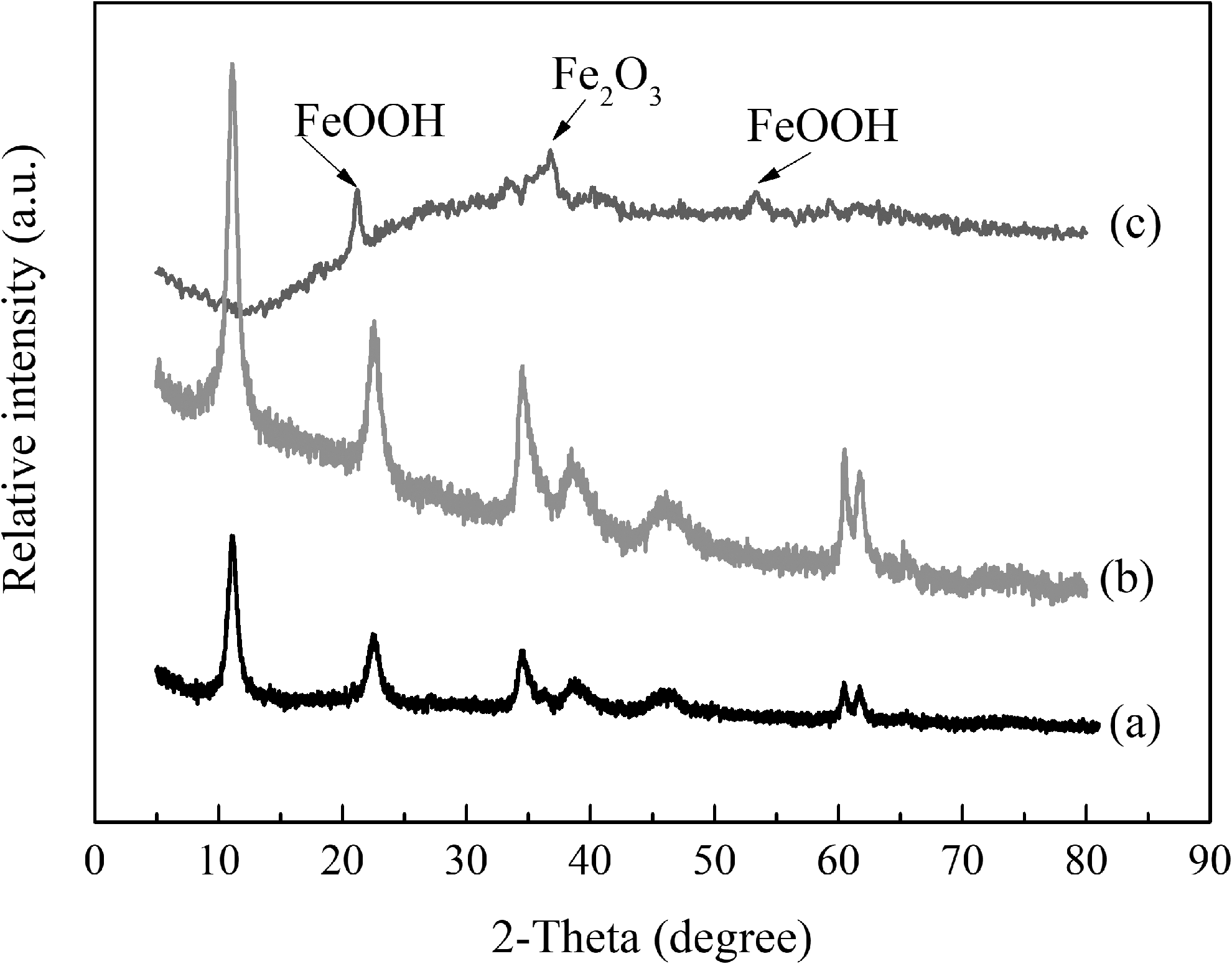
The XRD patterns of
The XRD patterns of the LDH after Fe(II) sorption are depicted in Supplementary Fig. S5 and are similar to the results of the LDH-G. Compared with contact oxidation filtration, Fe2O3 or FeOOH was formed on the surface of the LDH or LDH-G, which was more stable than Fe(OH)3. Hence, the removal of Fe(II) by adsorption and oxidation onto LDH or LDH-G was more effective and can avoid secondary pollution.
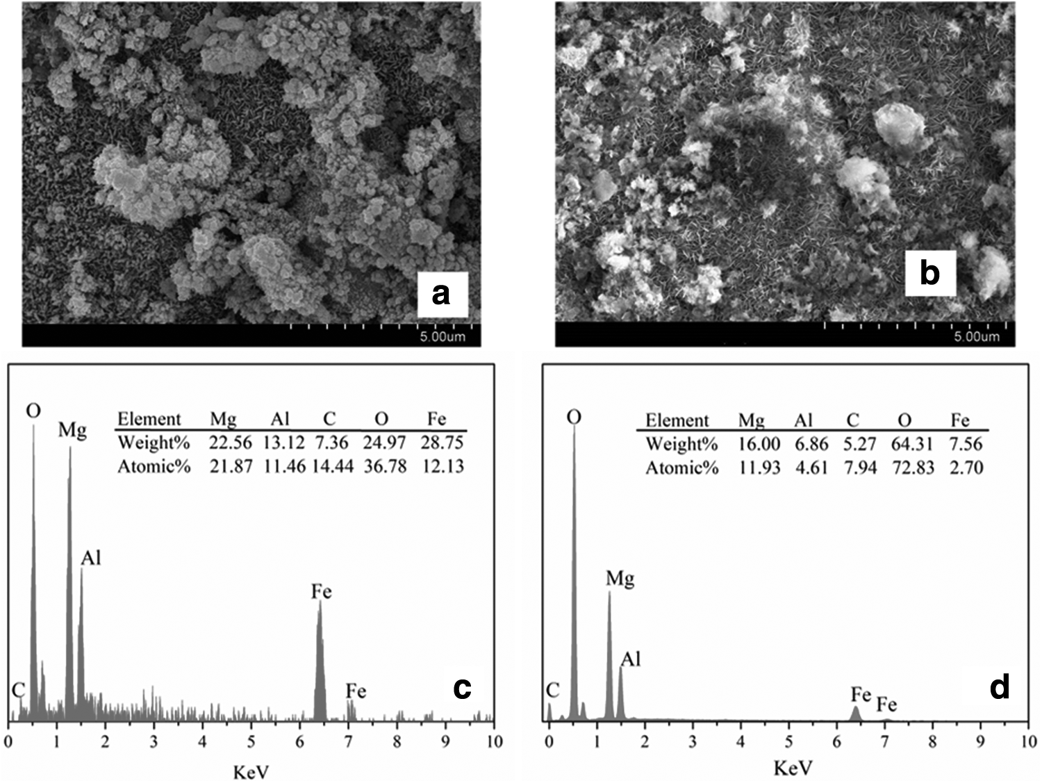
The SEM images of the LDH and LDH-G after Fe(II) sorption at an initial concentration of 100 mg/L are shown in Fig. 6. It is obvious that the morphologies were clearly different from the originals. The surface of LDH-100 was covered by iron compounds. The rod-like FeOOH and other agglomerates of iron compounds were observed on the surface of LDH-100. However, it was different from the case of LDH-G, and the morphology of the LDH-G-100 (Fig. 6c) had an obvious alteration. The flakes in the substrates were uniform, and most of the flakes were upright after reacting with Fe(II) at a concentration of 100 mg/L. The adsorption and oxidation of Fe(II) altered the structure and formed large pores, which were beneficial for mass transfer and chemical reactions on the surface of the absorbents.
The SEM of
The difference between LDH and LDH-G was due to the LDH-G composites having a larger pore volume and specific surface area, so Fe(II) was easily transferred into parallel lamellae and stretched the distance between each flake. Moreover, the surface functional groups of the LDH-G possibly formed intensive electrostatic and chemical interactions with the metal ions. These interactions dragged Fe(II) into the inner space and formed iron compounds between the LDH-G flakes, which loosened the parallel flakes and allowed them to become upright in orientation.
In addition, some agglomerated flocs were visualized, which suggested iron compounds. The EDS spectrum showed that the atomic content of Fe on the surface of the LDH (12.13%) was much higher compared with LDH-G (2.7%). This phenomenon that was interpreted as SEM/EDS revealed the morphology and element contents on the surface of the materials. Iron compounds were formed on the surface of the LDH, while they intercalated into the inner space of the LDH-G. The EDS spectra of LDH and LDH-G demonstrated that the main elements on the surface were still Mg, Al, and O, which confirmed that the main phases were still LDH and LDH-G. Moreover, XRD was used to confirm the crystal structure, but the quantity of iron compounds formed on the LDH and LDH-G was so low at an Fe(II) concentration of 100 mg/L that the reflection from the iron compounds on the LDH and LDH-G could not be identified in the XRD patterns.
The morphology of LDH-G-N2 (Supplementary Fig. S6) was similar to that of the pristine LDH-G, and the EDS spectrum showed only 0.6% (atomic%) of Fe loaded on the surface of the LDH-G. This indicated that LDH-G-N2 remained in its structure after Fe(II) sorption under N2 protection. The SEM image of the LDH-G-1000 (Supplementary Fig. S6) implied that the intersection structure was completely destroyed and replaced by iron compounds packed closely together, which was consistent with the XRD patterns. The atomic percentage of Fe increased from 2.70% to 13.87% when the initial concentration of Fe(II) increased from 100 to 1,000 mg/L. The atomic percentage of Mg decreased dramatically, which suggested that isomorphic substitution of Mg occurred when the composites were exposed to Fe(II) solution. Replacement ions must have the same total ionic charge and approximately the same size as those replaced (Liang et al., 2013). Fe(II) ions have the same total ionic charge and similar particle radius as Mg(II) ions, so the replacement occurred between Fe(II) and Mg(II).
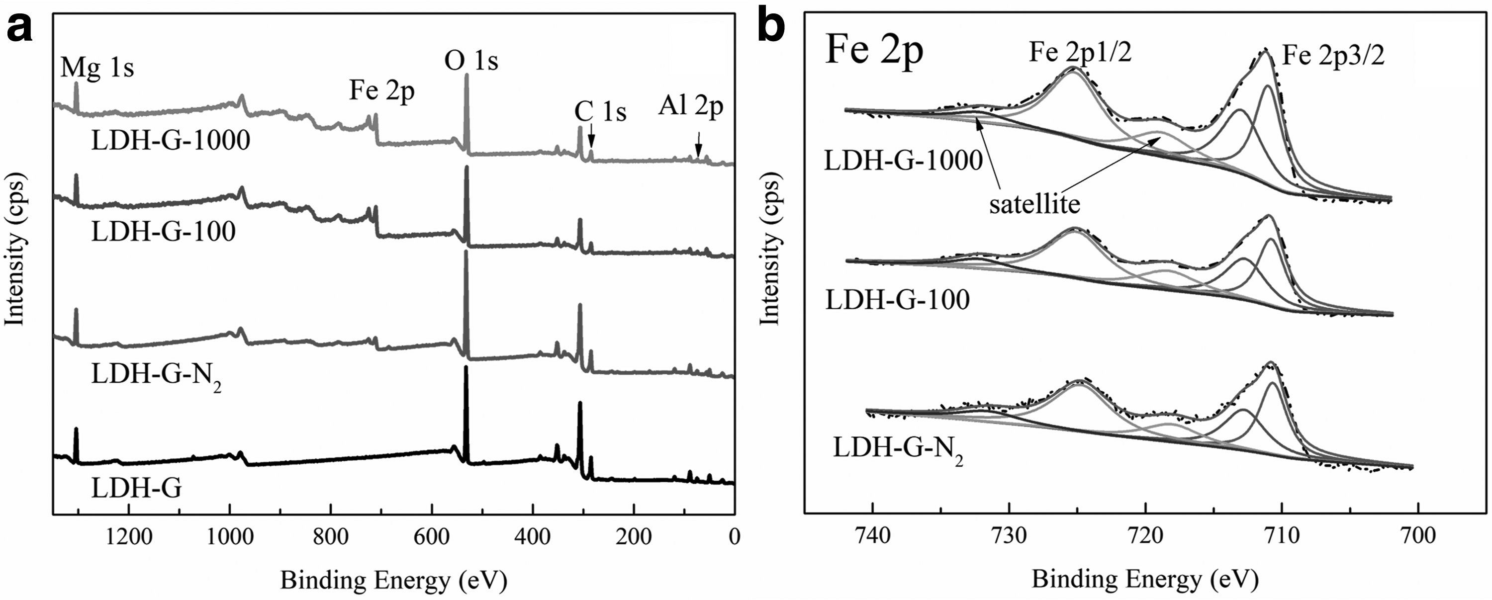
XPS is a surface characterization technique that provides qualitative and quantitative information from the outermost atomic layers. The XPS survey spectra of the LDH-G after Fe(II) sorption are depicted in Fig. 7a, and the element compositions determined by XPS are summarized in Supplementary Table S4. The surface content of Fe increased and the content of Mg decreased after adsorption of Fe(II), which confirmed the conclusion on the mechanism of isomorphic substitution between Fe and Mg. The most significant features in these spectra were the Fe 2p, O 1s, and C 1s signals. Therefore, the fitting results of the XPS spectra of Fe 2p, O 1s, and C 1s with the Shirley method of background subtraction are shown in Fig. 7b and Supplementary Fig. S6. All binding energies and Full Width at Half Maxima (FWHMs) are summarized in Table 1.
X-Ray Photoelectron Spectroscopy Results for Layered Double Hydroxide-Graphene Surface Before and After Removal of Fe(II) Under Different Conditions
FWHM, full width at half maxima; LDH-G, layered double hydroxide-graphene.
Figure 7a shows the wide scan XPS spectrum of the original LDH-G composites, and the photoelectron peaks at the binding energies of ∼293, ∼536, ∼1,307 and ∼80 eV are attributed to C 1s, O 1s, Mg 1s, and Al 2p, respectively. Both the O 1s and C 1s regions were deconvoluted into three peaks (Supplementary Fig. S7), which suggested the presence of oxygen-containing functional groups. The relative contents of the oxygen-containing functional groups were 45.72%, 38.15%, and 16.13% for 284.6 eV C-C, 285.1 eV C-O, and 288.9 eV C = O, respectively (Yang et al., 2013; Yu et al., 2017).
The wide scan XPS spectrum of the LDH-G after reacting with Fe(II) indicated the presence of Fe. The spectra of Fe 2p detected in the survey scan provided direct evidence for Fe(II) removal by the LDH-G. The peaks of Fe 2p1/2 and Fe 2p3/2 were observed at ∼725 and ∼711 eV, respectively. In addition, the satellite peak of Fe 2p1/2 at ∼733 eV and the satellite peak of Fe 2p3/2 at ∼719 eV were also seen (Wu et al., 2016). For a stoichiometry evaluation of Fe, only Fe 2p3/2 was analyzed, and Fe 2p1/2 was used as a reference for the fitting procedure based on previous literature (Volgmann et al., 2012; Li et al., 2014). The signal of Fe(II) peak contribution was found at ∼711.0 eV with different FWHMs obtained under different conditions. Another peak observed at a binding energy of 713.0 eV was assigned to Fe(III) (Yin et al., 2014). The presence of Fe(III) in the LDH-G-N2 was due to Fe(II) oxidation during the sample preparation process, which also indicated that the adsorbed Fe(II) was easily oxidized.
The detailed spectra of the O 1s are illustrated in Supplementary Figure S7. The peak located at ∼530 eV corresponded to an oxide-type oxygen (O2−), and the O 1s component at ∼531 eV accounted for a hydroxide (OH)-type oxygen (Yin et al., 2014; Wu et al., 2016). The binding energy side at ∼532 eV of the O 1s peak was assigned to C-O bonds and adsorbed water and oxygen (Li et al., 2014; Kwan et al., 2015). The amount of the oxide-type oxygen (O2−) increased and shifted to a lower binding energy with the enhancement of initial Fe(II) concentration, which indicated a larger amount of the oxide form. Combined with the Fe 2p line, it was concluded that Fe2O3 and FeOOH formed mainly on the surface of the LDH-G (Yin et al., 2014; Wu et al., 2018; Li et al., 2019).
Based on the present experiments and previous studies, it was concluded that the main Fe(II) removal mechanism by LDH-G was adsorption through isomorphic substitution under the protection of N2. Combined with the results of the XRD pattern, SEM/EDS, and XPS, it was concluded that both Fe2O3 and FeOOH formed on the surface of LDH-G after removal of Fe(II) in the presence of oxygen. The possible interaction mechanisms between Fe(II) and the LDH or LDH-G in the presence of oxygen included the following steps. First, Fe(II) in the aqueous solution was adsorbed onto the surface of the LDH or LDH-G through isomorphic substitution of Mg(II). Then, oxidation of the adsorbed Fe(II) occurred by aeration. GO played an important role in the Fe(II) immobilization due to the aggregate-preventive function for the LDH sheets and its intense inherent adsorption affinity for metal ions. In addition, the oxidation products of Fe(II) provided additional sites and further adsorbed and oxidized Fe(II) in the solution.
Larese-casanova and Scherer (2007) demonstrated that an Fe(II)-Fe(III) interfacial electron transfer occurred during the sorption of Fe(II) on hematite, and the Fe(II) was oxidized by the structural Fe(III) in hematite. In this study, electron transfer was not observed under the protection of N2. It was believed that Fe(II) was oxidized by aeration in the system, and the adsorbed Fe(II) quickly underwent electron transfer with oxygen. Fe(II) removal by the LDH-G composites was controlled by surface adsorption through isomorphic substitution, oxidation, and surface complexes as vividly illustrated in Fig. 8, along with the LDH composites.
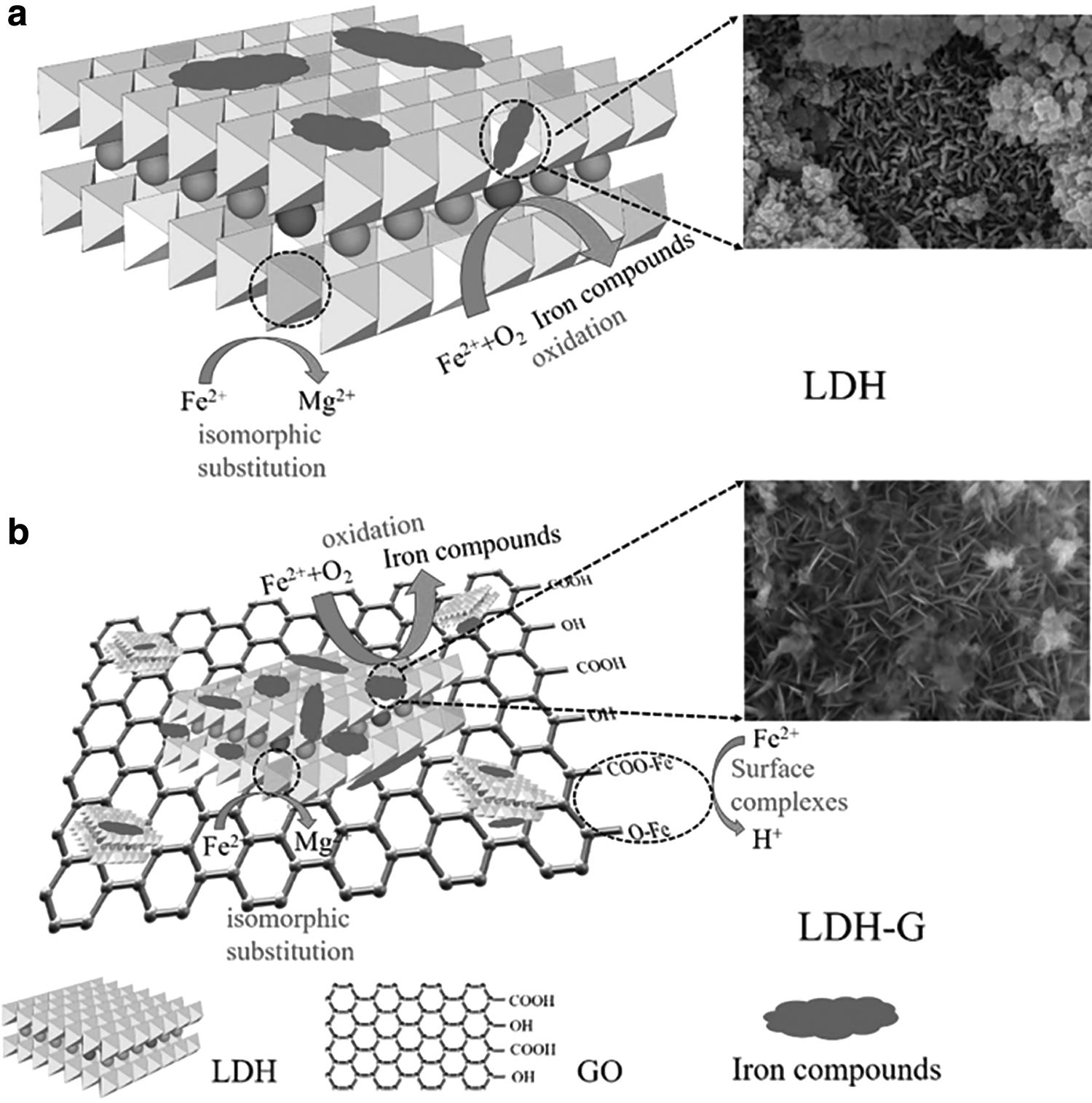
Schematic illustration of the probable mechanism for the removal of Fe(II) on
Conclusion
Highly efficient performance for Fe(II) elimination can be realized using LDH-G, and the maximum adsorption capacity is 662.9 mg/g. The obtained hierarchical LDH-G composites exhibit significant Fe(II) removal performance that is accredited to the combination of several advantageous features compared with the LDH. First, the higher surface area and abundant surface active sites of the LDH-G result from the ultrathin sheets and hierarchical structure, which are beneficial for the enhanced adsorption and oxidization toward Fe(II), especially under high Fe(II) concentrations. In addition, the functional oxygen-containing groups of the composites make the interlayer negatively charged, which can attract the positively charged Fe(II) into the inner structure of the LDH-G composites. In summary, the GO-supported LDH is a potential application for Fe(II)-containing wastewater treatment.
Footnotes
References
Supplementary Material
Please find the following supplemental material available below.
For Open Access articles published under a Creative Commons License, all supplemental material carries the same license as the article it is associated with.
For non-Open Access articles published, all supplemental material carries a non-exclusive license, and permission requests for re-use of supplemental material or any part of supplemental material shall be sent directly to the copyright owner as specified in the copyright notice associated with the article.