




Abstract
The biological response of Chlorella to nanometal oxides is the basis for expanding the engineering application of Chlorella and inorganic metal oxide nanoadsorbents in the treatment of nitrogen and phosphorus wastewater. The objective of this study was to ascertain the response of Chlorella to nano-ZrO2 and nano-Mg(OH)2 either free or immobilized to anion exchange resin D301. Our results show that immobilization of ZrO2 and Mg(OH)2 to D301 was significantly less harmful (p < 0.05) to Chlorella than free nano-ZrO2 and nano-Mg(OH)2. Chlorella has greater tolerance and positive biological response to fixed nanometal oxides, especially to D301-ZrO2 (nano-ZrO2 immobilized on D301), which can significantly stimulate the growth of Chlorella. Scanning electron microscopy observations indicated that most of algal cells treated with free nano-Mg(OH)2 and nano-ZrO2 were entrapped in the aggregates of nanoparticles and were both deformed and atrophied in appearance. In contrast, D301-Mg [nano-Mg(OH)2 immobilized on D301] immobilized beads demonstrated relatively few malformed algal cells on the surface, and D301-Zr showed no indications of deformed or atrophied cells either within or on the immobilized beads. Therefore, immobilization appears to have obvious effect on reducing the toxicity of powder nanoparticle and carrier, and the immobilization of ZrO2 and Mg(OH)2 on D301 might provide a novel application to Chlorella wastewater treatment to improve algae wastewater treatment efficiency, as well as these sorts of immobilized nanoadsorbent might be regenerated using Chlorella.
Introduction
Inorganic metal oxide nanoparticles have been developed as adsorbents due to their higher surface area to volume ratio and high selectivity (Singh et al., 2018), and are commonly used in the wastewater treatment to remove phosphorus (Zhang et al., 2013a, 2016; Wang et al., 2015a), heavy metals (Pan et al., 2007; Zhang et al., 2009; Teng et al., 2015; Rachna and Rimmy, 2019), and other pollutants (Qiao et al., 2019; Gusain et al., 2019). To solve the application defects of powder (free) inorganic metal oxide nanoparticles, such as pore plugging, aggregation, pore collapse, difficult solid-liquid separation and so on, the immobilized nano-oxide/hydroxide is then prepared and applied (Zhang et al., 2013b, 2015; Wang et al., 2015b).
In addition, researchers have begun to pay attention to the potential adverse effects of powder inorganic nanoparticles on organisms for the concerns that large amounts of these inorganic nanoparticles were either purposefully or inadvertently released to the environment during their applications (Moore, 2006). The toxic effects of metal oxide nanoparticles on microorganisms have been reported (Lin et al., 2005; Ji et al., 2011; Quigg et al., 2013; Iswarya et al., 2015). CuO, ZnO, TiO2, Ag, and Silica nanoparticles all have been shown to inhibit the Chlorella growth (Ji et al., 2011). However, numerous studies have also demonstrated that nanometal oxides in a suitable concentration range have a biopromoting effect on some bacteria (Gong et al., 2011). Nanometal oxides can stimulate the physiological activity of bacteria (Kuang et al., 2013), thereby accelerating the environmental bioremediation (Wannoussa et al., 2015).
It is well known that the main nutrients required by Chlorella are NO3−, PO43−, Fe3+, and so on, and so Chlorella can be used to remediate nitrogen and phosphorus wastewater (Wang et al., 2019). In addition, Chlorella contains protein, vitamins, unsaturated fatty acids, active polysaccharides, oils, fats, and so on, and it is the bait for fish, shrimp, and other aquatic organisms (Gladue and Maxey, 1994) and also is a feasible feedstock for biofuel production (Khoo et al., 2019). Therefore, a series of interesting speculations have been triggered. For example: Could adding nanometal oxides improve the efficiency of Chlorella in wastewater purification? Could the replacement of free metal nano-oxides with supported metal nano-oxides avoid the damage to Chlorella caused by the agglomeration of free metal nano-oxides? Could the inorganic metal oxide nanosorbents be regenerated by Chlorella after they were exhausted in the treatment of nitrogen and phosphorus wastewater? Could the damage of nanomaterials to Chlorella be reduced due to the immobilization? Could the bioregeneration of the immobilized material by Chlorella be realized? The above all scientific problems need to observe the biological response of Chlorella at different concentrations of nanometal oxides before and after the immobilization. In other words, the biological response of Chlorella to nanometal oxides before and after immobilization should be the basis for expanding the engineering application of Chlorella and inorganic metal oxide nanoadsorbents in the treatment of nitrogen and phosphorus wastewater.
In view of this, the present study focused on the biological response of Chlorella to some common nanometal oxides and their corresponding carrier-supported composites, and also explore whether Chlorella shows greater tolerance and positive biological response to the fixed nanometal oxides. The results should provide necessary information for improving algae-wastewater treatment technology, upgrading of nanometal oxides, and the bioregeneration of immobilized composites.
To achieve the aim stated above, free nano-ZrO2, nano-Mg(OH)2, and their immobilized composites D301-Zr and D301-Mg were first prepared as the models of free and immobilized inorganic metal oxide nanoparticles, respectively, since these inorganic metal oxide present different zeta potential (ZP), solubility, and stability (Parks, 1965). Afterward, the response of Chlorella was evaluated and compared to either free ZrO2 and Mg(OH)2 particles or the polymeric support D301 in multiple biological end-points, including growth rate, chlorophyll fluorescence, and photosystem II quantum efficiency. In addition, the observation of scanning electron microscopy (SEM) was also performed as the evidence for the evaluation.
Materials and Methods
Materials
All reagents were pure and of analytical grade and purchased from Tianjin Reagent Station Co. (Tianjin, China). D301, a macroporous anion exchange resin with total capacity of 4.0 meq/g was purchased from Ningbo Glory Resin Co. (Zhejiang, China). D301 particles were purified by sieving (0.6 mm), washed with 1% sodium hydroxide and 1% hydrochloric acid, and finally with deionized water to remove the residue impurities until neutral pH (7.0). Purified D301 was then vacuum-desiccated at 60°C until reaching the constant weight for further experiments.
The unicellular green algae, Chlorella sp. used in this study were kindly provided by College of Marine and Environmental Science, Tianjin University of Science and Technology, China. Chlorella was cultured in f/2 medium at 25°C, under an 4000 lx of 12 h light/12 h dark cycling illumination (GXZ Intelligent light incubator; Jiangnan CO., Ningbo, China), with intermittent shaking (three times daily). Chlorella response experiments were carried out as described by Lin et al. (2005). The Chlorella cells, in midexponential growth phase, were harvested by centrifugation at 4000 g for 10 min, washed three times with 0.2 mg/L phosphate buffer solution, and resuspended in sea water (f/2 medium) at a concentration of 106 cell/mL for response experiments. The algal cell concentration was determined by measuring the absorbance at 680 nm according to calibration curves obtained previously.
Nanoparticles preparation
Nano-Mg(OH)2 was prepared by first dissolving 25 g magnesium nitrate into 100 mL deionized water, to which 400 mL 2% NaOH was added to the solution and stirred over night at room temperature. Nano-ZrO2 was prepared by first dissolving 15 g zirconyl chloride into 100 mL alcohol, to which 1500 mL 2% NaOH was added to the solution and stirred over night at room temperature. Following the overnight stir at 250 rpm, respective solutions were centrifuged at 1600 g for 10 min (80-2 Electric Centrifuge, Chang Zhou, China) to remove the respective solvents. Nanoparticles were then washed and centrifuged several times with deionized water until the pH of effluent water reached 7.0. Finally, the purified Mg(OH)2 or ZrO2 nanoparticles were vacuum-desiccated at 60°C to constant weight for subsequent use.
The hybrid materials (D301-Mg, D301-Zr) were fabricated by precursor diffusion and an in situ precipitation process. D301-Mg was prepared by immersing 5 g of treated D301 into 100 mL of a 1 M magnesium nitrate solution and stirred at 250 rpm at room temperature for water evaporating off to ensure the dispersion of magnesium nitrate into the inner pores of D301. The resulting beads were then added to 400 mL of a 2% NaOH solution and stirred at 250 rpm for 16 h. These products were desiccated at 60°C in a vacuum oven for nano-Mg(OH)2 immobilization, resulting in the successful yield of hybrid D301-Mg. Final D301-Mg was screened with a 0.6-mm-diameter sieve and rinsed with deionized water until neutral pH before experimental use.
Likewise, preparation of D301-Zr, the procedure was similar with that of D301-Mg, with the exception of using 15 g of ZrOCI2 as the precursor dissolved into anhydrous ethanol. This solution was then stirred at 250 rpm until dissolved, at which point 1500 mL 2% NaOH was added and stirred for 16 h. The resulting solution was vacuum desiccated. Before use, they were screened with a 0.6 mm diameter sieve and washed with deionized water until neutral pH.
To calculate the content of supported nanoparticles on D301, 1000 beads of treated D301 were randomly selected, weighed (W0), and used for immobilization. The final immobilized beads were screened with 0.6 mm diameter sieve to remove any crushed beads, the intact immobilized beads were weighed (Wt) and counted (n). Three replicates were conducted, then the nanoparticle content [Mg(OH)2 or ZrO2] loaded on the D301 was calculated by the following formula:
Characterization of nanomaterials and surface topography of algal cells
Algal cells destined for ultrastructural and morphological analyses were harvested at the 48 h time point. All algal samples were fixed in 2.5% glutaraldehyde in phosphate buffer at 4°C for at least 24 h after incubation. The specimens were then dehydrated in an ethanol series (30%, 50%, 70%, 80%, 90%, and 100%, 15 min in each treatment) at room temperature. SEM (S-4800 II; Hitachi, Japan), coupled with an energy dispersive X-ray spectrometer (EDS; Horiba, Japan), was used to observe the morphology of algal cells treated by each nanomaterial as well as the surface morphology and element analysis of Mg(OH)2, ZrO2, D301, D301-Mg, and D301-Zr. Besides, the crystalline pattern of nanoparticles was analyzed by X-ray diffraction (XRD) using an X-ray Diffractometer (Rigaku, Inc., Tokyo, Japan).
Algal response tests
Response tests were performed by adding nano-Mg(OH)2, nano-ZrO2, D301, D301-Mg, or D301-Zr in their respective concentrations to sterilized Erlenmeyer flasks, each containing 200 mL algal suspension (4 × 10⁶ cell/mL). According to the lethal concentration of each material for Chlorella tested in the preexperiment, the concentration gradient was set on both sides of the lethal value. Nano-Mg(OH)2 concentrations were prepared as: 0, 0.5, 1.0, 2.0, 3.0, 8.0, and 10.0 (g/L); nano-ZrO2 concentrations were prepared as: 0, 2.5, 5.0, 7.5, 10.0, 12.5, 15.0, 20.0, and 25.0 (g/L); D301 concentrations were prepared as: 0, 5.0, 6.0, 7.0, 8.0, 10.0, 15.0, 25.0, and 30.0 (g/L); D301-Mg concentrations were prepared as: 0, 2.0, 4.0, 8.0, 16.0, 32.0, and 45.0 (g/L); and D301-Zr concentrations were prepared as: 0, 1.0, 2.0, 4.0, 8.0, 16.0, and 32.0 (g/L). The experiment was conducted in three replicates.
Serial samples were taken from each conical flask at intervals of 0, 24, 48, 72, and 96 h for determination of the algal cell density, soluble protein content, and chlorophyll-a content. Each experiment was conducted in triplicate. Algal cell number was determined using a hemocytometer (Kaihong, Beijing, China) under a microscope (E600; Nikon, Tokyo, Japan), and the 96-h growth rate (%) was calculated using the formula:
To measure soluble protein content, 8 mL algae sample of each serial test was centrifuged at 1600 g at 2°C for 5 min to obtain the cells fraction and washed with ice-cold phosphate buffer solution three times and restored to their initial volumes (8 mL) with ice-cold phosphate buffer solution. Cell suspensions were homogenized with ultrasonic cell disruptor (Ningbo Xin Zhi Biological Technology Co., China) on ice at 400 W for 15 min, and then, homogenates were centrifuged at 1600 g at 2°C for 5 min to obtain the supernatant fractions. One milliliter Coomassie brilliant blue solution was added to 5 mL of the algal supernatant and incubated for 5 min, upon which time absorbance was measured using a spectrophotometer (595 nm). Soluble protein content (mg/L) was calculated by the following formula:
C was determined by the absorbance according to calibration curves obtained previously; VT: volume of extracting solution; VS: volume of sample.
The total 96-h soluble protein variation (%) of Chlorella cells response to nanoparticles either free or after being immobilized was calculated using the following formula:
Chlorophyll-a content as the main index of photosynthesis was extracted by the hot-ethanol method (Feng et al., 2013). Ten milliliters of each algal culture was sampled and extracted using 90% ethanol extracted at 75°C in water bath, and measured by spectrophotometer at 630, 643, 664, and 750 nm, respectively. The chlorophyll-a content was calculated by the following formula:
where D is the absorbance at different wavelength.
The total 96 h chlorophyll-a variation (%) of Chlorella cells response to nanoparticles either as free or immobilization was calculated using the formula:
Statistical analysis
Statistical analysis was performed using SPSS 11.0 statistical software with significant differences (p < 0.05) among means being tested by one-way analysis of variance followed by Duncan's multiple range tests. Data are reported as mean with standard error.
Results and Discussion
Characterization of D301-Mg and D301-Zr composite materials
The synthesized composite materials, D301-Mg and D301-Zr, were well characterized by SEM, EDS, and XRD analysis. Figure 1 illustrates the SEM images and their spectrograms of SEM-EDS for the carrier D301 (Fig. 1A, B), D301-Mg (Fig. 1C, D), and D301-Zr (Fig. 1E, F). SEM images revealed that the formed nano-Mg(OH)2 and nano-ZrO2 particles successfully deposited on the pore surface of D301 (Fig. 1C, E). As depicted in Fig. 1C and E, the encapsulated Mg(OH)2 exhibits a deciduous leaf-like thin flake structural morphology (Fig. 1C), and the encapsulated ZrO2 exhibits an irregular protuberance structural morphology (Fig. 1E), both structures indicated the potential larger surface area. Moreover, the spectrograms of SEM-EDS further demonstrated the successful Mg and Zr implantation (Fig. 1D, F), the intensity for Mg and Zr element indicate the high level of the deposited nanoparticles. In contrast, the carrier D301 demonstrated no detectable Mg and Zr element (Fig. 1B).

SEM-EDS images of materials.
The XRD pattern (Fig. 2) also indicates the successful Mg(OH)2 and ZrO2 nanoparticle immobilization by D301. The diffraction pattern for nano-Mg(OH)2 displayed eight obvious peaks at 18.5°, 32.9°, 38.0°, 50.8°, 58.7°, 62.1°, 68.2°, and 72.1° (Joint Committee on Powder Diffraction Standards [JCPDS] no. 44-1482) (Fig. 2A). The XRD pattern of D301-Mg presented a single broad peak near 18.5° and two smaller peaks at 38.0° and 58.7° (JCPDS no. 44-1482) (Fig. 2A), which indicates the presence of fewer low-crystalline Mg(OH)2 nanoparticles on the surface of D301. The XRD pattern of nano-ZrO2 presented a single broad peak at 29.7° and three small sharp peaks, but the broad peak and the small sharp peak at 59.2° were not observed in the XRD pattern of D301-Zr. In fact, all the ZrO2 peaks of D301-Zr were weak (JCPDS no. 37-0031), which indicates that the nanoparticles of ZrO2 immobilized on D301 were all amorphous crystals.
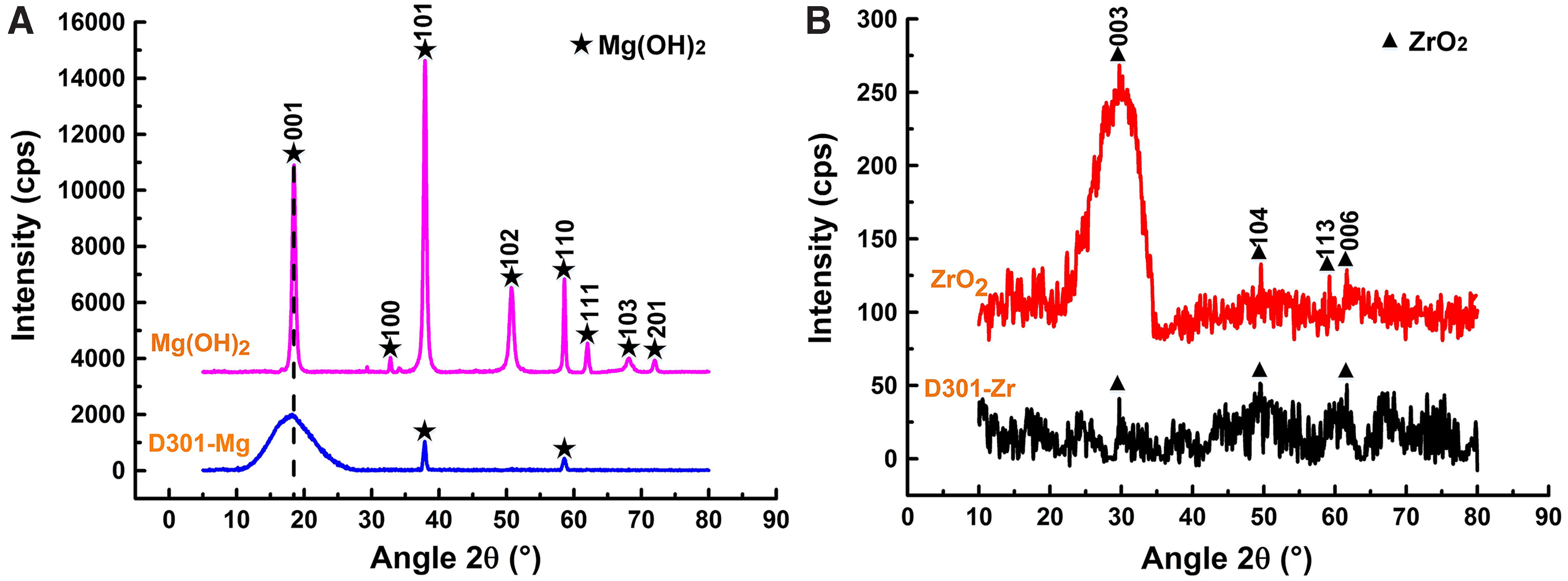
XRD patterns of Mg(OH)2 and ZrO2 loaded into the porous surface of D301.
Algal response tests to carrier, nanoparticles, and composite materials
For the algal response to nano-Mg(OH)2, nano-ZrO2, D301, D301-Mg, and D301-Zr, cell growth, soluble protein, and chlorophyll-a were measured using the cells in midexponential growth phase.
The algal cell numbers measured are plotted in Fig. 3. The individual effects of nano-Mg(OH)2, nano-ZrO2, and D301 on cell growth were depicted in Fig. 3A–C. The addition of D301 (Fig. 3A), Mg(OH)2 (Fig. 3B), or ZrO2 (Fig. 3C) at all the concentrations inhibited the growth of Chlorella to varying degrees compared with that of the control (blank) group, and the lethal effect occurred for these three materials at the different concentrations (D301 > 8.0 g/L, Mg(OH)2 ≥2.0 g/L, ZrO2 >10.0 g/L). These data indicate that free nanoparticles [Mg(OH)2 and ZrO2], as well as D301, are toxic to algae.
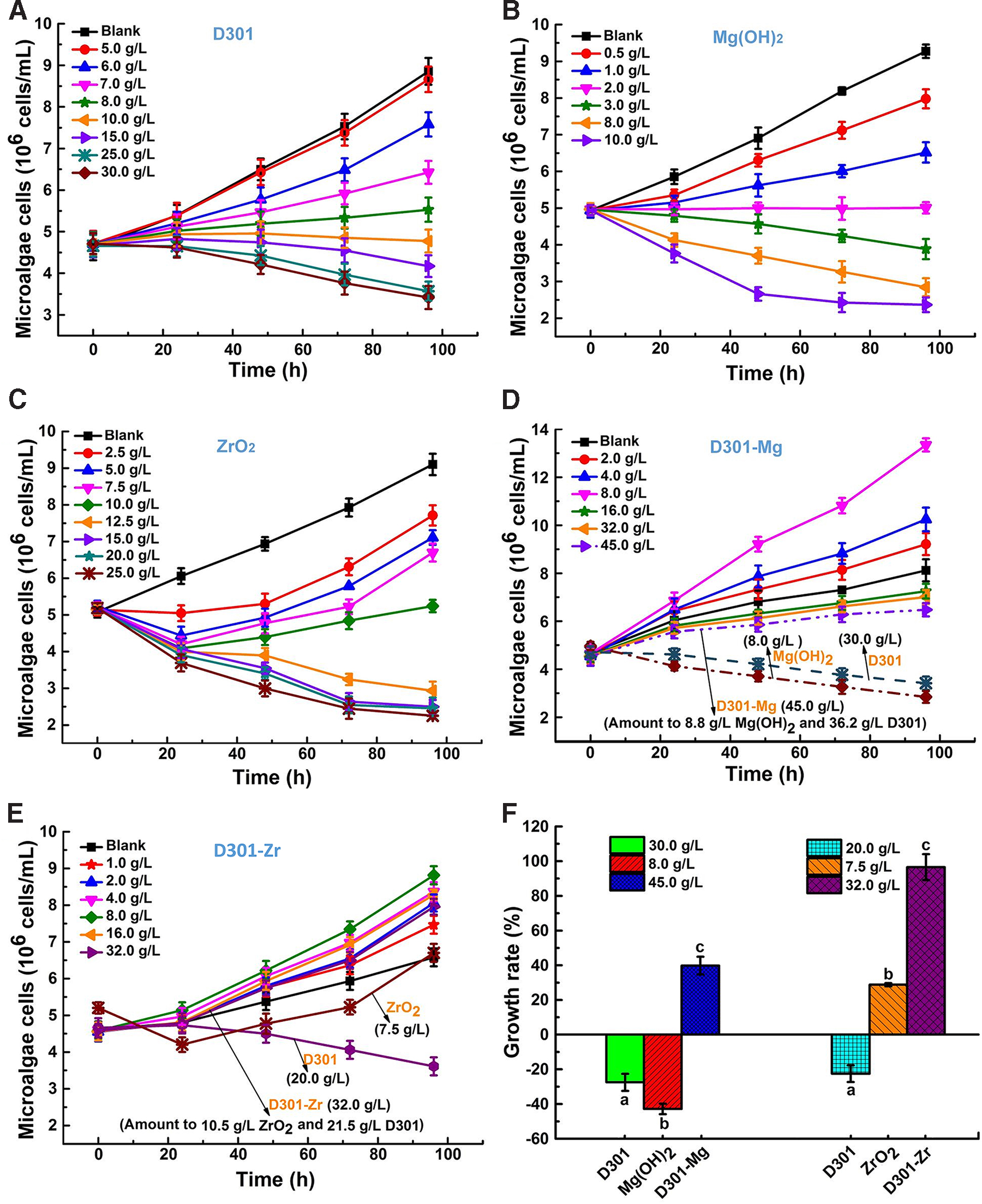
The effects of materials on Chlorella growth and growth rates versus time in algae exposed to different concentrations of materials.
In contrast to free form, the D301-Mg inhibited algal cell growth at much higher concentrations (16.0, 32.0, and 45.0 g/L; Fig. 3D). Remarkably, the concentration of D301-Mg, 45.0 g/L [45.0 g D301-Mg = 8.8 g Mg(OH)2 immobilized within 36.2 g D301] had much less adverse effect than that of these ingredients separately at 8.0 g/L Mg(OH)2, or 30.0 g/L of D301 (Fig. 3D). The addition of D301-Zr, at all the concentrations, did not show any adverse effects on the growth of Chlorella compared with that of each control group, where D301-Zr at 32.0 g/L solution (32.0 g D301-Zr = 10.5 g ZrO2 immobilized within 21.5 g D301) permitted more cell growth than that of suspension containing the equivalent weight of free ZrO2 (7.5 g/L) and D301 (20.0 g/L; Fig. 3E). It seems that D301-Zr, at all the concentrations, can significantly stimulate the growth of Chlorella compared with that of each control group.
In addition, the growth rates (%) in the presence of materials also showed significant effect (Fig. 3F). When compared to control treatments at the 96-h time point, the growth rates of algal cells treated with 30.0 g/L D301 was −27.5%, 8.0 g/L Mg(OH)2 was −42.8%, and 45 g/L D301-Mg [45 g D301-Mg = 8.8 g Mg(OH)2 immobilized within 36.2 g D301] was 39.8%. Similarly, the algal cell growth rates treated with 20.0 g/L D301 was −22.5%, 7.5 g/L ZrO2 was 28.8%, and 32.0 g/L D301-Zr (32.0 g D301-Zr = immobilizing 10.5 g ZrO2 with 21.5 g D301) was 96.6% compared to controls. These data indicate that the toxicity of free nanoparticles of Mg(OH)2 or ZrO2 was significantly higher (p < 0.05) than after being immobilized onto D301 and that immobilization of these nanoparticles to carrier can reduce their toxicity.
As reported in prior article, soluble proteins are involved in the process of exposure of toxic substances, and are often used as indicators for resistance (Gong et al., 2011). Chlorophyll, primary photosynthetic pigment necessary for the algal cell function, is also used as an indicator for toxicity (Iswarya et al., 2015). Figures 4 and 5 illustrate the effects of nanoparticles on soluble protein and chlorophyll-a content in algal cells. Obviously, increasing soluble protein and chlorophyll-a concentration were positively correlated to the duration of treatment with free D301, Mg(OH)2, and ZrO2 below the minimum lethal concentrations [D301: 8.0 g/L; Mg(OH)2: 2.0 g/L; ZrO2: 10.0–12.5 g/L] (Figs. 4A–C and 5A–C). However, beyond 48 h of exposure, the soluble protein and chlorophyll-a content decreased as nanoparticle concentrations reached or exceeded the minimum lethal concentration (Figs. 4B, C and 5B, C). In contrast, cells treated with D301-Mg or D301-Zr demonstrated a positive correlation that continued throughout all test concentrations (Figs. 4D, E, and 5D, E). At the 96-h time point, soluble protein in algal cells treated with 8.0 g/L Mg(OH)2 was −41.9%, and that measured in cells exposed to 45 g/L D301-Mg [45 g D301-Mg = immobilizing 8.8 g Mg(OH)2 to 36.2 g D301] was 380.3% over the 96-h time course (Fig. 4F). Algal cell chlorophyll-a also showed a similar pattern, chlorophyll-a of cells exposed to 8.0 g/L Mg(OH)2 was −90.0%, but that of cells exposed to 45 g/L D301-Mg was 243.5% over the 96 h time course (Fig. 5F).

Soluble protein content of Chlorella exposed to different concentrations of materials.
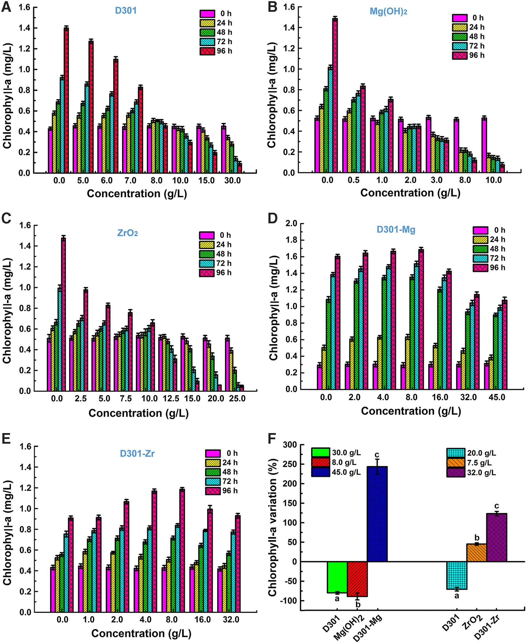
Chlorophyll-a content of Chlorella exposed to different concentrations of materials.
This trend was mimicked by algal cells exposed to ZrO2, where at the 96 h time point, the increased rate of soluble protein was 34.8% and chlorophyll-a was 45.2% in cells treated with 7.5 g/L concentration ZrO2, respectively, far less than the 332.2% increase rate in soluble protein and 123.3% increase rate in chlorophyll-a in algal cells treated with the 32.0 g/L D301-Zr (32.0 g D301-Zr = immobilizing 10.5 g ZrO2 to 21.5 g D301) (Figs. 4F and 5F).
These results further verified the above test results on algal growth rates, which demonstrated that immobilizing Mg(OH)2 and ZrO2 to D301 significantly reduces the toxicity of these nanoparticles. Of particular note, our data demonstrate that immobilization appears to have an obvious effect on reducing toxicity. The experimental data provided the best evidence for this conclusion: expose algal cells to 45 g/L D301-Mg, which is the equivalent of 8.8 g/L Mg(OH)2 and 36.2g/L D301 (Fig. 3D). This amount is four times of the minimum lethal concentration of free Mg(OH)2 (2.0 g/L) (Fig. 3B) and over four times of the minimum lethal dose of D301 (8.0 g/L) (Fig. 3A). A similar effect with immobilized D301-Zr compared to the individual ingredients (Fig. 3A, C, E). Chlorella has greater tolerance and positive biological response to fixed nanometal oxides, especially to D301-ZrO2. It can be concluded that immobilization of nanometal oxides could assist Chlorella in wastewater treatment to improve algae wastewater treatment efficiency.
SEM (Fig. 6) showed the observations of Chlorella cells exposed to both free nanoparticles at their lethal concentration and when these particles were immobilized to D301 at the 48-h time point. Chlorella cells cultured in medium without nanoparticles were spherical shaped with a smooth and intact outer surface (Fig. 6A). In contrast, Chlorella cells cultured with D301 (Fig. 6B) nanoparticles of Mg(OH)2 (Fig. 6C) or ZrO2 (Fig. 6D) showed obvious surface deformation (Fig. 6B-b, C, D-d). Cells treated with free nanoparticles were encased by and showed entrapment of aggregates of these free nanoparticles, with significant cellular deformation and indications of cell atrophy (Fig. 6C, D-d). In comparison, these aggregates were rarely found in Chlorella cells exposed to D301-Mg (Fig. 6E) or D301-Zr (Fig. 6F), and especially cells (intact cell and irregular cell) were rarely observed on or within D301-Zr (Fig. 6F), only several malformed cells were found on the surface of D301-Mg (Fig. 6E-e). Besides, these phenomena were also observed under the microscope (Supplementary Fig. S1). The aggregates enveloping algae cells were found in the algae suspension with free nanomaterials (Supplementary Fig. S1A-a, B-b), but not be found in the cells suspension with immobilized materials (Supplementary Fig. S1C, D). Also, there is an obvious space between D301-Zr and the algae cells (Supplementary Fig. S1C), which should be caused by mutual repulsion between the same surface charges of D301-Zr and algae cells (Supplementary Table S1). The data of ZP and hydrodynamic diameter in Supplementary Table S1 showed that aggregate blocks appeared while free nanomaterials were added to the culture medium, and aggregate diameters were much larger than their particle size in deionized water. The results were consistent with the observation results of SEM and microscope (Supplementary Table S1). Therefore, the SEM and microscope observations provide evidence that free nanoparticle materials were more harmful to Chlorella cells than that of immobilized on D301 under our test conditions.
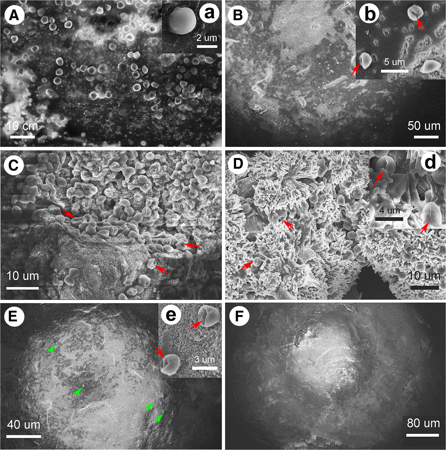
SEM observation of Chlorella exposed to different materials for 48 h.
One of the main reasons for the different responses of algae to free and fixed nanoparticles is probably due to the fact that the carrier blocked the nanoparticles escaping into the culture medium, and did not cause a large amount of particles to besiege the algae cells, and then influence the reproductive rate of algae, since nanoparticles are easy to agglomerate in water (Zhang et al., 2015). The different responses between D301-Mg and D301-Zr may be attributed to that nano-ZrO2 immobilized on D301 is more stable than the nano-Mg(OH)2 immobilized on D301 relatively (Zhang et al., 2013b, 2015; Wang et al., 2015b). Besides, the aggregation mechanism of nanoparticles may be closely related to the particle size, zeta potential, surface charge, and wastewater composition (Zhang et al., 2008; Wang et al., 2011; Mukhopadhyay et al., 2013; Karunakaran et al., 2015), as well as the reason for the stimulation of D301-Zr to algae growth perhaps related to its adsorption of phosphorus (Wang et al., 2015a), which was one of main nutrients of algae. All these guess and hypothesis for the present results still need further research.
Conclusion
A systematic research on the evaluation of response of Chlorella to Mg(OH)2 and ZrO2 nanoparticles before and after immobilization on D301 confirms that the physical restraint by immobilization to D301 can reduce the toxicity of these nanoparticles toward algae. Namely, the replacement of free metal nano-oxides with supported metal nano-oxides can avoid the damage to Chlorella caused by the agglomeration of free metal nano-oxides. Adding immobilized stable nanometal oxides with appropriate concentration can improve the efficiency of Chlorella in wastewater purification. The results of present study will contribute to the bioregeneration for immobilized nanoadsorbent by Chlorella.
Footnotes
Acknowledgment
We thank Prof. Brian Michael Chung (Weber State University, Ogden, UT) for grammar revision.
Author Disclosure Statement
No potential conflict of interest was reported by authors.
Funding Information
We greatly acknowledge the financial support from the Natural Science Foundation of Hebei Province (Grant No. D2017203317) and Key Project of Hebei Education Department (Grant No. ZD2019071).

References
Supplementary Material
Please find the following supplemental material available below.
For Open Access articles published under a Creative Commons License, all supplemental material carries the same license as the article it is associated with.
For non-Open Access articles published, all supplemental material carries a non-exclusive license, and permission requests for re-use of supplemental material or any part of supplemental material shall be sent directly to the copyright owner as specified in the copyright notice associated with the article.
