
Abstract
Benzoic acid (BA) is potentially toxic to humans and recalcitrant in the environment. Although it may be degraded by fungi, potential degradation pathways has received limited study. In the present work, concentrated BA degradation by Hypocrea lixii AH, a type of soil-borne or wood-decaying fungi, was investigated. The degradation products were characterized by UV-vis, Fourier transform-infra red (FT-IR), and gas chromatography-mass spectrometer (GC-MS) and the degradation routes were evaluated with a quantum mechanical method. Results showed that up to 29% of the BA could be degraded by H. lixii AH with lignin peroxidase (LiP) and laccase (Lac) playing a central role. FT-IR results showed that the degradation mechanism involved cleavage of the benzene ring and removal of the carboxyl group to form aliphatic alkenes. GC-MS analysis confirmed the presence of alkanes, esters, and carboxylic acid in the final products. Using quantum mechanical analysis, it appears that BA is reduced to benzoic alcohol through electrophilic attack by the hydroxyl group on the para- and meta-carbons in the benzene ring. The attack by LiP and Lac may also render the C–C bonds adjacent to substituents (C3–C4, C4–C5) in the benzene ring prone to cleavage.
Introduction
Benzoic acid (BA) is a prevailing by-product of chemical and petrochemical processing and an intermediate in the commercial production of plastics, textile, food processing industries, and agricultural and industrial wastewater (Gao et al., 2006; Garg and Prasad, 2017). The chemical structure of BA is closely related to several phenolic compounds, such as vanillin, syringic acid, hydroxybenzoic acid, and resveratrol. BA and its derivatives are persistent (Keshavarz et al., 2012; Garg and Prasad, 2017; Huang et al., 2018), and have been reported to be detrimental to human health and the environment (Malakahmad et al., 2016). For example, BA is required to be removed from sewage before discharging it to the receiving body of water (Santos et al., 2019).
Although traditional methods have not been shown to be particularly effective (Zhu et al., 2012), it has been reported that BA can be effectively removed using biological treatments such as activated sludge or membrane bioreactors (Lu et al., 2010; Zhu et al., 2015; Alneyadi et al., 2018). Of the microbes involved in biological treatment, white-rot fungus (WRF) has received the most attention due to its ability to degrade a wide range of organic pollutants (Asif et al., 2017; Grandclément et al., 2017; Huang et al., 2018; Jambon et al., 2018). However, a majority studies related to the degradation of organic pollutants have been of relatively low concentration (ranging from a few nanograms to a few hundred milligrams per liter) while studies with concentrated organic contaminants have been rarely reported (Wang et al., 2010; Jeswani and Mukherji, 2013). This is primarily due to the general toxicity of BA to microorganisms (Del Olmo et al., 2017). To address toxicity effects, it can be advantageous to isolate more tolerant microbial strains found in many industrial and agricultural applications. Pathways of microbial degradation for BA include ortho, meta, gentilic, and protocatechuic acid and have been identified as a result of genes and gene cluster analyses (Peng et al., 2003; Cao et al., 2008). However, degradation pathways using the quantum mechanical method have not been fully explored.
In this study, WRF Hypocrea lixii AH was used to degrade BA at high concentrations (>2,000 mg/L) with the aim of investigating the extent of biodegradation, characterizing the specific enzymes secreted, and investigating the mechanisms of biodegradation.
Materials and Methods
Microbe and culture medium
Previous work has shown that H. lixii AH can degrade coal composed primarily of polycyclic aromatic hydrocarbons (Tao et al., 2010). H. lixii AH (Genbank accession number: FJ45728) was isolated from subsurface soil in a coal mine and preserved at the Bioengineering Department at the China University of Mining and Technology located in Xuzhou, China (Tao et al., 2010). The strain was cultivated at 30°C in a constant temperature shaker before use. The composition of the Czapek-Dox modified culture medium was 10.0 g/L glutamic acid 10.0, 2.0 g/L sodium glutamate, 3.0 g/L sodium nitrate, 2.0 g/L dipotassium hydrogen phosphate, 0.5 g/L magnesium sulfate heptahydrate, 0.5 g/L potassium chloride, and 0.25 g/L sulfate pentahydrate. The pH of the medium was adjusted to 7.0. H. lixii AH was allowed to grow and was filtered (pore size of 10 μm; Double Circle, China) following the exponential growth phase to remove mycelia. The filtrate was then centrifuged at 10,000 rpm for 20 min to collect spores and spores pellets were resuspended with the DOX culture medium for further study.
Measurement of fungal growth, enzyme activity, and degradation rates
The spore suspension was inoculated into a 250-mL flask containing 100 mL sterile medium to achieve a final spore concentration of 1.0 × 10⁶ spores/mL. BA was added to the culture medium at final concentrations of 1, 2, and 3 g/L. Triplicate culture tests were set up unless otherwise stated. The culture solutions were incubated at 30°C in a rotary shaker at 170 rpm until they reached the stationary phase (∼10 days). The culture solutions were filtered (pore size of 10 μm; Double Circle) and the fungal mycelia were washed with pure ethyl alcohol several time to remove any residual organic compounds then rinsed with distilled water. The mycelia were then freeze-dried and weighed. The filtrate was transferred to new tubes for enzyme activity measurements, degradation studies, and intermediates analyses. The activity of enzymes (lignin peroxidase [LiP], laccase [Lac], and MnP) was determined according to Wang and Ming (2000) and the activity of the LiP, Lac, and MnP was determined by measuring absorbance at wavelengths of 310, 420, and 240 nm. An enzyme unit (U) is defined as the amount of enzyme that catalyzes the conversion of one micromole of substrate per minute. BA was extracted with benzene from the culture media as described by Wang and Ming (2000). The extracted samples were measured using a UV-vis spectrometer (UV-2802 PCS Unico, China) at 190–450 nm (accuracy of 1 nm) with benzene as the baseline reading. The maximum absorption peak of BA occurs at 285 nm, respectively. The extent of BA degradation was determined by analyzing the absorption values of initial and final culture solutions.
Analysis of intermediates
The organic functional groups of the degradation products were analyzed by Fourier transform-infra red (FT-IR, Nicolet 380; Nicolet, Inc.) with a scanning range of 4,000–450 cm−1 and a resolution of 4 cm−1 using a diffuse reflectance attachment. The data were processed with a peak-fitting program (Peakfit v4.12; Systat Software, Inc.) to deconvolute the peaks. The composition of the degradation intermediates was analyzed with a Hewlett-Packard 6890/5973 gas chromatography/mass spectrometer (GC/MS) equipped with a capillary column coated with HP-5 (cross-link 5% phenyl methyl siloxane, 30 m length, 0.25 mm inner diameter, and 0.25 μm film thickness) and a quadrupole MA detector and operated in electron impact (70 eV) mode. The chromatographic conditions were as follows: starting temperature was 150°C for 1 min, rising to 250°C at 15°C/min for 5 min, rising to 300°C at 15°C/min for 8 min. The gasification chamber was 300°C, the split was 10:1, and the helium flow rate was 1.2 mL/min. The mass spectrometry conditions are as follows: mass selective detector, electron ionization mode, mass scanning range of 25–600 amu ionization voltage 70 eV, quadrupole temperature 150°C, ion source temperature 230°C, transmission line temperature 300°C, and electron multiplier voltage It is 2.565 V. Compounds were identified by comparing mass spectra with the NIST05 library data.
Degradation pathway of BA by the quantum mechanical method
The pathway of BA degradation was speculated by analyzing the intermediate products in the culture solutions using a dual descriptor (DD) calculation methodology. The method has been reported to be a robust and accurate predictor of reactive sites for electrophilic or nucleophilic substitutions (Morell et al., 2005; Fu et al., 2014). The nucleophilic and electrophilic attack sites can also be revealed using the DD calculation methodology. For quantitative analysis, the condensed dual descriptors (CDD) of carbon atoms were also determined. In general, more positive DD values indicate a greater likelihood of electrophilic and nucleophilic attack. The strength of the chemical bonds was also determined by calculating the Laplacian bond order (LBO), which is consistent with bond dissociation enthalpies (Lu and Chen, 2013). The M062X/6-311G**//M062X/def2-TZVP method was utilized in this research for both calculations (DD and LBO) using Multiwfn (Lu and Chen, 2012).
Results and Discussion
Cell growth in BA and degradation rate
Although the data clearly show that the fungus grows much better without BA, as shown in Fig. 1, H. lixii AH can grow in the presence of BA at concentrations up to 3 g/L. The mass of the fungus with BA was ∼30% of that compared to control. BA clearly suppressed the growth of the fungus, which agrees with the results from other studies (Varsha et al., 2014). Moon et al. (2018) showed that BA reduces antiaflatoxin activity by inhibiting aflatoxin biosynthesis-related genes. Whether the inhibition of BA on the growth of H. lixii AH is related to the gene regulation is yet to be determined. After 10 days of growth, the degradation rate of BA was 29% (only 1 g/L BA data was shown here), which is comparable to other studies using Candida sp. S1 to degrade polycyclic aromatic hydrocarbons (Hadibarata et al., 2017). In general, compounds with abundant electron donating functional groups such as hydroxyl groups (–OH) are more susceptible to electrophilic attack by oxygenase. In contrast, compounds containing strong electron-withdrawing functional groups such as carboxylic acids (–COOH) are less susceptible to oxidative catabolism (Tadkaew et al., 2011), which is one of the contributing factors related to BA degradation.
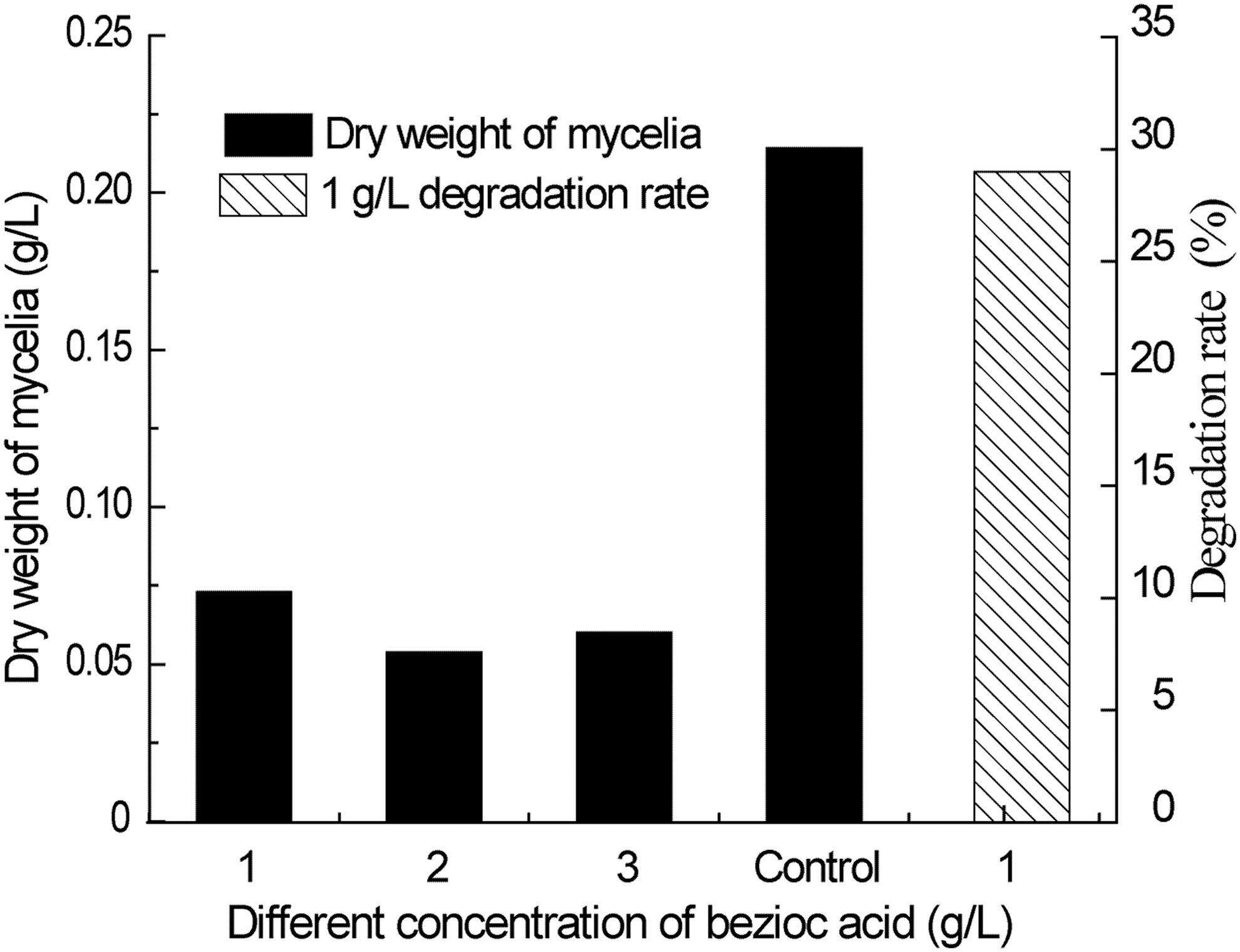
Weight of biomass in the presence of BA and percentage of degradation. BA, benzoic acid.
LiP, MnP, and Lac activity
Figure 2 shows the enzymatic activities of three ligninolytic enzymes (LiP, MnP, and Lac) in different concentrations of BA. The Lac activity is much greater than the other enzymes for each of the BA degradation systems, suggesting that Lac plays an important role in the degradation of BA. BA may also act as an inducer to stimulate the production of the enzyme because the activity of Lac appears to be positively correlated to the concentration of BA in the medium. Other studies have also demonstrated that other aromatic compounds such as gallic acid can induce the production of Lac (Bettin et al., 2014). Lac is reported to degrade a number of environmental contaminants including per- and polyfluoroalkyl substances and bisphenol A, which are persistent to the environment (Haritash and Kaushik, 2009; Luo et al., 2015; Barrios-Estrada et al., 2018a, 2018b). In contrast, the activity of LiP is negatively correlated to the concentration of BA (14 U at 1 g/L BA to 0 at 3 g/L BA) suggesting that high BA concentrations can completely inhibit the production of LiP. As shown, the activity of MnP is the lowest among all three ligninolytic enzymes and it was not detectable in the 2 g/L BA culture.
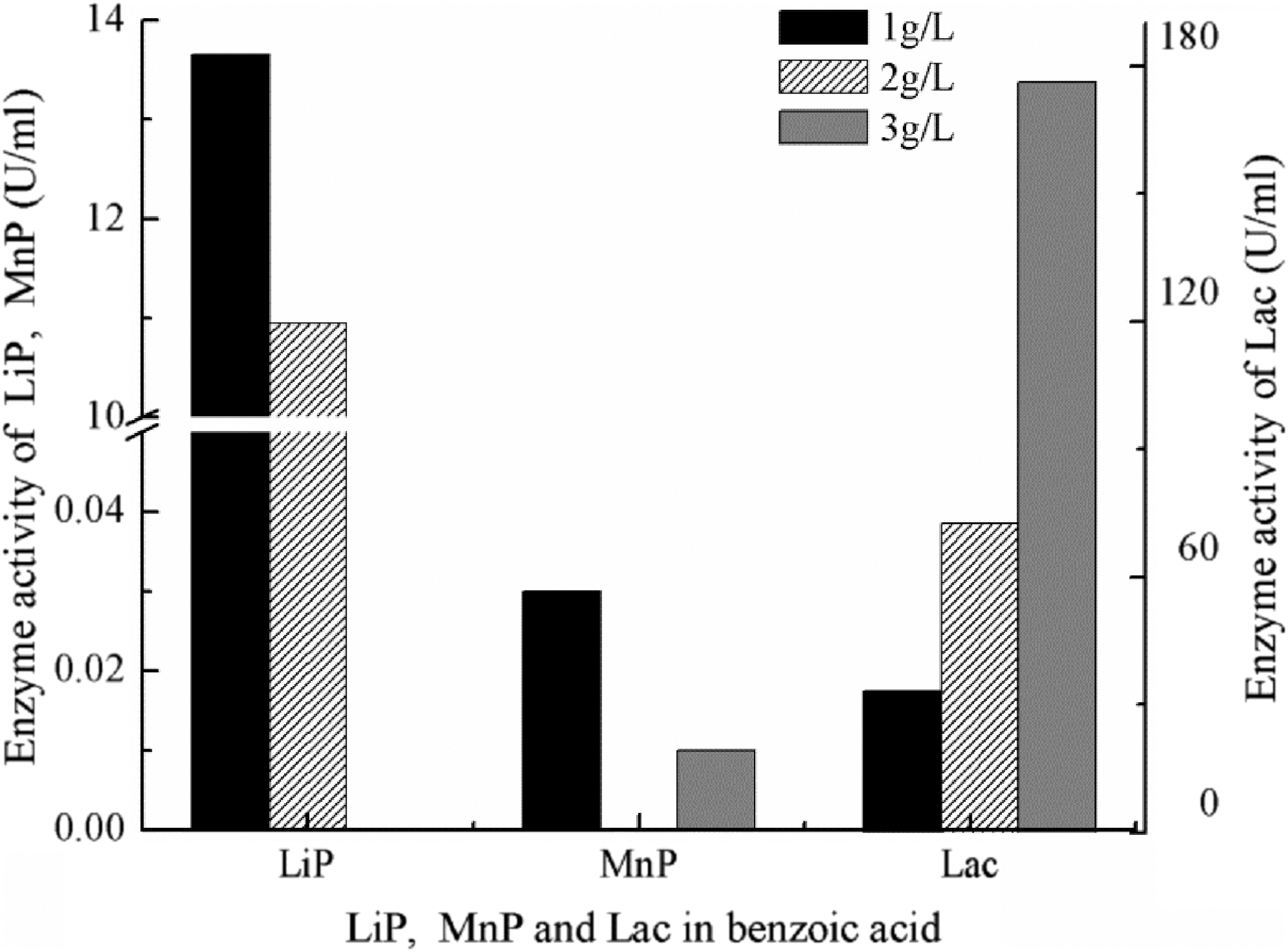
Enzymatic activities of three ligninolytic peroxidases (LiP, MnP, and Lac) in different concentrations of BA. LiP, lignin peroxidase; Lac, laccase.
Analysis of products after degradation of BA
UV-Vis spectral analysis
Figure 3 shows the UV-Vis absorption spectra of BA before and after degradation by the fungus. BA has been reported to have two adsorption bands between 200 and 300 nm (Deng et al., 2006). The adsorption of BA has a slight shift, from 280 nm before degradation to 283 nm after degradation. The intensity of the peak increased after the treatment, suggesting the structures of the degradation products are similar to that of BA or that there may be interferences between the extracellular polymers and the sample (Deng et al., 2011).

UV-Vis spectra of BA before and after degradation.
FT-IR analysis
FT-IR was used to analyze the functional group changes in the BA reaction system before and after the reaction to facilitate the probe of possible biodegradation process of BA. The infrared spectra before and after degradation of BA are shown in Fig. 4. Comparing the infrared transmission spectrum of the reaction solution before and after degradation indicates that differences in functional groups before and after degradation occur mainly at 1,500–500 cm−1, 1,750–1,500 cm−1, and 4,000–2,800 cm−1. The 1,500–500 cm−1 range is primarily composed of C–O stretching vibration, C–H bending vibration of alkanes, and C–H bending vibration of substituted benzenes.
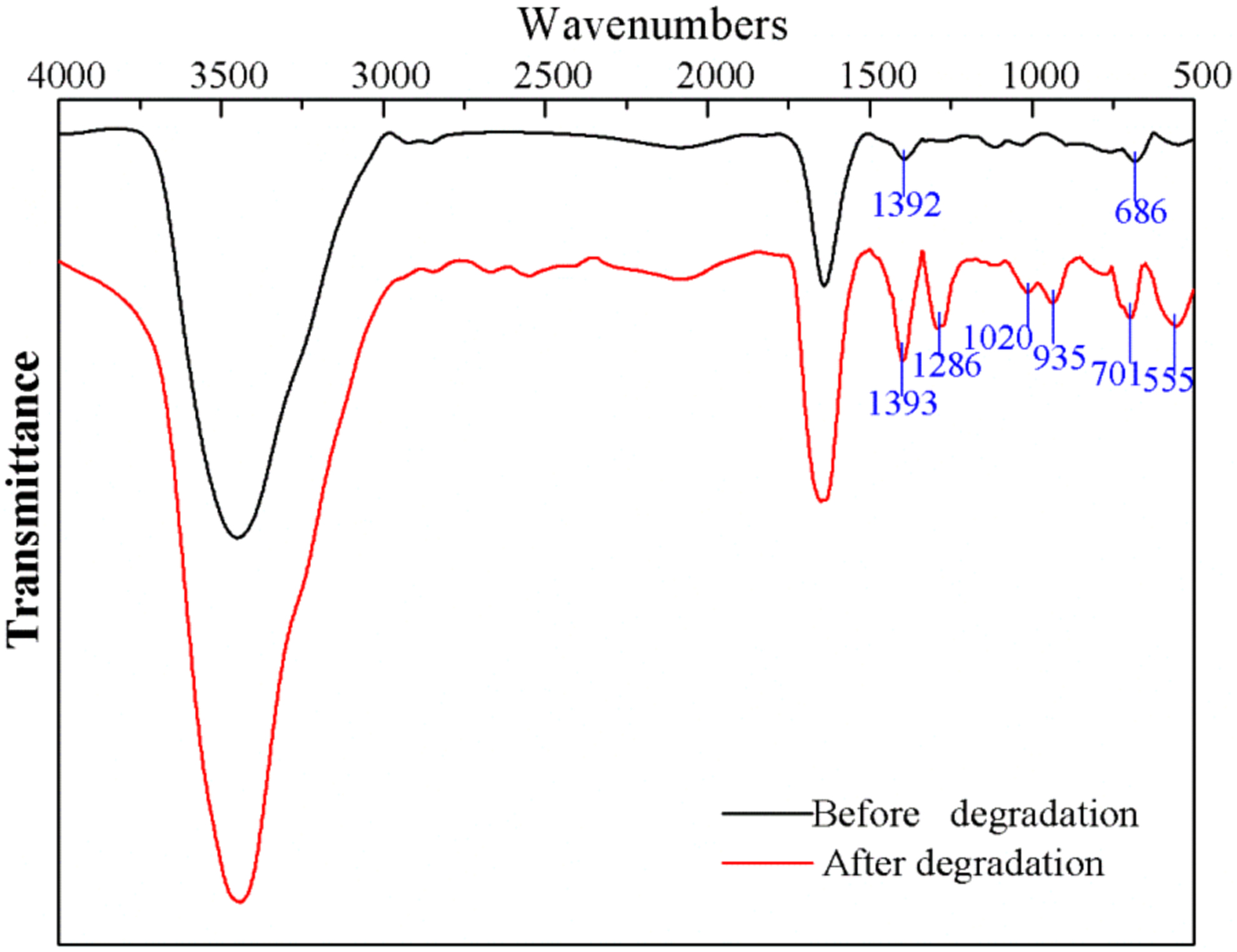
FT-IR spectra before and after degradation of BA. FT-IR, Fourier transform-infrared.
The peak assignment results for each location are listed in Tables 1 and 2. The number of substituted aromatic rings in the reaction liquid after degradation increased, and C–H bonds of saturated alkane, C–O single bonds of phenol, and C–O–C single bond of ester or ether appear in the reaction liquid. These results indicate that in the process of degradation of BA by white-rot fungus H. lixii AH the aromatic ring of BA underwent a substitution reaction and that alkanes, phenols, esters, and ethers may be present in the degradation products. Of the four types of BA degradation pathways reported in the literature, three involve the opening of the benzene ring to form aliphatic compounds (Milovac et al., 2014; Mohite et al., 2015).
Infrared Peak Assignment Before Benzoic Acid Degradation
Infrared Peak Assignment After Degradation of Benzoic Acid
The peak range at 1,750–1,500 cm−1 and 4,000–2,800 cm−1 is wider due to the superposition of various substances. To find out the corresponding peaks and their intensities corresponding to various functional groups, the height of the peaks was determined using Peakfit V4.12 and the results are shown in Fig. 5. The spectral region of 1,750–1,500 cm−1 was used to study the stretching vibration of C = O and the stretching vibration of C = C (Krimm and Bandekar et al., 1986). There are two main absorption peaks near the wavenumbers of 1,639 cm−1 and 1,633 cm−1 (Fig. 5[A-1], [A-2]), which can be assigned to the stretching vibration of C = O. In addition, the medium-intensity peak at 1,685 cm−1 (Fig. 5[A-2]) can be attributed to the stretching vibration of C = C indicating that olefins are also produced as a result of BA degradation.
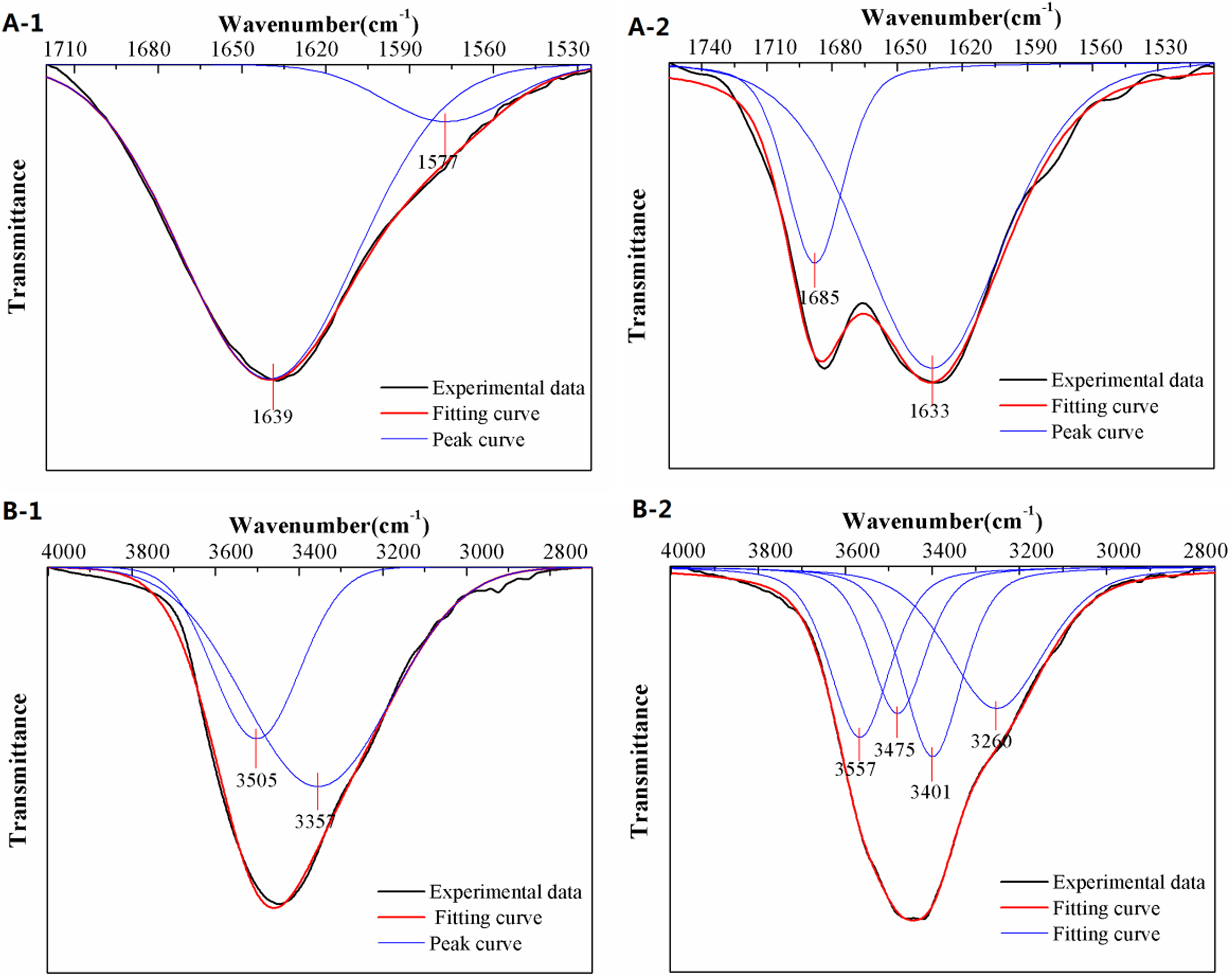
Deconvolution of FT-IR peaks before and after BA degradation at 1,750–1,500 cm−1
The vibration frequency range between 4,000 cm−1 and 2,800 cm−1 is assigned to the hydroxyl-OH (Fig. 5[B-1], [B-2]). As shown, the number of characteristic peaks of the hydroxyl group after degradation has significantly increased (Fig. 5[B-2]), which illustrates the formation of hydroxyl groups in different chemical environments during the degradation of BA. This finding combined with the previous results strongly suggest that the white-rot fungus H. lixii AH produced phenolic substances in the process of degrading BA and it has been shown that phenolic substances are produced as a result of the ortho-metabolic and meta-metabolic pathways (Neidle et al., 1991; Ampe and Lindley, 1996).
GC-MS analysis
The total ion chromatograms for the degradation products of BA are show in Fig. 6. The related peaks were identified by comparing mass spectra with the NIST05 library data (Data not shown here). In most of the previous WRF studies the focus has been in relation to the disappearance of the substrate compound not the degradation pathways and intermediates. Dittmann et al. (2002). reported that the factors influencing the degradation of aromatic compounds and the fungal biomass development included compound structure, external concentration, and the fungal species. In this study, BA was shown to be nearly completely degraded by the AH. By comparing to the NIST05 library data, three types of alkane (2,3,6-trimethyl decane, 2,3-dimethylun decane and 2-methylun decane), one type of phenol(4-(hydroxymethyl)phenol), one ester (2-methyl-3- butanoate), and one carboxylic acids(2-hydroxybenzenecarboxylic acid) were detected as degradation products, respectively.
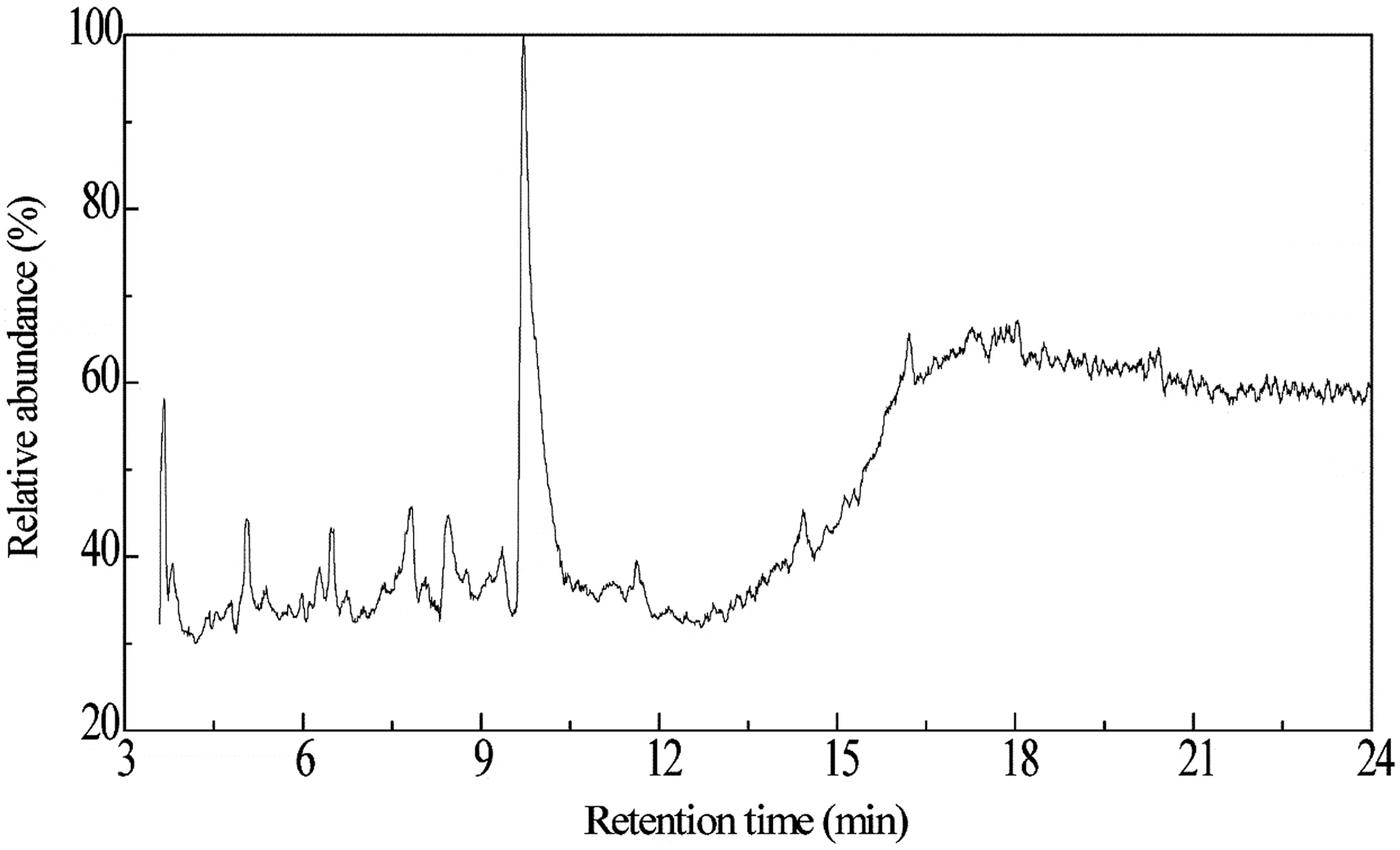
Total ion chromatogram of degradation product of BA.
Proposed degradation pathway of BA
The DD of BA (Fig. 7a) and benzoic alcohol (Fig. 7b) were calculated and their isosurface are shown in Fig. 7. Green and blue isosurface represent positive and negative region of DD, respectively. For BA, its C3, C4, C5, and C6 showed negative value of DD except the C1 (not participate in reaction). In addition, the result of the CDD was consistent with DD isosurface. Thus, para- and meta-carbons were activated for electrophilic attack by the hydroxyl group. However, the CDD of C3 and C4 were two evident negative sites, so both were more likely to be attacked, generating 3-hydroxybenzoic acid or 4-hydroxybenzoic acid via the hydroxylation induced by LiP. For the positive value, C12 was determined to be the nucleophilic attack site. Consequently, BA must have been first reduced to benzoic alcohol.

DD isosurface and LBO of BA
As shown in Fig. 7b, C4 was the negative site, which means that the benzoic alcohol further reacted to produce 4-(hydroxymethyl) phenol. In addition, LBO calculations reveal that the C–C bonds adjacent to substituents (C1–C2, C1–C6) were less than those of the other benzene ring sites. Under the catalysis of LiP and Lac, ring opening reactions were likely to occur at this position, followed by polymerization generating 2,3,6-trimethyldecane, 2,3-dimethyl-undecane, and 2-methylundecane that could be converted to 2-methyl butyl-3-acid, which was further esterified to 2-methyl-3-butenoate catalyzed by LiP and other unknown esterase. Based on above analysis, a possible degradation route has been proposed and is shown in Fig. 8.

Proposed transformation pathway of BA (1) 2,3,6-trimethyldecane, (2) 2,3-dimethyl-undecane, (3) 2-methylundecane,(4) 4- (hydroxymethyl) phenol, (5) 2-methyl-3-butenoate, (6) 2-hydroxybenzenecarboxylic acid.
Conclusion
A fungus H. lixii AH was used to biodegrade concentrated BA. Although fungi growth was inhibited by high concentrations of BA, BA degradation was still able to reach 29% after 10 days. Lacs were shown to play an important role in BA degradation. Conversely, the activity of MnP and LiP were inhibited by BA. Based on the product analysis, a possible degradation route was proposed with BA first being reduced to benzoic alcohol. The para- and meta-carbons in the benzene ring are then activated for electrophilic attack by the hydroxyl group with further attack by LiP and Lac rendering the C–C bonds adjacent to substituents (C3–C4, C4–C5) in the benzene ring prone to cleavage.
Footnotes
Author Disclosure Statement
No competing financial interests exist.
Funding Information
This work was supported by the Fundamental Research Funds for the Central Universities (No. 2017XKQY037).