
Abstract
Here, a facile, efficient, and sensitive method is reported for detecting malathion based on ligand exchange between the surface capping molecules of CdSe quantum dots and malathion in organic solution. Under optimal conditions, there was a linear relationship between the relative fluorescence intensity (Imax/I) and the logarithm of malathion concentration ranging from 9.08 × 10⁻¹⁰ to 3.03 × 10⁻⁷ mol/L (Signal to Noise ratio = 3), and the detection limit was 3.03 × 10⁻¹⁰ mol/L. A relative standard deviation of 1.25% was detected for 9.08 × 10⁻⁹ mol/L malathion (N = 5). The method was finally validated by the detection of malathion in emulsifiable concentrate formulation and vegetable samples, and the obtained results were highly consistent with those of gas chromatography.
Introduction
Organophosphates (OPs) have been commonly used as pesticides in agricultural production for controlling various insects on vegetables, fruits, shrubs, and landscaping plants (Kim et al., 2011; Vikrant et al., 2017). More than two million tons of OP pesticides are applied in agriculture worldwide each year (Jayaraj et al., 2016). According to the World Health Organization, >200,000 deaths are related to OPs every year owing to occupational exposure or self-poisoning (Liu et al., 2015). Due to their high and acute toxicity to acetylcholinesterase, OP pesticides can cause serious damage to human cardiovascular system, respiratory tract, and nervous system (Kanagasubbulakshmi et al., 2017; Venkidasamy et al., 2021).
Moreover, the relatively high persistence of OPs can result in large amounts of residues in the environment to affect the balance of aquatic systems, and indirectly pose threats to the environment and human health through contamination and residual in farm products (Jayaraj et al., 2016; Shieh et al., 2019). Among various OPs, malathion is the most frequently used, which is the main cause of most acute pesticide poisonings (Bray et al., 2021). Currently, some analytical methods are used to detect malathion, including gas chromatography and mass spectrometry (GC-MS), high-performance liquid chromatography and tandem MS (Hua et al., 2020; Sirintorn et al., 2020; Mashaalah et al., 2021).
These technologies are sensitive and can discriminate different compounds, but are costly, cumbersome, and require pretreatment of the samples by skilled technicians, which largely hinder their actual application in field analysis (Kanagasubbulakshmi et al., 2017; Mashaalah et al., 2021). Therefore, it is highly necessary to develop a simple, rapid, and sensitive method to detect malathion for environmental protection and food safety.
Recently, highly sensitive and selective fluorescent sensors have been extensively reported (Bala et al., 2018; Jia et al., 2021). As semiconductor luminescent nanomaterial, quantum dots (QDs) are frequently employed in multiple detection techniques for their superior fluorescence (FL) features, such as stable, narrow, and tunable emission spectra with broad excitation spectra. The sensing of QDs is based on the physical or chemical reactions between QDs and the target analyte, which can result in photoluminescence enhancement or quenching (Liu et al., 2019; Wang et al., 2021). Hence, QDs have been widely applied as FL probes in analytical chemistry for easy and in situ detection of OP pesticides through cooperative recognition and signaling, which are apparently advantageous over those traditional cumbersome off-line approaches.
For instance, Zhang et al. (2010) constructed an easy, efficient, and specific method for the detection of chlorpyrifos, which considers that chlorpyrifos induce the changes in surface-bound organic molecules of QDs. More recently, thiol-capped CdS QDs were proposed as FL probes to detect malathion (Kanagasubbulakshmi et al., 2017). These findings indicate that QDs can serve as FL probes to detect drug molecules according to changes in their luminescent properties.
Here, we propose a facile method for malathion detection (structure as shown in Fig. 1). CdSe QDs were applied as highly selective and sensitive FL probes based on the ligand exchange between surface capping ligand hexadecylamine (HDA)/dioctylamine (DOA) and malathion without any complex functional modification. The proposed method is facile and rapid, and exhibited high sensitivity and reliability for real samples, which may open a novel pathway for OP pesticide detection.

The chemical structure of malathion pesticide.
Experimental
Materials
Selenium powder (200 mesh), CdO, hexane, methanol, chloroform, and stearic acid were purchased from Shanghai Reagent Factory (Shanghai, China), which were used without any further purification. DOA, dimercaprol, trinoctylphosphine, trinoctylphosphine oxide (TOPO), and HDA were obtained from Aldrich. All other chemicals were of analytical grade. Malathion standard (99.0%) was purchased from Dr. Ehrenstorter. Ten percent and 80% malathion emulsifiable concentrate (EC) were provided by the Xiaodongmen (Wuhan) market. Pesticide emulsifier alkyl aryl polymeric polyoxypropylene polyoxyethylene ether (33#), calcium 12 alkylbenzene sulfonic acid (QD#), phenethoxy polyoxyethylene ether (601#, Hydrophile-Lipophile Balance [HLB] 13–14; 602#, HLB 15–16; 603#, HLB >17), were purchased from Alibaba.
Apparatus and methods
The ultraviolet and visible (UV-vis) absorption spectra were obtained with a 1.00 cm quartz curette on a Thermo Nicolet Corporation Model Evolution 300 Spectrophotometer (Nicolet, UK). All of the FL spectra were measured and recorded by a Perkin-Elmer LS-55 fluorescence spectrometer (Perkin Elmer, USA) equipped with a 20 kW xenon discharge lamp as a light source.
High-resolution transmission electron microscope images were obtained on a JEM2010FEF field-emission transmission electron microscope at an acceleration voltage of 200 kV (Jeol, Japan). The Raman spectra were acquired with an inVia micro-Raman spectroscopy system (Renishaw, UK) equipped with a He–Ne laser source operating at 633 nm. GC-MS was obtained using a Varian CP-3800 gas chromatography/saturn 2200 MS system (Varian, USA).
Preparation of CdSe QDs
CdSe QDs were generated with the method of Dong et al. (2008). We calculated the particle size and concentration of the obtained CdSe QDs using the following equations:
In the equations, D (nm) represents the size of CdSe QDs; λ (nm) stands for the wavelength of the first excitonic absorption peak of the corresponding sample (Yu et al., 2003; Shang et al., 2009); A represents the absorbance at the peak position of the first exciton absorption peak; C is the molar concentration (mol/L); L stands for the path length (cm) of the radiation beam used to record the absorption spectrum; and ɛ is the extinction coefficient per mole of nanocrystals (L/mol). The particle size and concentration of the CdSe QDs were ca. 2.91 nm and 9.06 × 10⁻⁴ mol/L, respectively.
Sample pretreatment
For analysis of 10% and 80% malathion EC, malathion EC was diluted with chloroform/hexane (1:9, v/v) to 7.3 × 10⁻⁹ and 8.9 × 10⁻⁹ mol/L directly (Garcia-Ruiz et al., 2005). For the analysis of cabbage leaves, cabbage leaves were collected within 12 h after treatment with 1.7 × 10⁻⁸ and 3.4 × 10⁻⁹ mol/L malathion EC. Then, the leaves were subjected to chloroform extraction, centrifugation, and evaporation to dryness under nitrogen. The extract was then dissolved in 2 mL of chloroform/hexane (1:9, v/v) for analysis.
Results and Discussion
Interaction between CdSe QDs and malathion pesticide
The interaction between CdSe QDs and malathion pesticide was examined through UV-vis absorption spectra and FL spectra. Figure 2 shows that there was no absorption band at the wavelengths of 450–650 nm for malathion (Fig. 2A-a). The excitation absorption peak of the as-prepared CdSe QDs was centered at 543 nm (Fig. 2A-b), and the addition of malathion resulted in no obvious change in the absorption spectra of CdSe QDs (Fig. 2A-b and -c).
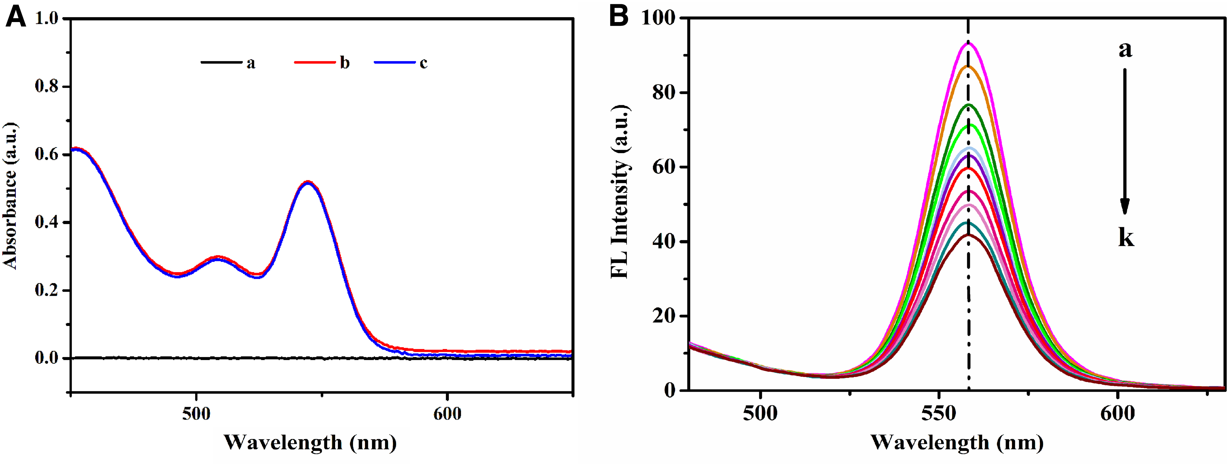
These results indicated that CdSe QDs would not aggregate or decrease in size under the induction of malathion (Dong et al., 2008). The absorption spectra of the mixed solution of CdSe QDs and malathion pesticide were different from the sum of their respective absorption spectra. In addition, malathion pesticide could effectively quench the FL intensity of CdSe QDs in a dose-dependent manner (Fig. 2B). These results indicated that CdSe QDs and malathion had a strong interaction with each other.
Optimization of the experimental conditions
Pre-experiments demonstrated that the reaction of CdSe QDs with malathion was finished within 5 min; however, the FL signals could remain constant for >30 min, indicating that the reaction was rapid. Thus, we recorded the FL signal after the addition of malathion for 8 min (Lin and Hee, 1998).
Calibration curve and liner range
Under optimal conditions, the relative FL intensity (Imax/I) showed linear changes with malathion concentrations ranging from 9.08 × 10⁻¹⁰ to 3.03 × 10⁻⁷ mol/L (Fig. 3), and the correlation coefficient was 0.991. The detection limit (Signal to Noise ratio = 3) was 3.03 × 10⁻¹⁰ mol/L, which is sufficient for analysis of real food and environmental samples. A relative standard deviation (RSD) of 1.25% was detected for 9.08 × 10⁻⁹ mol/L malathion (N = 5).
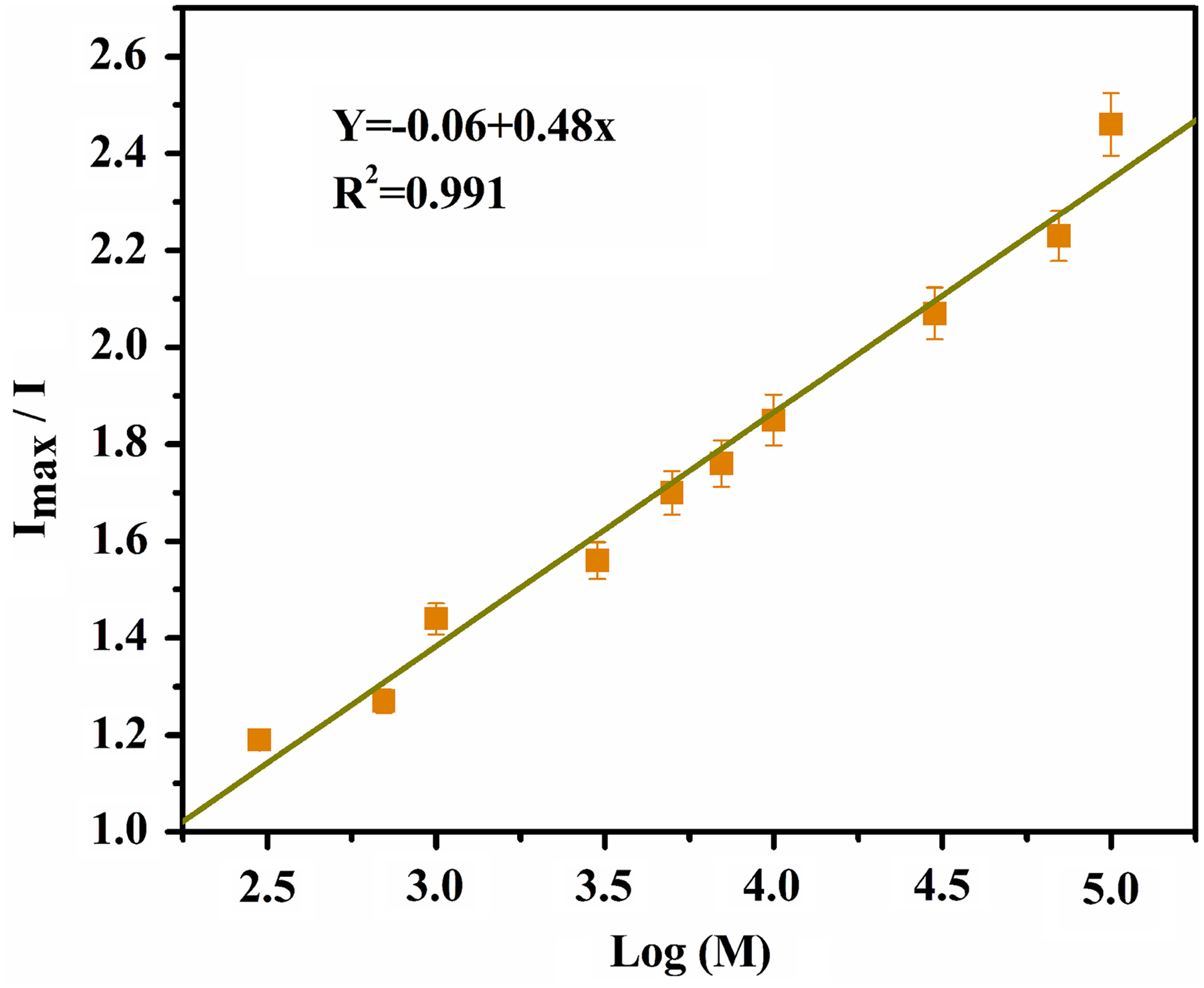
The linear correlation existing between Imax/I (Imax is the recovered FL intensity of QDs in the presence of malathion and I is the FL intensity of QDs at 560 nm) and the logarithm of the malathion concentration.
Selectivity of ligand-exchange probes
QD FL emissions can be potentially quenched by many compounds (Ji et al., 2005). EC often contains the following excipients: dichloromethane, acetonitrile, benzene, pesticide emulsifier alkyl aryl polymeric polyoxypropylene polyoxyethylene ether (33#), calcium 12 alkylbenzene sulfonic acid (500#), phenethoxy polyoxyethylene ether (601#, 602#, 603#), xylene, and toluene (Thacker and Young, 1999). Accordingly, we determined whether and how these excipients affect the FL emissions of QDs.
Generally, the content of organic solvents in 80% EC will not exceed 5% of the total volume, and that of pesticide emulsifier will not be >10% of the total volume. Therefore, 5% solutions of dichloromethane, acetonitrile, benzene, xylene, and toluene and 10% solutions of pesticide emulsifier 33#, 500#, 601#, 602#, and 603# in chloroform/hexane (1:9, v/v) were used to examine the effects. Figure 4 shows that the organic solvents and pesticide emulsifier exhibited little effect on the FL of CdSe QDs under the given conditions. If the relative error caused by the coexisting compounds was lower than ±5% in the FL intensity of the QDs, it was considered that they do not interfere with the detection of malathion (Yang et al., 2019).

Effect of several excipients and concomitants on the FL of CdSe QDs. (1) Control, (2) dichloromethane, (3) acetonitrile, (4) benzene, (5) 33#, (6) 500#, (7) 601#, (8) 602#, (9) 603#, (10) xylene, and (11) toluene. The excipients and concomitants are prepared in chloroform/hexane (1:9, v/v) solution. The concentrations of CdSe QDs are 6.04 × 10⁻⁶ mol/L.
Detection of pesticide residue in cabbage leaves
The proposed method was further validated through the detection of malathion in EC and cabbage leaves, respectively. As shown in Table 1, the obtained results are highly similar to those detected by GC, with recovery rates ranging from 86.25% to 102.89%. The RSD for EC and cabbage leaves were 1.45% and 1.31%, respectively. Overall, our results indicate that the method is highly accurate, precise, and reproducible. In comparison with other previous methods, the proposed method can produce satisfactory results as well as exhibit a higher sensitivity for the detection of malathion (Table 2).
Determination of Malathion in Emulsifiable Concentrate and Cabbage Leaves with the Present Method and Gas Chromatography
EC, emulsifiable concentrate; GC, gas chromatography; RSD, relative standard deviation.
Comparison of the Present Method with Published Results for Malathion Determination
CE, capillary electrophoresis; FL, fluorescence; FT, fourier transform; GC-MS, gas chromatography and mass spectrometry; HPLC, high-performance liquid chromatography.
Ligand displacement reaction
The quenching mechanism of CdSe QDs was investigated with Transmission Electron Microscope (TEM) and Raman spectra. In Fig. 5, TEM images reveal that the as-prepared CdSe QDs were of monodisperse and uniform particles, whose average size is 2.9 ± 0.2 nm (Fig. 5A). Neither obvious change nor obvious aggregation was observed when malathion was added to the CdSe QD solution (Fig. 5B). These results indicate that the ligand exchange with thiol-containing malathion results in the release of capping ligands from the CdSe QD surface, resulting in more surface defects and simultaneously a decrease in FL, which is not due to CdSe QD aggregation. Therefore, the major reaction mechanism should not be aggregation-dependent FL quenching.
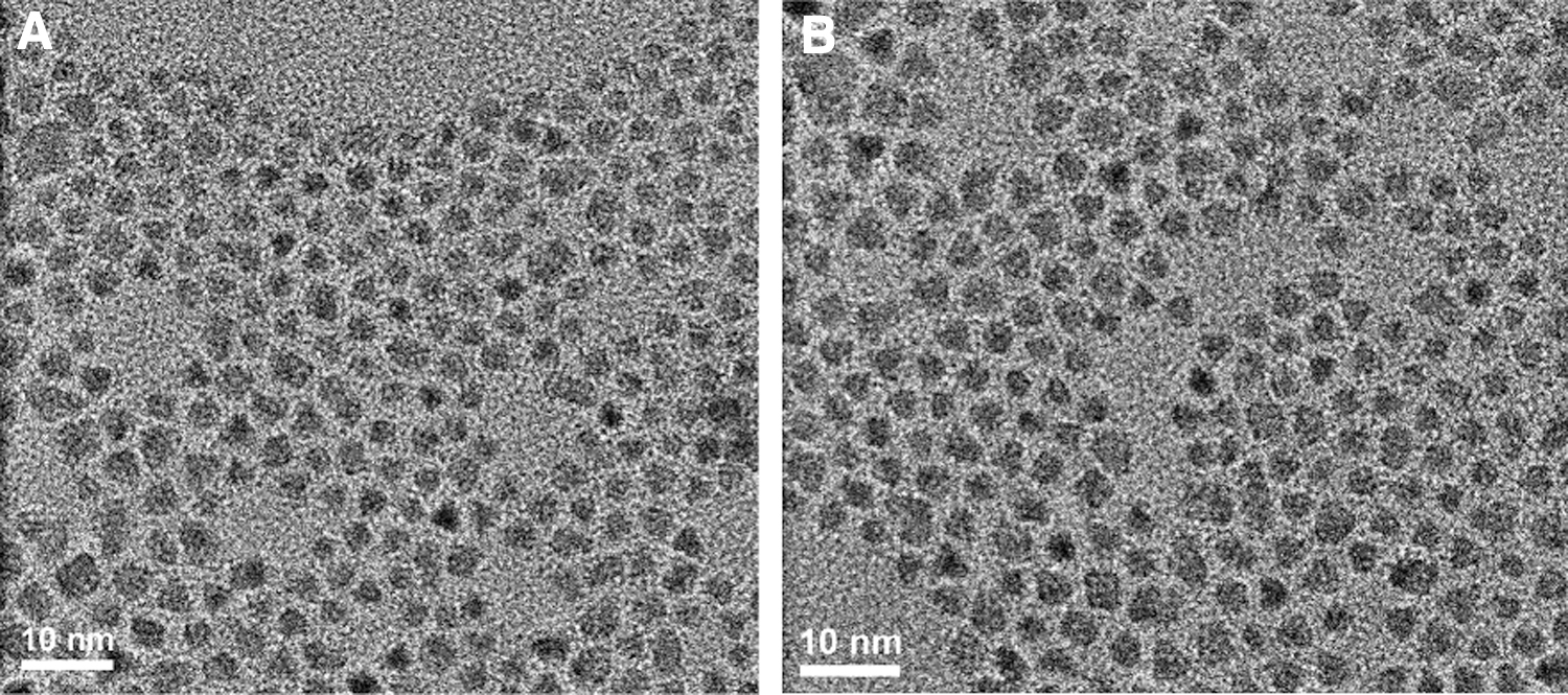
TEM image of CdSe QDs
Raman spectroscopy is usually employed to study the changes in the properties of nanoparticles (Landi et al., 2006; Dong et al., 2008, 2009; Liang et al., 2008). To further elucidate the reaction mechanism, the Raman spectra of CdSe QDs (a), malathion (b), and CdSe QDs with malathion added (c) were observed (Fig. 6).

Raman spectra of CdSe QDs
The assignment of bands in Raman spectra is shown in Table 3. The bands at 854, 1,060, 1,306, 1,456, and 1,685 cm−1 in the spectra (a) are related to normal CdSe QDs. The bands at 854 cm−1 are assigned to the CH3 wagging modes, and 1,060, 1,685 cm−1 correspond to the C–N stretch vibrations from HDA and DOA, respectively (Landi et al., 2006). The peaks at 1,306 and 1,456 cm−1 are associated with P = O stretch vibration and (CH)n torsional vibrations, respectively (Yu et al., 2003).
Band Ascription for Raman Spectra of CdSe Quantum Dots Malathion and Malathion and CdSe Quantum Dots
DOA, dioctylamine; HAD, hexadecylamine; QDs, quantum dots.
As a comparison, the Raman peaks of CdSe QDs at 1,060 cm−1 nearly disappear upon the addition of malathion (spectrum c), suggesting the partial replacement of HDA on QD surface (Dong et al., 2009). Besides, the peaks at 854, 1,060, 1,306, and 1,456 cm−1 become weak, possibly due to a decrease in long-chain alkyl compounds.
Normal malathion exhibited five strong bands at 656, 854, 1,456, 1,599, and 1,740 cm−1 (spectrum b). The peak at 656 cm−1 is ascribed to C–S–H in-plane bending vibration, and the peaks at 854 and 1,456 cm−1 belong to CH3 stretching. The last two bands (1,599 and 1,740 cm−1) both correspond to C = O stretching modes. Compared with those in spectra (b) and (c), the peaks at 854 and 1,599 cm−1 nearly disappear, and some new peaks appear at 656, 695, 773, 830, and 1,203 cm−1 in the spectrum (c).
It has been demonstrated that the Cd–S bond is much stronger than the Cd–N or Cd–P bond (Qu and Peng, 2002; Zhou, 2019). Hence, the disappearance of peaks at 854 and 1,599 cm−1 could be due to the binding of malathion to the CdSe QD surface. This result implies that there are interactions between malathion and the surface capping CdSe QDs, which might also lead to ligand exchange on the surface of QDs (Yang et al., 2019).
It has been revealed that the passivation of surface capping molecules can improve the FL efficiency of QDs (Mao et al., 2005). Therefore, changes in the surface situation can greatly affect the FL properties of QDs, possibly due to the alteration of previously perfect surface passivation of QDs, which leads to more surface defects and then a significant FL quenching of QDs following the changes in surface layer (Donega et al., 2013; Adegoke et al., 2021; Vu et al., 2021).
In this study, a very high FL efficiency was observed for the surface capping organic molecules (HAD and DOA) of CdSe QDs. However, when malathion was added to the CdSe QD solution, the surface ligands of CdSe QDs were exchanged with those of malathion, which may disrupt the previously perfect surface passivation resulting from the capping of a TOPO/HDA layer and lead to more surface defects (Fig. 7). Therefore, the exchange of surface capping organic molecules of CdSe QDs after the addition of malathion to the QDs solution may be the possible quenching mechanism.
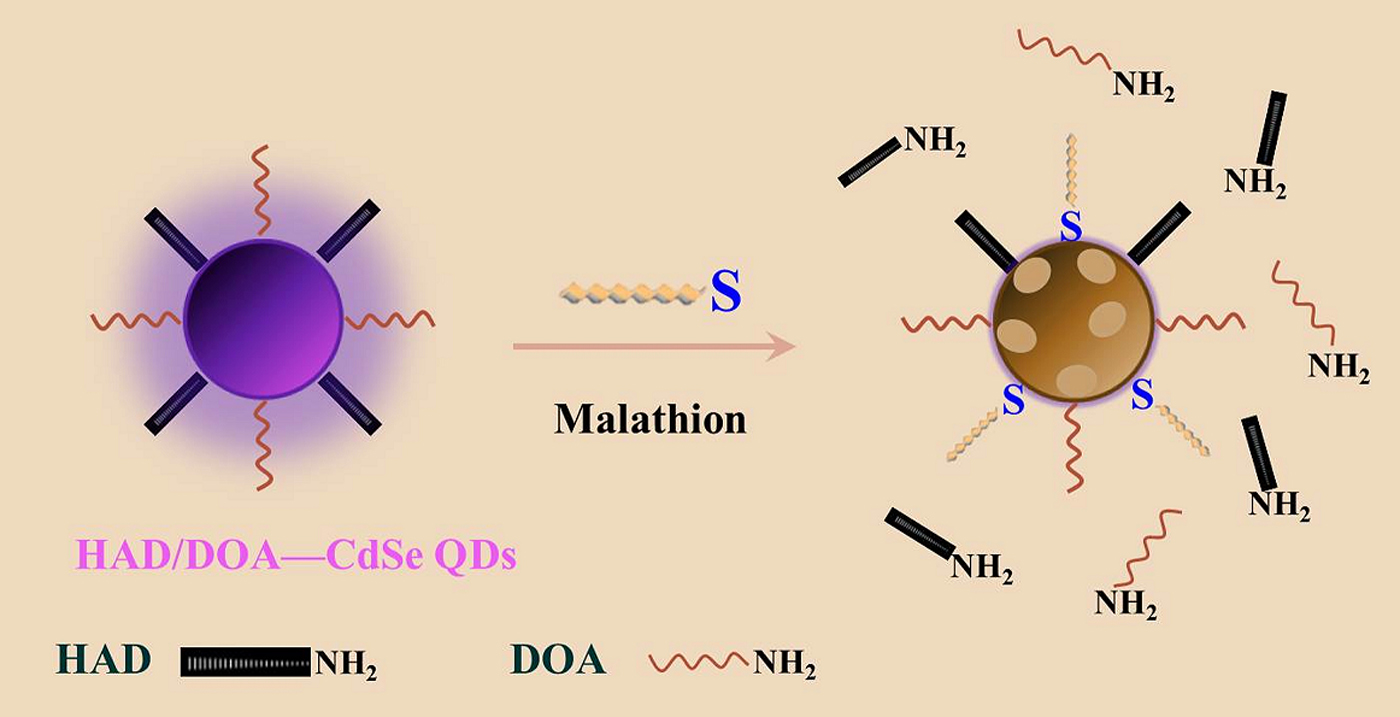
A schematic illustration of CdSe QDs before and after the ligand exchange.
Conclusions
In summary, a facile, efficient, and sensitive method was developed to detect malathion residue in vegetables by using CdSe QDs as ligand-exchange probes. An analysis of the interaction between QDs and malathion revealed that the thiol moiety of malathion could effectively displace the capping ligands from QD surface, resulting in the quenching of FL, which can be used for quantitative estimation of various pesticide molecules. In addition, we applied the proposed method to detect malathion in EC and cabbage leaves without complex pretreatment, and the obtained results were highly consistent with those detected by GC. Based on the ligand displacement reaction, CdSe QDs probes exhibited the potential to simplify the analysis of pesticide residues in food and environmental samples, and might open new avenues in the construction of optical sensors by using QDs.
Footnotes
Author Disclosure Statement
No potential conflict of interest was reported by the authors.
Funding Information
This work was supported by the PhD Research Startup Foundation of Hebei Normal University of Science and Technology #1 under Grant (No. 2013YB005), National Natural Science Foundation of China #2 under Grant (Nos. 31501680 and 31600240), and Natural Science Foundation of Hebei Province #3 under Grant (No. B2018407058).