
Abstract
ZnO has been known to be an excellent photocatalyst and production of suitable morphology by cheap methods to increase catalytic efficiency is important in practical applications. In this study, a fast precipitation method was used to produce the carnation-like morphology to improve the performance of ZnO in the removal of tetracycline hydrochloride (TCH). The precursor of zinc nitrate, precipitation agents of sodium hydroxide, and trisodium were used to control the morphology of ZnO. The characterization of as-prepared material was confirmed by X-ray diffraction, scanning electron microscopy, Fourier transform infrared spectroscopy, diffuse reflectance ultraviolet-visible, and N2 adsorption/desorption isotherms. As a result, the ZnO appeared with a regular and uniform three-dimensional carnation-like morphology and consisted of numerous nanosheets with a thickness of 0.013 μm. The average size of the ZnO flowers was 5 μm. In addition, the plausible formation mechanism of nanoflower with time was proposed. The effects of reaction conditions on the catalytic performance of ZnO were thoroughly examined, and its performance was compared with those of other ZnO forms and other materials. As-prepared ZnO showed to efficiently decompose TCH with the decompose efficiency and rate constant of 89.92% and 0.038 min−1, respectively. Moreover, the •O2− and h+ were key active species in the decomposition of the TCH process.
Introduction
ZnO is recognized as one of the most important II–IV semiconductor oxides because of its superior physicochemical properties and environmental friendliness (Shenoy et al., 2021). Hence, over the past decade, it has been intensively investigated and applied in photocatalysts (Kahsay et al., 2019; Rupa et al., 2019), antifungal activity (Chai et al., 2019), antibacterial activity (Khan et al., 2020), biosensing (Das et al., 2018; Das et al., 2017), and Li-ion battery (Palaniyandy et al., 2018). Moreover, the optical and electronic properties of ZnO remarkably depend on its shape, crystal size, and morphology. Among the 1D and 2D structures, the three-dimensional (3D) hierarchical structure made up of nanosheets, nanowires, and nanorods has attracted great interest due to its high specific surface, unique shape, and outstanding optical property (Boccalon et al., 2020).
Flower-like morphology is the most common 3D structure and is used for many potential applications. For example, the different nanoflower was synthesized by microwave, hydrothermal, and microwave-assisted hydrothermal methods and exhibited high photodegradation with methylene blue (Das et al., 2021). 3D ball-flower ZnO was synthesized by hydrothermal method and used to sense NOx with excellent performance (Li et al., 2019). 3D fluffy ZnO nanoflowers were prepared by different methods and showed efficient decomposition of methyl orange (Qu et al., 2020). These studies have shown that ZnO nanoflowers are effective in the treatment of toxic organic substances in aquatic environments.
Some morphologies of ZnO also exhibit distinct properties when doped with metals, oxides, or salts. ZnO nanorod array loaded with Bi2WO6 had high capacity in the photocatalytic fuel cell; the maximum power density of the Bi2WO6/ZnO nanorods array was 2,707 μW/cm2 (Yong et al., 2022). The 0-D/3-D heterojunction composite constructed by decorating transition metal oxide nanoparticles on peony-like ZnO hierarchical microstructure had the high photodegradation of palm oil mill history, the DE was 91.7% and the rate constant was 0.0102 min−1 after 240 min irradiation (Chin et al., 2022b). The composite formed by marimo-like Bi2WO6 and mammillaria-like ZnO for expeditious sunlight photodegradation of dimethyl phthalate; the photodegradation rate constant was 0.0259 min−1 (Chin et al., 2022a).
Water pollution is always a hot problem in the world, which is becoming more serious. In recent years, antibiotic pollution has been recognized as water pollution. Notably, in surface water and groundwater domestic water, more than dozens of antibiotics sulfonamide, tetracycline, and fluoroquinolone were detected (Pei et al., 2021). As a typical antibiotic, tetracycline hydrochloride (TCH) has high quality, low price, and broad-spectrum characteristics. Therefore, it has been used widely in human treatment, animal husbandry, and agriculture. However, the indiscriminate use and the excessive consumption of tetracyclines caused many threats to the whole ecosystem and humans (Zhong et al., 2022). It is found that regular penetration of tetracycline will cause damage to the human liver and kidney. And pregnant women are more susceptible to tetracycline hepatotoxicity.
Much data have shown that long-term and repeated use of tetracycline is harmful to teeth because it can affect the growth and formation of teeth and cause teeth to turn yellow (Yan et al., 2019). Tetracycline residues left in the environment can cause the development of antibiotic-resistant pathogens and cause serious problems for human health. As a result, it imperatively needs techniques to remove pharmaceutical antibiotic-containing wastewater. There have been various methods developed by researchers to eliminate antibiotics such as adsorption (Zhang et al., 2023), advanced oxidation process (Cheng et al., 2016), and photocatalytic techniques (Li et al., 2022; Zhu et al., 2022). Among these methods, photodegradation has been intensively used in recent years due to eco-friendly operation, inexpensive, and nontoxicity.
To improve the photocatalytic degradation of organic substances and antimicrobials, composite materials from many components were developed (Lam et al., 2022; Md Rosli et al., 2022). However, this hindered the practical application of catalysts because it required many chemicals and many steps to produce them. This study used the fast precipitation method to prepare the carnation-like ZnO. X-ray diffraction (XRD), field emission-scanning electron microscopy (SEM), diffuse reflectance ultraviolet-visible (UV-Vis DR), N2 adsorption/desorption isotherm, and Fourier transform infrared spectroscopy (FT-IR) methods were used to analyze as-prepared ZnO samples.
It was successfully used for photocatalytic degradation of TCH in an aqueous medium under visible light. The effect of time in the formation of flower ZnO was clarified by a series of experiments with different precipitation times. In addition, the influences of reaction parameters on the degradation performance of materials were comprehensively investigated. Moreover, the performance of as-prepared ZnO was compared to the samples in works of literature; also, the stability and photocatalytic mechanism of ZnO were studied.
Materials and Methods
Materials
Zinc nitrate hexahydrate (Zn(NO3)2.6H2O, 99%), sodium hydroxide (NaOH, 96%), and trisodium citrate dihydrate (C6H5Na3O7.2H2O, 99%) were purchased from Xiong Scientific Co., Ltd, China. Isopropanol alcohol (IPA), ethylenediaminetetraacetic acid disodium salt (EDTA), silver nitrate (AgNO3), and ascorbic acid (AA) were purchased from Merck. TCH (C22H24N2O8.HCl, 99%) was purchased from HeFei BoMei Biotechnology Co., Ltd, China.
Methods
The powder XRD was carried out on a Bruker D8 Advance diffractometer (Germany) with Cu Kα irradiation (λ = 1.54060) operating at 35 mA and 40 kV. The crystal structure was analyzed with the 2θ range from 20° to 80°. SEM analysis was practiced with a JEOL series 7,600 F, which was used to explore the surface morphology and size of the sample. The optical properties were determined by a UV-vis-NIR spectrometer Cary 500. Thermos Scientific—NICOLET iS50FT-IR was used to explore the changes in functional groups of the material. The wavenumber region was investigated from 4,000 to 500 cm−1; the absolute threshold is 100.376 with a sensitivity of 60.
The preparation of material
The synthesized procedure of the carnation-like ZnO was referred from the previous article with some modifications (Vu Anh et al., 2021). In the typical synthesis, a mixture containing 60 mL of distilled water, 3 mmol of Zn(NO3)2.6H2O, and 7.2 mmol of water was stirred at 250 rpm to form a transparent solution. Then, 15 mmol of NaOH was added to the mixture directly under stirring for 2 h. After filtering and washing with distilled water and ethanol, the ZnO sample was collected by drying at 80°C overnight.
Photocatalysis procedure
Fifty milligrams of catalyst was dispersed in a beaker of 250 mL containing 100 mL of 20 mg/L TCH, pH = 4.5. This beaker was stirred at 250 rpm continuously for 30 min in dark to get an adsorption equilibrium. Subsequently, a 250 W Hg lamp was used to irradiate this mixture. After 10 min, a certain suspension was filtered through a Millipore filter. The residual TCH was measured by a UV-Vis spectrophotometer (Agilent 8453) at a maximum wavelength of 375 nm. The decompose efficiency TCH (DE) was calculated by Equation (1), and the rate constants were calculated according to the first-order Equation (2) and second-order Equation (2) (Nguyen and Vu, 2022) as follows:
where Co and Ct are the initial concentration and time concentration of TCH (mg/L) and k is the rate constant (min−1).
Results and Discussion
Characterization of material
Structure properties
Figure 1a shows the XRD pattern of the as-prepared sample. It can be seen all samples have the hexagonal wurtzite structure of ZnO (the standard data JCPDS file no. 36-1451) (Ganesh et al., 2018). The sharpness of peaks and the absence of impurity peaks demonstrated the high crystallinity of ZnO. In addition, the average crystalline size of the sample was determined through X-ray line broadening of the diffraction peaks by using the following Debye–Scherrer equation (Nguyen and Vu, 2022):

where K is a dimension shape factor, λ is the wavelength of X-ray used, β is the angular peak width at half maximum in radians, and θ is the Bragg's diffraction angle. Hence, the average crystalline size was 19.59 nm.
Morphological properties
The surface morphology structure of ZnO was exhibited in Fig. 1b and c. SEM images showed that the ZnO appeared with a regular and uniform 3D carnation-like morphology and consisted of numerous nanosheets with uniform shape and rough surface. The thickness of sheets was 0.013 μm and the average diameter of the ZnO flowers was 5 μm.
To investigate the light absorption and optical properties of the catalyst, the DR/UV-Vis of the ZnO sample was carried out, as shown in Fig. 1d. It exhibited a strong absorption of radiation in the region of 250–375 nm; these demonstrated photocatalytic ability in the UV region.
Furthermore, the bandgap energy (Eg) of the catalyst was also evaluated by Tauc's method (Cheng et al., 2018):

where α is the absorption coefficient, hν is the photon energy, A is a constant, and Eg is the band gap energy. As shown in Fig. 1e, the band gap energy of ZnO is 3.19 eV.
Fourier transform infrared spectroscopy
The FT-IR spectrum of the ZnO sample is shown in Fig. 1f. In this study, the range of wavenumber was investigated from 500 to 4,000 cm−1. The bands at 3,152 and 1,713 cm−1 correspond to the IR spectra of the covalent activity of O-H in adsorbed water (H–O–H) on ZnO (Ahmed et al., 2020). Two peaks were observed in 1,713, and 1,574 cm−1 which correspond to the C = O bonds of the carboxylic groups (–COOH) (Hong et al., 2006). The small bands at 910 and 1,383 cm−1 of ZnO are thought to arise from the oscillations of the O-H bond in ZnO-O-H (Mai et al., 2018). Finally, the strong peak at 565 cm−1 is assigned to the Zn-O bond (Ren et al., 2010).
Textural properties
Figure 1g and h show the typical N2 adsorption/desorption isotherm and the pore size distribution curve ZnO sample. The isotherm of ZnO was defined as the combination of type III and IV with a small hysteresis loop (H3) according to IUPAC classification, which was characteristic to material, including mesopore and macrospore. This was consistent with the result of pore size distribution, showing in three regions, 7–22, 25–45, and 60–120 nm. Also, the specific surface area of ZnO was 16.1 m2/g.
Formation mechanism of carnation-like ZnO
In the previous study, temperature did not significantly affect the 3D structure of ZnO (Mac and Vu, 2022). The thickness and tightness of the petals increased as the temperature increased from 25°C to 75°C, while the flower structure was retained. The experiments were conducted to study the effect of reaction time on the morphology of ZnO. When the reaction time was 2 min, uniform ZnO nanoparticles were generated with an average size of 200 nm (Fig. 2a).
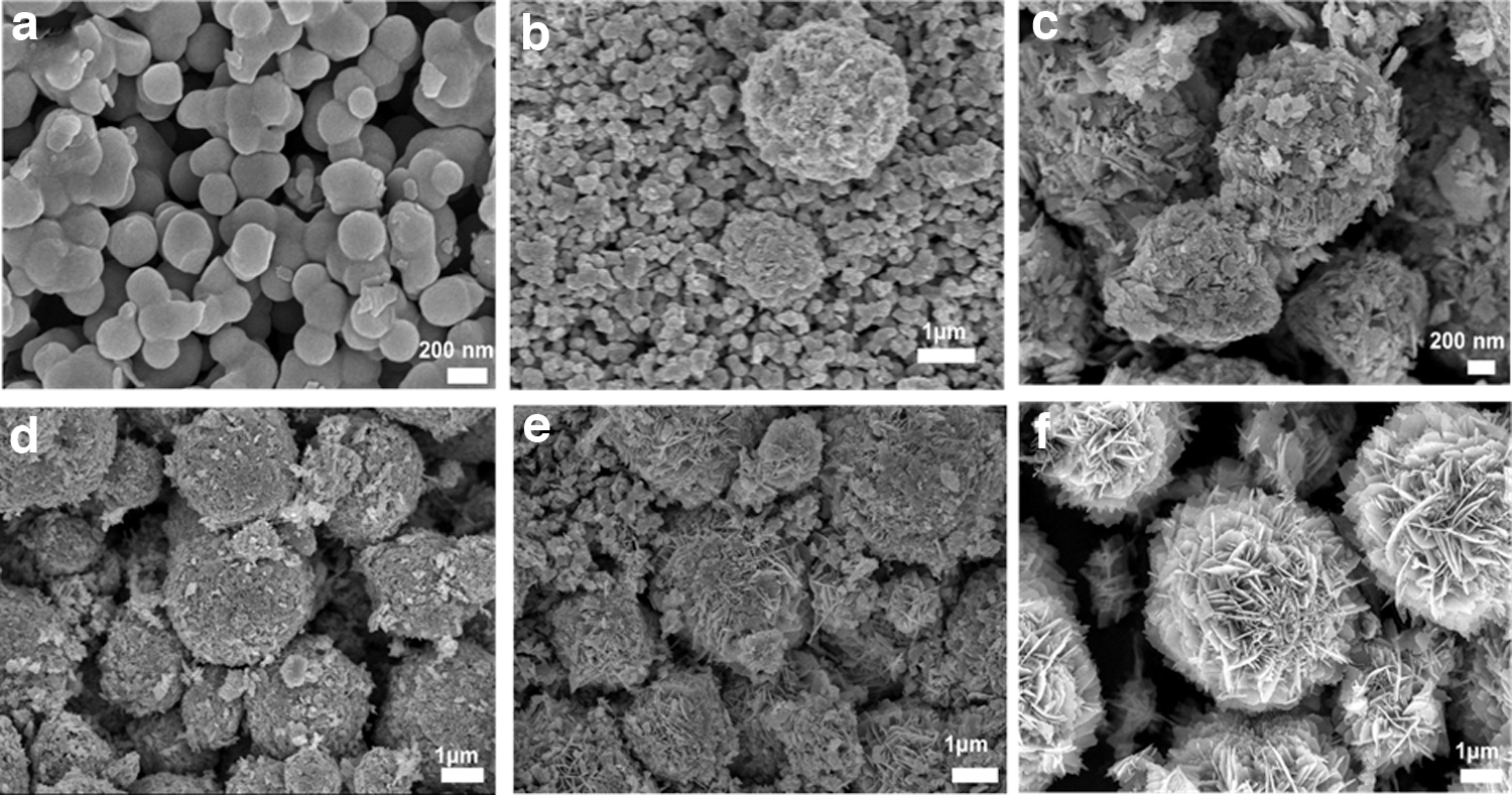
FE-SEM images of ZnO formation at different reaction times
As the reaction time increased to 3 min, nanoparticles began to conglomerate and larger irregular spherical nanoparticles were presented (Fig. 2b). As shown in Fig. 2c, after 4 min, spherical nanoparticles vanished, as well as many small nanosheets were developed and covered on the surface of nanoparticles. The surface of the sample obtained after 5 min became smoother, and its average diameter was increased. After 60 min, the nanoflower structure was obtained by forming and arranging the petals. However, many small nanosheets exited on the surface. When increasing the time reaction to 120 min, the flower structure was composed of well-arranged petals, in Fig. 2f.
Based on the above analysis, the reasonable formation mechanism for carnation-like ZnO structure could be speculated. In the early stage, the explosion of homogeneous nucleation occurred to lead to numerous ZnO nanoparticles being formed. Then, to minimize the surface energy, the oriented aggregation of the initially formed ZnO seeds started to generate the spherical ZnO. At the second growth stage, the structure-oriented agent C6H5Na3O7 was preferentially absorbed into the [0001] plane along the c-axis. Thus, restricting the growth crystal in this direction led to ZnO crystals developing in other directions.
As a result, many nanosheets have formed. The defects and thermodynamically unstable regions inevitably appeared on the surface of the primary nanosheets formed during this period. This would be a good condition providing active sites for the development of secondary nanosheets. The second nanosheets would preferentially grow along the (0001) and (0110) directions in the [2110] plane and lead to the formation of nearly vertical nanosheets on the side surface (Lu et al., 2008). Finally, more and more third or fourth-branched nanosheets were formed interlaced and superimposed on the developed nanosheets, when increasing reaction time, leading to the formation of carnation-like ZnO with structure stratification, as shown in Fig. 2f.
Photocatalytic activity
Figure 3 shows the decomposition of TCH on carnation-like ZnO, the reaction conditions set at the TCH concentration of 20 mg/L and catalyst dose of 0.05 g/L, and pH solution of 7.0. After 30 min of adsorption, the DE of TCH was 15.83%. Then, the TCH was almost degraded within 60 min under Hg lamp, showing a DE of 89.92%. Under UV lamp, the DE achieved 73.66% with a constant rate of 0.018 min−1.

The DE and rate constant under the Hg lamp were higher than that under the UV lamp, in Fig. 3a and b. This could be explained by the light intensity of the Hg lamp (13350 LUX) being higher comapred with the UV lamp (471 LUX). In addition, the photocatalysis of TCH could be studied using the second-order kinetic model, which is expressed in section 2.4. The results are shown in Table 1. According to the first-order kinetic plot, the rate constant was 0.038 min−1 with an R2 of 0.998. According to the second-order kinetic plot, the R2 of 0.943 was lower compared with the first-order kinetic plot. This revealed that the first-order kinetic model was in good relation to the photodegradation process and could be employed to study the kinetics.
The Kinetic Parameters of the Photocatalysis of Tetracycline Hydrochloride on ZnO Materials
One of the crucial factors determining the economic efficiency of materials in industrial applications is the stability of catalytic. Therefore, in this study, ZnO was used to run through four successive recycles, and the result is shown in Fig. 4a. The DE was slightly decreased to 82.65 and 70.51% in the second and third cycles, respectively. However, the DE significantly declined in the fourth cycle, reaching 56.43%. The decrease in removal efficiency of TCH can be attributed to the loss of active sites after regeneration, the reduction of the interaction surface, and the increase in intermediate products on the surface after each recycling.

A series of experiments were carried out to examine the role of active species in the photodegradation of TCH by ZnO, including IPA (2 mM), AA (1 mM), EDTA (1 mM), and AgNO3 (1 mM), and the results are exhibited in Fig. 4b. These scavengers were used to trap corresponding to •OH, •O2−, h+, and e−. According to Fig. 4b, with additions of AgNO3 and IPA, the DEs corresponded to 87.75 and 75.50%, which were slightly decreased compared to the absence of scavengers. However, with the addition of AA and EDTA, the DEs were significantly decreased, showing 7.18 and 4.03%, respectively. This demonstrated that in the decomposition of the TCH process, •O2− and h+ were key active species. Furthermore, the valence band (EVB) and conduction band (ECB) energy were calculated according to the following equations (Thi and Vu, 2022):
where χ is the absolute electronegativity of ZnO, and Eg and Ec are the band gap energy of ZnO and the energy of free electrons in the hydrogen scale, respectively. According to Equations (5) and (6), the EVB and ECB values were determined at 2.88 and −0.31 eV, respectively.
From there, the TCH decomposition mechanism can be drawn as follows (Fig. 4c): when the light was irradiated, the electron in the VB is excited and jumps to the CB with a higher energy level. At the same time, in the region, valence appears holes. Then, the photo-induced electron could react with O2 molecules in the air to form superoxide anion radicals (•O2−). Similarly, the hydroxyl radical (•OH) was generated by a reaction between the OH- ions and the holes (Chin et al., 2022a). Finally, the •O2− and •OH agents reacted with TCH to generate CO2, H2O, and other intermediate products.
Investigation of factors affecting the photocatalysis process
Initial antibiotic concentration
The initial antibiotic concentration is an essential factor that directly affects the photocatalysis process. This study investigated the initial antibiotic concentration at about 15–30 mg/L. The reaction conditions were set at the catalysis dose of 0.5 g/L, light source of 250 W Hg lamp, and pH solution of 7.0. The results are shown in Fig. 5a1–a2. It showed that the photocatalytic efficiency decreases as the initial antibiotic concentration increased. The removal efficiency decreased from 96.08% to 54.43% when the concentration increased from 15 to 30 mg/L. It could be explained that as the concentration of TCH increase, the generation of •OH− and •O2− radicals is decreased because the photons are intercepted before they can reach the catalyst surface. It leads to the removal efficiency of TCH decreasing. In addition, the active site on the catalyst surface is occupied by a few adsorbent TCH (Nguyen et al., 2020).

Catalysis dose
To study the effect of catalysis dose on the photocatalysis, experiments were conducted with ZnO from 0.25 to 1.25 g/L at fixed conditions (TCH concentration of 20 mg/L, the light source of 250 W Hg lamp, and pH solution of 7.0); the results are presented in Fig. 5b1–b2. The higher the catalysis dose, the higher the DE and the rate constant. At the catalysis dose of 0.25 g/L, the DE was 78.62%. It significantly increased at higher catalysis dose; the DE achieved 97.39% at 1.0 g/L. As the catalysis dose increased from 0.25 to 1.0 g/L, the rate constant increased from 0.028 to 0.087 min−1.
At 1.25 g/L, the DE was 98.84% in 60 min, but the rate constant (0.066 min−1) was lower than those at 0.75 and 1.0 g/L. The influence of catalysis dose on the decompose efficiency of TCH can be explained by the following reasons: an increase in catalysis dose leads to an increase in the number of active sites available on the catalyst surface and an increase in the density of the catalyst particles in the illuminated area. Therefore, the photocatalytic ability of the material is better, leading to a rapid increase in the decompose efficiency TCH (Qu et al., 2018). However, the catalysis dose is too high, which leads to the increasing light scattering effect, causing a decrease in the constant rate (Pham et al., 2020).
The pH of the initial TCH solution
The pH of the initial solution is also an important parameter in the photocatalysis process. In this study, the effect of initial solution pH on TCH depletion was investigated in the pH range from 5 to 11 at fixed conditions (catalysis dose of 0.5 g/L, TCH concentration of 20 mg/L, and the light source of 250 W Hg lamp), and the results are shown in Fig. 5c1–c2.
It was found that in the pH range from 5 to 9, the rate constants were almost the same (k = 0.018 min−1). The optimal pH for the decompose efficiency of TCH was 11. After 60 min, 90.01% of TCH had been removed with the rate constant of 0.037 min−1. At pH = 11, the decompose efficiency of TCH increased. This can be explained by the large amount of OH− ions on the surface of the nanoparticles as well as in the reaction medium, facilitating the formation of •OH. As a result, the removal efficiency of TCH and the rate constant increased (Vu and Pham, 2023).
Effect of morphology
The photocatalysis activity of as-prepared ZnO samples with different reaction times was investigated at the catalyst dose of 0.5 g/L, TCH concentration of 20 mg/L, Hg lamp, and pH of 7.0. The removal efficiency of TCH is shown in Fig. 5d. It can be seen that both the nano and flower ZnO had similar TCH degradation although the reaction time affected the morphology of ZnO. However, the photocatalytic efficiency increased if the reaction time was 120 min. This proved that the complete 3D ZnO structure had a higher ability to degrade TCH than its intermediate structures, in reaction times of 2 to 60 min.
Comparison of ZnO nanoflower with other ZnO samples
Many scientists in many different fields have applied ZnO nanoparticles. Primarily, ZnO nanoparticle is used as a photocatalyst to treat polluted water environment. Some research results on ZnO nanoparticles are presented in Table 2. ZnO nanoparticle was used to treat various pollutants such as Methyl Blue (Kaliraj et al., 2019; Sahu and Kar, 2019; Tripathi et al., 2014; Yang et al., 2010), Congo Red (Mohammad et al., 2016), Metamitron (Xu et al., 2016), Nitro Benzene (Swarnavalli et al., 2019), and so on (Qu et al., 2020; Wang et al., 2008; Zhang et al., 2015; Zhou et al., 2016).
Some Research Results on ZnO Samples
CP, cholrophenol; CR, congo red; MB, methylene blue; MO, methyl orange; NB, nitrobenzene; NF, norfloxacin; TCH, tetracycline hydrochloride; UV, ultraviolet.
It is difficult to evaluate the removal efficiency of different pollutants of ZnO due to the application of varying reaction conditions. This study used ZnO to remove antibiotic contaminants that had not been studied before. TCH was almost degraded within 60 min, showing a DE of 89.92%. The easy synthesis, cheap, and being environmentally friendly makes ZnO nanoflower a potential catalyst for the effective treatment of TCH and other organic matter in aquatic environments.
Comparison of the TCH degradation ability of different catalysts
Since tetracycline is a broad-spectrum antibiotic found in surface water, groundwater, and demotic water, many materials have been studied to remove TCH in water. Some research results are shown in Table 3. It can be seen that decompose efficiency of TCH on ZnO at the catalyst dose of 1.0 g/L, TCH concentration of 20 mg/L, under a 250 W Hg lamp, and pH of 7.0 was higher than those of Ag/AgIn5S8 (Deng et al., 2018), B/ZnO (Nguyen and Vu, 2022), and CuAl2O4, g-C3N4, CuAl2O4/g-C3N4 (Chen et al., 2021). It still was lower than those of MIL-88A (Zhang et al., 2019) and CuO/B2O3 (Zhu et al., 2022). However, the catalytic process that used MIL-88A required the support of the strong oxidizing agent S2O82− and the catalyst process that used CuO/B2O3 required the support of the strong oxidizing H2O2.
Some Research Results on Tetracycline Hydrochloride Degradation
Conclusion
The complete carnation-like ZnO structure has been successfully prepared by the fast precipitation route at room temperature. The hexagonal wurtzite structure ZnO was confirmed by XRD analysis. SEM images showed a regular and uniform 3D carnation-like ZnO with an average diameter of 5 μm. The 3D flower ZnO consisted of numerous nanosheets with uniform shapes and rough surfaces. The mean thickness of the sheets was 0.013 μm. The formation of nanoflower was proposed with secondary nanosheets growing along the (0001) and (0110) directions in the [2110] plane. As a result, the band gap energy of ZnO is 3.19 eV.
The effects of reaction parameters on catalytic performance were comprehensively studied. Based on both decompose and rate constant, ZnO exhibited the best catalytic performance at the TCH concentration of 15 mg/L, catalyst dose of 1.0 g/L, and pH of 11. At the first use, ZnO showed to efficiently degrade TCH with the DE and rate constant of 89.92% and 0.038 min−1, respectively. After four successive recycles, the DE value significantly decreased to 56.43%. The decrease in photocatalytic activity can be speculated to be due to the loss of active sites, the reduction of the interaction surface, and the increase in the intermediate products on the surface after an experiment.
Footnotes
Authors' Contributions
T.H.N.: research ideas, synthesis of ZnO, studying the effects of reaction conditions on TCH decomposition, collection of results, and writing the article; T.A.N.C.: analysis of N2 adsorption/desorption isotherm and studying of TCH decomposition under UV irradiation; A.-T.V.: experiment setup, material characterization, review and editing, revision, supervision, and project administration.
Author Disclosure Statement
No competing financial interests exist.
Funding Information
The authors are grateful for the financial support from Vietnam National Foundation for Science and Technology Development (NAFOSTED) under grant no. 104.05-2018.333. The work was performed at the Vietnam Germany Catalysis Centre—RoHan Project funded by the German Academic Exchange Service (DAAD, no. 57315854) and the Federal Ministry for Economic Cooperation and Development (BMZ) inside the framework “SDG Bilateral Graduate school program.