
Abstract
Flexible cystoscopy is well established in urological practice. We present a unique image obtained during bladder inspection, illustrating visual refractive distortion, which highlights the potential difficulty in optical interpretation.
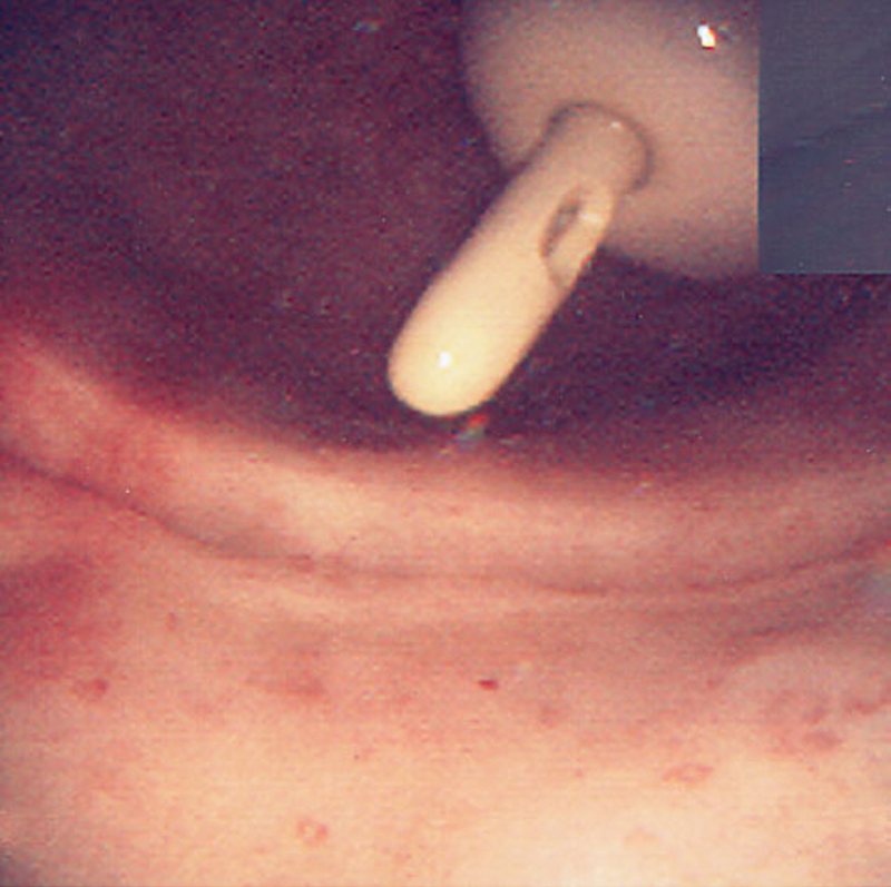
Figure 1 obtained at the beginning of cystoscopy raised concerns as the catheter tip is clearly seen entering and exiting the bladder mucosa, giving the appearance of bladder perforation. On continued bladder filling the catheter reassuringly lifted away from the bladder wall as shown in Figure 2. The distorted image was due to the edge of the air bubble resting on the catheter tip, thereby warping the image. During cystoscopy, while we instill fluid into the bladder, we often insert air trapped from within the given set tubing. The collected air forms a bubble that usually migrates to the dome of the bladder.

Initial appearance from flexible cystoscope, showing suprapubic catheter apparently traversing the bladder mucosa (arrow).
The same study with further bladder filling, showing how the catheter balloon lifts away and is no longer traversing the gas–fluid interface.
Light travels at varying speeds in different media, and consequently, our eyes may be deceived at the air–fluid interface as light is refracted unevenly, thereby potentially distorting the appearance of the underlying bladder mucosa. Further, the edge of the air bubble provides a reflective surface, which may simply display the adjacent tissues, not the mucosa beyond. Although we may be careful to prevent producing an air bubble during cystoscopy, some insufflation may occur inadvertently. The endoscopist should be aware of the potential for mucosal distortion at the air–fluid interface and tailor the examination accordingly. To our knowledge this phenomenon of refractive image distortion has only previously been reported in the medical literature in the context of fiberoptic intubation laryngoscopy 1 and never in clinical urology.
Footnotes
Disclosure Statement
No competing financial interests exist.