
Abstract
Hexylaminolevulinate (hexa ALA) “blue light” fluorescence for detecting cellular changes in mucosal epithelium is used in urology to detect transitional-cell carcinoma and carcinoma in situ in the bladder. It is is also being studied for the detection of precancerous/cancerous lesions in the colon. We present a case in which hexa ALA fluorescence cystoscopy was used in the evaluation of cecal mucosa that was incorporated into the bladder as an augmentation cystoplasty. To our knowledge, this is the first time the imaging appearance of an augmented bladder under Hexa ALA fluorescence cystoscopy is described.
Introduction
We present a case in which hexa ALA fluorescence cystoscopy was used to evaluate large bowel mucosa that has been incorporated into the bladder as an augmentation cecocystoplasty. This is the first time the imaging appearance of an augmented bladder under hexa ALA fluorescence cystoscopy is described.
Case Report
A 45-year-old Caucasian woman with spina bifida had an augmentation cecocystoplasty during her teenage years. She had previously undergone left nephroureterectomy for chronic pyelonephritis. She had a right groin dissection for an enlarged lymph node that had been found to contain squamous-cell carcinoma; this was managed with radical radiotherapy. Despite full investigation, no primary had been found, and this prompted cystoscopy when the patient had been found to have a trace of blood on urine dipstick evaluation.
The patient underwent a hexa ALA fluorescence cystoscopy with biopsies in our center on an urgent basis after the bladder was found to have an abnormal appearance at flexible cystoscopy. The appearance of the augmented part of the bladder was described as looking red with inflamed areas of white patches.
Hexa ALA cystoscopy of the augmented bladder was performed using standard technique. Under white light, the junction between the bladder and bowel mucosa looked normal. The bowel mucosa looked generally inflamed, with white patches scattered over the mucosa (Fig. 1). Blue light was then used, and the whole of the bowel mucosa looked pink. The white patches were also seen to be pink (Fig. 2). Biopsy specimens were taken from the augmented bowel mucosa and the white patches. Histologic examination demonstrated chronic inflammation of the bowel mucosa, but showed no evidence of malignancy (Fig. 3).
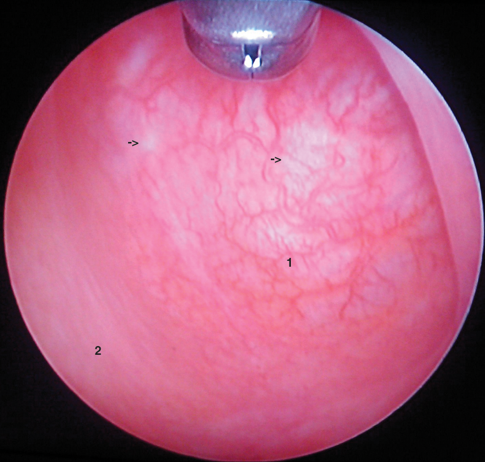
The augmented bladder under white light cystoscopy. Arrows are pointing toward the "white patches" seen. 1 = cacal mucosa; 2 = transitional zone.

The augmented bladder under blue light cystoscopy. A generalized pink appearance can be seen. Arrows point to the area of white patches. 1 = cecal mucosa.

Histology of the cecal part of the augmented bladder showing chronic inflammatory changes. 1 = chronic inflammatory cell infiltrate; 2 = crypt hyperplasia.
Discussion
Hexa ALA is incorporated at the start of the haem biosynthesis pathway and is not itself photochemically active. When instilled into the bladder, it undergoes a series of biochemical reactions, leading to the formation of the active photosensitizing compound, protoporphyrin IX. A negative feedback mechanism normally prevents the accumulation of protoporphyrin IX in the presence of adequate haem molecules. The use of hexa ALA, however, bypasses this control, causing a temporary accumulation of protoporphyrin IX.
The mechanism that allows increased selective uptake into rapidly proliferating cells is not fully understood, but there are several theories. Some are related to the structural changes in the cell membranes, causing increased permeability, while others relate to intracellular biochemical factors such as the iron pool.
Augmentation cystoplasty is a well-established procedure for the management of congenital and acquired bladder instability and is commonly used in patients with spina bifida. The concern of long-term risk of malignancy developing in augmented bladders is a subject of ongoing debate but is generally considered to be modest. The types of tumor reported vary, but most are adenocarcinomas arising at the junction of the vesicoenteric anastomosis. 2
In augmented bladders, the bowel mucosa is continually exposed to urine. It is well recognized that the chronic exposure of bowel mucosa to urine induces histologic appearances that are suggestive of chronic inflammation, such as crypt hyperplasia and chronic inflammatory cell infiltrates. 3 Some studies done demonstrate that normal large bowel mucosa shows no fluorescence under hexa ALA. 1 The exact reason for the generalized pink appearance of the cecal mucosa under blue light (Fig. 2) in our case cannot be ascertained. It could be related to the chronic inflammation in the mucosa from constant urine exposure, or related to some form of different enzyme activity of the cecal mucosa. It can also be because of precancerous changes in the mucosa, warranting repeated check cytoscopies in this patient. The specificity/sensitivity of hexa ALA detection of malignant cells in augmented bladders is unknown and should be assessed with a prospective, randomized controlled trial.
Conclusion
We conclude that augmented bladder mucosa looks pink under blue light cystoscopy, and hexa ALA use in similar cases should be questioned until more evidence is available.
Footnotes
Disclosure Statement
No competing financial interests exist.