




Abstract
Background and Purpose:
Cryptorchidism is an ubiquitous feature in prune belly syndrome (PBS). Laparoscopic orchidopexy allows dissection of the spermatic cord with minimal morbidity. We discuss the technical difficulties and outcome of three boys with PBS who underwent two-stage laparoscopic Fowler-Stephens orchidopexy (F-SO).
Patients and Methods:
Three boys, ages 1, 2, and 4, underwent laparoscopic F-SO. All boys had viable testes that were found within 3 cm of the deep inguinal ring. The testicular vessels were either ligated bilaterally with 4/0 polyglactin or monopolar diathermy was used and the vessels divided. Bilateral second-stage F-SO was performed within 6 months in two boys and limited to one side in the third boy. One boy awaits the contralateral second stage.
Results:
All three boys have adequately sized gonads. Based on our experience, the port incisions should be smaller than routine practice to prevent air leak in PBS. Although the intra-abdominal pressure of 12 mm Hg did not vary from our normal practice, a high flow rate is necessary after initial insufflation (6 L/min) to compensate for inevitable gas leaks because the abdominal wall is so thin. Risk of diathermy injury to the thin abdominal wall and the vessels is significant. Laparoscopy enables easy visualization of the ureter, testes, and testicular vessels and permits complete dissection of testicular vessels. It is easier to maintain integrity of spermatic vessels.
Conclusion:
Use of radially expanding trocars, small incisions, and high gas flow rates permit this procedure to be performed safely with good outcome and cosmetic results in this challenging group of boys.
Introduction
We discuss the technical difficulties and outcome of three young boys with PBS who underwent laparoscopic FS-O.
Patients and Methods
Three boys, ages 1 (Patient 1), 2 (Patient 2), and 4 (Patient 3) with antenatal diagnosis of prune belly syndrome, chronic renal failure, and bilateral cryptorchidism were referred to the urology clinic for evaluation. The 1-year-old boy also gave a history of posterior urethral valve ablation and recurrent urinary tract infections. The 4-year-old boy also had associated congenital talipes equinovarus and a history of multiple surgeries for closure of exomphalos minor, open reduction of congenital dislocated hips, and Nissen fundoplication. They underwent laparoscopic two-stage FS-O.
A Veress needle was inserted into the peritoneal cavity through the thin abdominal wall, and carbon dioxide was introduced to distend the abdomen insufflated to 20 mm Hg. The pressure was maintained at 14 mm Hg.
A 5-mm sheath and trocar were inserted using the same umbilical incision. Under laparoscopic vision, two smaller ports (3 mm) were inserted in the right and left anterior lumbar region (Fig. 1). Trocar placements were identical in the three patients and uneventful. All three had short tethered testicular vessels. Patients 1 and 3 had suture ligation of the testicular vessels, whereas Patient 2 had plasma kinetic occlusion to obtain sufficient length. Occlusion/ligation of the vessels was performed at 2 to 3 cm from the testes. The second stage was carried out 6 months later.
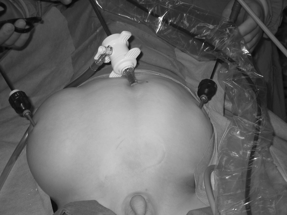
Port placement sites.
Results
Patient 1
Laparoscopic evaluation showed the left testis was situated lateral to the sigmoid colon and the right testis between the anterior abdominal wall and paracolic gutter. Mobilization on the vas and associated vessels enabled the testes to be brought to the inguinal canal. The second stage involved bilateral dartos pouch orchidopexy. He has adequately sized testes at 2-year follow-up.
Patient 2
This 2-year-old boy underwent the standard laparoscopic evaluation with both testes proximal to the internal inguinal ring. Both testicular vessels were severed with the PK scissors, and the testes were mobilized to the inguinal canals. He underwent second stage F-SO with adequate sized testes in the scrotum at 4-year review.
Patient 3
In this patient, the left testis was found proximal to the inguinal ring and the right farther up. Severing the short tethered testicular vessels enabled mobilization to the inguinal canal.
Six months later, the right testis was mobilized on the vas deferens and vessels to reach the right inguinal ring. An 11-mm short STER port was brought from the scrotum, directed medial to the epigastric vessels, and a grasper used to deliver the testis into the dartos pouch for fixation. This mobilization resulted in a stretched cord; a per-operative decision was made to delay the left second stage FS-O because the potential risk for testicular atrophy was considered to be significant. He has a sufficiently sized right gonad at 4 years, however, and awaits the left second stage, which has been delayed because of serious maternal illness.
Discussion
Laparoscopic orchidopexy has become the accepted norm in pediatric urology for intra-abdominal testis. In children with PBS, the expectant risks have resulted in surgeons being reluctant to undertake laparoscopic surgery in these patients. More so, management of the undescended testes in PBS is technically challenging with the high intra-abdominal testes and short vessels. Laparoscopic F-SO was the first therapeutic laparoscopic procedure performed on a child with PBS. 2
Laparoscopic F-SO is the current favored management technique. A staged procedure has been reported to enhance success rate. 3,4 Laparoscopic F-SO enables extensive dissection of the gonadal vessels. This can be performed with preservation of the distal triangle between the vas deferens and the vessels to maximize collateral vasculature development. 5
We raise the following technical points from our experience. The port incisions should be smaller than routine practice to prevent air leak in PBS. An intra-abdominal maintenance pressure of 12 mm Hg was necessary in our series to maintain adequate pneumoperitoneum. The usual flow rate was 1 to 3 L/min, but higher rates were used when there was an air leak. None of the patients had surgical emphysema or pulmonary compromise, despite this high flow. Risk of diathermy injury to the thin abdominal wall and the vessels is greater, and laparoscopic illumination should be adequately maintained at all times.
Laparoscopy enables easy visualization of ureter, testes, and testicular vessels. It permits complete dissection of testicular vessels and is easier to maintain integrity of spermatic vessels. The ureter can be dilated and tortuous, complicating mobilization. Saxena and Brinkmann 6 reported the need for longer instruments. We used standard pediatric laparoscopic instruments but can appreciate that some cases could need longer instruments. There is also minimal morbidity, with these patients going home after an overnight stay. Overnight stay was necessary to make sure none had respiratory compromise.
Docimo and coworkers 2 explain the creation of the neocanal retrograde from the scrotum. They describe an antegrade technique that includes passing a straight grasper from the ipsilateral laparoscopic port out through the scrotal incision. The canal is then dilated using a radially dilating trocar with the sheath placed retrograde over the grasper. We have used this technique with similar experience in safe scrotalization of the testes with no epigastric or iliac vessels injury.
This article is the second case series worldwide and the first from the United Kingdom that reports on the outcome of laparoscopic orchidopexy in PBS.
Conclusion
Laparoscopic orchidopexy should be the gold standard in the management of boys with cryptorchidism, more so in those with PBS. The technical points raised here should significantly improve surgical practice, advance the learning curve of pediatric surgeons who intend to undertake this procedure, and reduce the risk of associated injuries.
Footnotes
Disclosure Statement
No competing financial interests exist.