
Abstract

Case Presentation
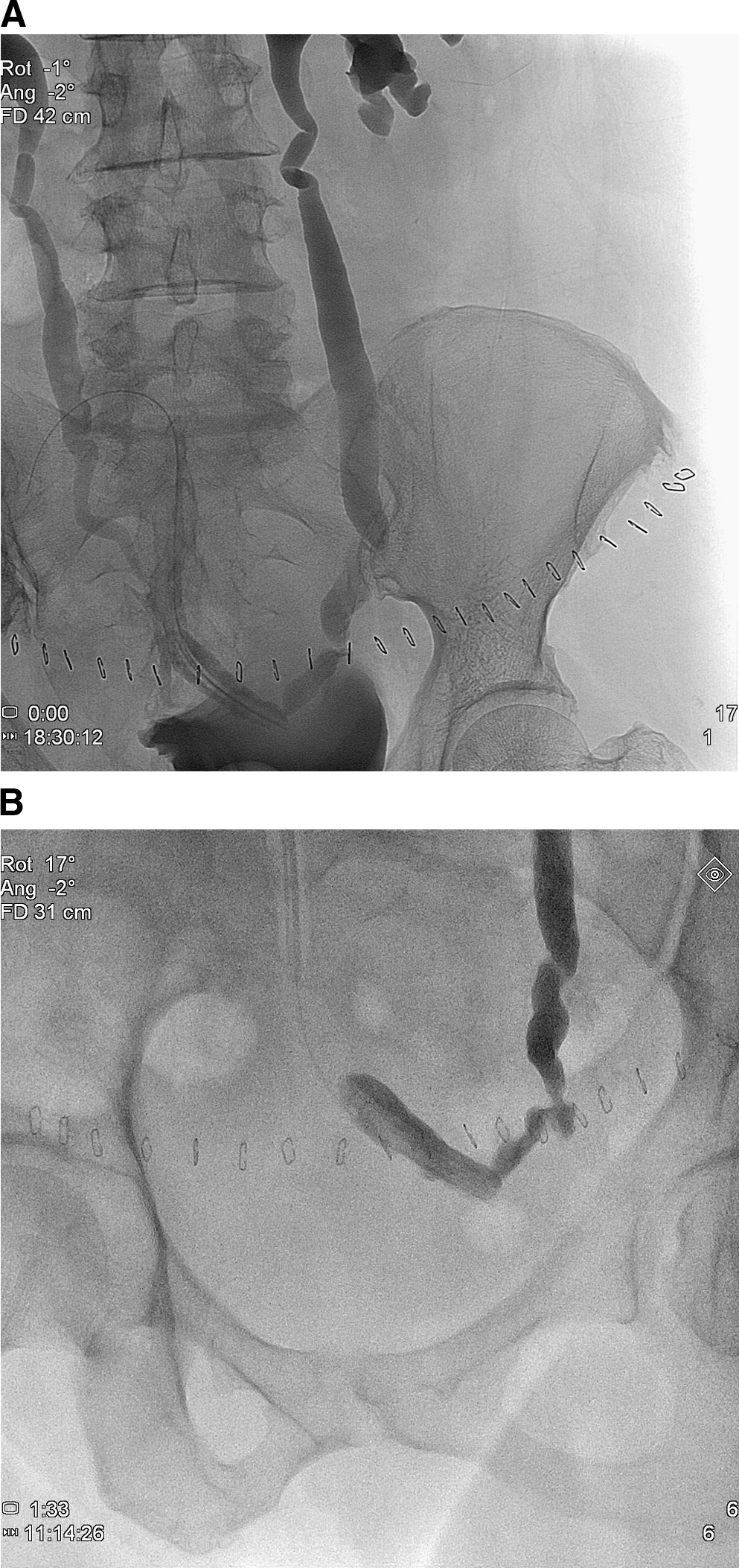
A 55-year-old woman underwent open radical hysterectomy without experiencing any intraoperative incident. On the second postoperative day, she complained of left flank and lower abdominal pain that subsided later while increased drain fluid output, high fever, and chills were observed. The drain fluid was analyzed and proved to be urine. Ultrasonographic imaging of the abdomen and pelvis showed minimal dilation of the left collecting system and normal right collecting system. Bilateral antegrade pyeloureterographies were performed and revealed complete left lower ureteral obstruction, leakage, and communication with the drain (Figures 1A and B). The right collecting system and ureter were normal. During the attempt to cross the site of the ureteral obliteration, the wire pass through the drain tube and the access of the wire to the bladder were not possible (Figures 2A and B). A nephrostomy tube was inserted for the drainage of the left collecting system.
(

(
Gynecologic injuries of the ureter are associated with incidence ranging between 0.5% to 30%, while the intraoperative recognition rate ranges from 11% to 33%. Moreover, a ureter accidentally ligated by suture is usually the cause of the obstruction. The attempt to manage the stricture with a percutaneous approach was not successful, and there is a standing question for the further treatment of the patient.
Expert Opinions
No. 1
The next step is to identify any other injuries in the genitourinary system with complete radiographic evaluation. Each ureter should be evaluated with either antegrade or retrograde pyelography, and the bladder should be evaluated for injury with standard cystography or the more sensitive CT cystography.
The patient already had bilateral nephrostomy tubes placed for bilateral antegrade pyelography. The left antegrade pyelogram (Fig. 1B) demonstrates extravasation of contrast into the abdominal drain without any antegrade flow into the distal ureter or bladder. The right ureter appears intact in Figure 1A. Right-sided contrast was administered after contrast from the left ureter extravasated into the pelvis, however, thus not completely confirming absence of injury.
Given the extent of the patient's left ureteral injury, she will need further intervention. The question of immediate vs delayed repair arises. If she is unable to tolerate an anesthetic or if she is clinically unstable, the patient can be temporarily treated with nephrostomy tube and Foley catheter drainage to divert the urine away from the site of extravasation. The abdominal drain should be left in place.
If the patient is stable enough for a general anesthetic, optimal management would be immediate surgical repair. The next decision relates to the surgical approach. There have been several reports of laparoscopic and robot-assisted ureteral reimplantation and Boari flap. 1 Because this patient underwent an open abdominal hysterectomy, she would be better suited for an open repair.
In the operating room, the patient should first undergo cystoscopy and left retrograde pyelography to determine whether there is any continuity between the proximal and distal left ureter as well as assess length of distal ureteral involvement. If contrast is visualized extending into the proximal ureter, a semirigid ureteroscope may be gently passed into the distal ureter if a wire cannot pass the obstructed segment. If the lumen of the ureter proximal to the defect can be seen or if a minor degree of extravasation is visualized, an attempt at placing a hydrophilic wire or sensor wire followed by a Double-J stent across the defect can be made. Stent placement in the acute setting may temporize the situation; however, definitive ureteral reimplantation will ultimately be necessary. If there is complete obliteration or disruption of the ureter, I would proceed with open ureteral reimplant with possible psoas hitch or Boari flap.
The surgical consent should include cystoscopy, bilateral retrograde pyelography, possible left ureteroscopy, left ureteral stent placement, possible open ureteral reimplant, possible psoas hitch/Boari flap. Reported open surgical repair success rates are excellent. 2,3
Although some surgeons may advocate delayed repair to allow time for postsurgical inflammation to subside, the presence of urine in the abdominal cavity will likely provoke a more intense inflammatory reaction over time. The patient may have more favorable tissue planes 3 days after her initial surgery. Assessment of baseline renal function should also be performed to counsel the patient as to success rates, because differential renal function > 25% predicts a better outcome.