
Abstract
Introduction:
Ureteroscopy is one of the therapeutic options in the management of urinary stone disease in children. Previous literature has described ureteroscopy primarily in mid to distal ureteral calculi. We report our experience with flexible ureteroscopy in the management of upper ureteral calculi.
Patients and Methods:
All children with upper ureteral stones were included prospectively in the study. Stone burden was measured in millimeters. Presentation, operative access, intraoperative complications, stone-free rates, and postoperative complications were evaluated.
Results:
A total of 80 children (69 boys and 11 girls) underwent 88 ureteroscopic procedures. In 72 (90%) children, complete stone clearance was achieved after a single ureteroscopic session. In 6 (7.5%) others, complete stone clearance was achieved after the second ureteroscopic session.
Conclusions:
Complete stone clearance after single ureteroscopy is possible if the calculi are single, small (<10 mm), and below the level of the pelviureteral junction.
Introduction
We have reported our experience with ureteroscopy for ureteral stones in 20 children. 6 The average age of the children was 5.2 years, and the mean size of the stone was 6 mm. Overall, 90% of the children were rendered stone free after one procedure and 100% after two procedures. With increasing experience and introduction of better flexible endoscopes, we have used flexible ureteroscopy as a first-line treatment in all children with upper ureteral stones and in whom conservative therapy failed.
Patients and Methods
All children with upper ureteral stones were prospectively included in the study. Ultrasonography of the kidneys, ureters, and bladder region was performed in all. Intravenous urography/CT urographies were performed to better image the stone and note the size, number, and site of stones (Figs. 1 and 2). Stone burden was measured in millimeters. All children were initially treated conservatively, which included hospitalization, intravenous fluids, and pain relieving drugs.

Radiography of the kidneys, ureters, and bladder shows an 8-mm calculus in the left upper ureter.
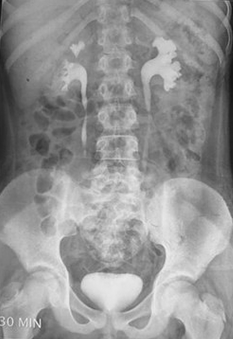
Intravenous urography demonstrates a left ureteral calculus at the junction of the upper and midureter.
Indications for ureteroscopic stone extraction included failed conservative management and persistence of pain. Ureteroscopy was performed under general anesthesia with the child in the modified lithotomy position. An initial cystourethroscopy was performed. A 0.035 safety guidewire was passed up the ureter. Over the safety guidewire, a double lumen ureteral catheter was passed up the ureter. A 0.025 inch working guidewire was passed alongside the safety guidewire. Active dilation of the ureteral orifice was performed using cone-tipped metal dilators. A ureteral access sheath (9.5/10 F) was used only in children who had undergone stent placement before definitive ureteroscopy. A flexible ureteroscope was passed over the working guidewire under both visual and fluoroscopic guidance. Whenever it was difficult to dilate the lower ureter or pass the double lumen ureteral catheter/flexible ureteroscope, the children underwent Double-J stenting placement before definitive ureteroscopy a week later. Calculi were fragmented with a 30 watt holmium: yttrium-aluminum-garnet laser. Fragments of 2 to 3 mm were extracted, and larger fragments were further lased into smaller fragments. A Double-J stent was placed at the conclusion of ureteroscopy in children who had a huge stone burden (10 mm or more), whenever ureteral injury was suspected, presence of infection, incomplete fragmentation, and presence of hematuria.
Stone clearance was assessed intraoperatively by direct ureteropyeloscopy and postoperatively by imaging that included ultrasonography, abdominal plain radiography, or noncontrast CT, depending on the discretion of the treating urologist. Stone free was defined as obvious stone seen on ultrasonography/abdominal plain radiography/noncontrast CT. Intraoperative complications were defined as evidence of ureteral injury, including ischemia, perforation, avulsion, extravasation of contrast material on ureteropyelography. Extravasation is described as the appearance of extravasation material outside the ureter into the retroperitoneal area. Postoperative complications were defined as worsening hydronephroureterosis, ureteral stricture, and other untoward events neccessitating additional surgical intervention.
All extracted stones were sent for chemical analysis, and all patients were evaluated for urolithiasis 6 weeks later. Double-J stents were removed using brief anesthesia 10 to 14 days after ureteroscopy.
Results
A total of 80 children (69 boys and 11 girls) underwent 88 ureteroscopic procedures during the study period (January 2002–December 2009). The mean age of the children was 9.5 ± 1.57 years (range 6–12 y). The mean stone burden was 10.18 ± 1.87 mm (range 7–16 mm). Eight (10%) children had two or more calculi, and the remaining had only one calculus in the affected ureter. Fifty-six (70%) children had calculi located in the upper ureter below the pelviureteral junction (PUJ), and the remaining 24 (30%) had calculi at the PUJ.
Sixty-three (78.75%) children presented to the hospital with pain alone, another 8 (10%) complained of hematuria with pain, and 5 (6.25%) children presented with fever and loin tenderness. Four (6%) children had a history of urolithiasis, and two (2.5%) of these had undergone ureteroscopy in the past. Three of five children who presented with fever and loin tenderness did not respond to antibiotics and underwent Double-J stent placement and catheterization to allow adequate drainage of the obstructed system. Retrograde access was successful in 52 (65%) of the children, and the remaining 25 (31.25%) children underwent stent preplacement. In 31.25%, the urethral dilatation was unsuccessful and, hence, these children underwent for stenting insertion before definitive ureteroscopy. There was no need of antegrade access in any of the children.
Ureteroscopy was performed using either a Storz flexible ureteroscope or an ACMI ureteroscope (Fig. 3). There was no difference between the type of ureteroscope used and the ability to gain retrograde access. In 72 (90%) children, complete stone clearance was achieved after a single ureteroscopic session. In 6 (7.5%) others, complete stone clearance was achieved after the second ureteroscopic session. Complete clearance of stone after the second ureteroscopic session was achieved in 97.5% of children (Table 1). In the remaining two children, residual stone fragments were seen in the lower and middle calix, and both needed an SWL session to clear these fragments.

Flexible ureteroscope is seen in the left ureter.
PUJ = pelviureteral junction.
The mean size of the stone burden in children who did not achieve complete stone clearance in one session was 13 ± 1.85 mm. Six of eight children had two or more calculi in the affected ureter, and all eight children had calculi situated at the PUJ. The mean follow-up was 20.4 months (range 3–42 mos).
No major intraoperative complications were noted in any of the children. Intraoperative bleeding was noticed in six (7.5%) children. Eight (10%) children had postoperative bleeding that settled on its own within 24 hours. Four (5%) children had postoperative fever that lastied for 24 to 48 hours and subsided with conservative treatment. Analysis of the extracted stone fragments revealed that 87.5% had pure calcium oxalate stones, 7.5% had mixed calcium oxalate and uric acid stones, and the remaining 5% had mixed calcium phosphate oxalate and urate stones.
None of the children had persistence or worsening hydronephrosis on ultrasonography performed at the 6-month follow-up.
Discussion
There has been a dramatic rise in the overall incidence of urolithiasis in children. 7 Although the exact incidence in India is not known, the incidence in the United States is reported to range between 0.1% and 0.5%, and the prevalence accounts for 1 in 1000 to 1 in 7600 hospitalizations. 8 The adoption of ureteroscopy in children with ureteral stones has been slow, because of concerns regarding ureteral ischemia, injury, perforation, ureteral stricture, and vesicoureteric reflux (VUR) after manipulation of small caliber pediatric ureters. Continued advances in endoscope technology, however, have allowed for the production of smaller, more flexible ureteroscopes with enhanced visual optics. Shepherd and associates 9 and Ritchey and colleagues 10 were the first to publish their respective studies on ureteroscopy in children in 1988, and since then, this technique has gained widespread acceptance among pediatric urologists. Several recent studies 6,11 –13 have confirmed the safety and efficacy of ureteroscopy in children, encouraging us to move toward ureteroscopy as first-line treatment of ureteral calculi.
Several studies have reported on the safety and success of ureteroscopy for mid to distal ureteral calculi in children. 3 –7 The great variability of reported instruments, techniques, and location of calculi, however, has clouded the efficacy of ureteroscopy in children. Kim and coworkers 14 reported on 170 ureteroscopic treatments in 167 children, of whom 101 had stones located in the proximal ureter, with a stone-free rate of 98%. This series 14 diverges from other ureteroscopy series in the fact that there were a significant number of proximal calculi (60%) that were successfully managed using flexible ureteroscopy.
The average diameter of the pediatric ureter varies from 2 to 5 mm; however, there is a wide range depending on the age and size of the child. There seems to be controversy in regard to active dilation of the ureteral orifice and the intramural ureter. Many authors are concerned and avoid any active manipulation of the ureteral orifice for fear of ureteral trauma and/or bleeding. Shepherd and associates 9 have shown that dilation of the ureter up to 12F did not result in the development of VUR postoperatively in their series of patients. Voiding cystography that was performed in pediatric patients after ureteroscopic procedures has shown that the incidence of transient low-grade VUR (grades 1–2/5) is as high as 15%. VUR resolved spontaneously with conservative management in almost all children. 1 Until long-term studies that evaluate active dilation are known, some authors believe that stent preplacement provides a safe and effective alternative in achieving access to the child's ureter. Rubenstein and colleagues 15 have suggested that the use of stent placement before ureteroscopy was associated with higher overall stone-free rates in adults.
Antegrade ureteral access may be considered in a child who has a percutaneous nephrostomy in place. It would be useful in a child with completely obstructed ureter. Reddy 1 reported using this technique in two children (<1 year of age) who had presented with complete ureteral obstruction from impaction of an 8-mm calculi. Similarly, Gupta and coworkers 16 reported the technique of supine antegrade flexible ureteroscopy in a 6-year-old child with right upper and middle ureteral calculus and inability to dilate the intramural ureter. An antegrade renal access was established in the supine position using ultrasonographic-guided puncture, serial dilation of the tract, and 22F Amplatz. The stone was disintegrated using a holmium laser.
Over the last few years, ureteroscopic disintegration of ureteral calculi has gained acceptance. Several studies 11 –14 have reported on the safety and effectiveness of this technique. The outcomes of ureteroscopy in terms of efficacy and rate of complication in prepubertal children were similar if not better than those reported in the adult literature. 17 Our study reveals that complete stone clearance after the first session of ureteroscopy is possible if the stone is small (<0 mm), solitary, and below the level of the PUJ. Similarly Kim and colleagues 14 reported that the stone clearance was 100% for stone burdens 10 mm or less and 97% for stone burdens greater than 10 mm after a single ureteroscopy.
Conclusions
Pediatric ureteroscopy is a safe and efficacious modality in the treatment of all upper ureter calculi. Complete stone clearance after single ureteroscopy seems more effective if the calculi is single, small (<10 mm) and below the level of PUJ.
Footnotes
Disclosure Statement
No competing financial interests exist.