
Abstract
Background and Purpose:
We have developed novel iron-based microparticles (Fe-MP) that bind to calcium oxalate stone fragments, rendering them paramagnetic. Previously, we demonstrated enhanced efficiency of stone extraction in an inanimate model using magnetic instrumentation. Before in vivo stone extraction studies, we sought to further characterize Fe-MP with regard to cellular toxicity and to assess the influence of biologic fluids on binding performance.
Materials and Methods:
Toxicity: Monolayers of murine fibroblasts, human urothelium, and human transitional-cell carcinoma cells were exposed to 1 mg/mL of Fe-MP or saline via an agarose overlay. Cellular viability was assessed using neutral red staining and densitometry. Biologic functionality: Human calcium oxalate stone fragments were incubated with a solution of 1 mg/mL of Fe-MP containing varying concentrations of urine (10%–50%) or blood (0.5%–2%) for 10 minutes. Fragments were then extracted using an 8F magnetic tool. Assays of 10 stone fragments categorized as small (3–3.9 mg) or large (6–6.9 mg) were run in quadruplicate at each concentration.
Results:
No toxicity was seen in any of the three cell lines after 48 hours of particle exposure, except in urothelial cells at the lowest cell concentration. Stone extraction success was 100% for all stones, regardless of concentration of urine or blood, and extractions were completed in less than 10 minutes.
Conclusions:
Preliminary toxicity testing revealed minimal to no cellular toxicity that was attributable to Fe-MP. The microparticles function well in the presence of clinically relevant concentrations of urine and blood that may be present during endoscopic stone surgery. Further toxicity and stone extraction testing in animal models is necessary.
Introduction
To this end, we developed iron-based microparticles (Fe-MP) that bind to human calcium oxalate kidney stone fragments, rendering them paramagnetic. These stone fragments can then be manipulated and extracted using novel magnetic instrumentation, thereby reducing extraction time and decreasing extraction attempts compared with standard nitinol basket extraction. 7 The ultimate goal is to suspend Fe-MP in irrigation fluid used during stone surgery so as to capitalize on this paramagnetization and improve extraction efficiency with novel magnetic tools. As with any developing medical technology, however, safety and physiologic feasibility must be assessed before in vivo evaluation. We examine the in vitro toxicity and magnetic binding performance of Fe-MP when exposed to surgically relevant biologic fluids, namely urine and blood.
Materials and Methods
Iron oxide microparticle composition and functionality
Superparamagnetic 1 μm diameter particles consisting of iron oxide (maghemite) embedded in a polystyrene matrix (Invitrogen, Carlsbad, CA) were coated with proprietary peptides that bind to calcium. These peptides contain amino acids with carboxyl chains that ionically bind to positively charged surface areas based on human calcium oxalate stone surface analysis. 7 The prepared particles were then suspended in physiologic saline.
Cell culture
Monolayers of murine embryonic fibroblasts (ATCC 3T3-L1, Manassas, VA), human urothelium (SVHUC-1 courtesy of Dr. Cathy Reznikoff, University of Wisconsin-Madison; ATCC SVHUC-1), and human transitional-cell carcinoma (TCC) (ATCC pT24-C3) were plated at concentrations of 2×10⁵, 5×10⁵, and 1×10⁶ cells/dish on p60 petri dishes in T medium (Invitrogen, Carslbad, CA) supplemented with 5% penicillin/streptomycin and 5% fetal bovine serum. Cells were cultured in a 37° C incubator at 5% CO2 for 24 hours to allow for cell attachment.
Indirect contact test and neutral red staining
International Organization for Standardization (IOS) protocols for in vitro toxicity were used. 8 An indirect contact assay for toxicity was selected because the rust-coloration of the Fe-MP interferes with the interpretation of the neutral red stain if a direct contact assay is used. Culture medium was aspirated and 5 mL of T medium containing 1% low melting agarose (LifeTechnologies, Carslbad, CA) were plated onto the cells. After cooling and solidification of the agarose, 100 μL of 1 mg/mL of Fe-MP microparticles were dispersed on the agarose surface. This concentration of Fe-MP was shown to efficiently magnetize stone fragments in our inanimate testing. Equivalent volumes of phosphate buffered saline (PBS) were placed on agarose plates as controls. After 48 hours of incubation, the agarose was aspirated, and each plate was washed with 100 μL of PBS to remove cellular debris.
A 1 mL T medium containing 0.0125% neutral red (Sigma Aldrich, St. Louis, MO) stain was added to each well and incubated for 3 hours in the dark at 37°C. Neutral red stain is a cationic dye that perfuses intact cell membranes and can subsequently be extracted to assess cell viability. Dead cells with disrupted cell membranes will not take up neutral red stain. Neutral red medium was aspirated and then 1 mL of washing buffer (10 mL of 4% formaldehyde, 10 mL of 10% anhydrous calcium chloride, and 80 mL of water) was added to each well for 2 to 3 minutes. After removing the washing buffer, 1 mL of extraction buffer (1 mL of glacial acetic acid and 99 mL of 50% alcohol) was added to each well for 10 minutes. A total of 200 μL from each well was plated and absorbance was measured at 540 nm using a BioTek ELx800 Absorbance Microplate reader (BioTek, Winooski, VT).
Experiments were performed six times for each cell line and respective cell concentration. Absorbance for the control and test groups was compared using Mann-Whitney tests for continuous, non-Gaussian distributed variables. Statistical significance was set at P≤0.05 and all P values are two-sided. Analyses were performed using SPSS v.17.0 (SPSS Inc, Chicago, IL).
Urine and blood interference tests
Institutional Review Board approval was obtained to use human stone samples for experimental purposes. Stone fragments composed of 100% calcium oxalate monohydrate derived from a single patient undergoing percutaneous stone removal were separated into groups of small (0.3–0.39 mg) and large (0.6–0.69 mg) fragments. Small and large fragments correspond to the average weights of stone fragments with diameters of 1 to 2 mm and 3 to 4 mm, respectively. Because magnetic attraction forces are heavily influenced by mass, the large fragment size represents the upper mass limit of current Fe-MP extraction ability. These large fragments should reveal any impact of biologic fluids on microparticle binding. Ten stone fragments of each weight were placed in a 2 mL Eppendorf tube, rinsed three times for 2 minutes with 1 mL saline, and then soaked in respective saline volumes for 2 minutes.
For urine tests, urine obtained from a single person was added to the fragments to create final concentrations of 5%, 20%, and 50% urine. For blood tests, blood from a single person was added to the fragments to create final concentrations of 0.5%, 1%, and 2% blood. These concentrations were selected by visual comparison of the redness and opacity of test tubes containing known concentrations of blood to the effluxing irrigant in PCNL. Namely, 2% blood was found to be much redder than the irrigant and, in fact, was so opaque as to be difficult to see through. A total of 100 μL of 10 mg/mL of Fe-MP were added to each of the tubes (final concentration of 1 mg/mL), gently agitated to suspend the particles in solution, and incubated for 10 minutes.
An 8F magnetic extraction device with strength of 0.35T consisted of 25 to 30 custom neodymium magnets (Grade N52, 0.1" diameter, 0.1" long, K&J Magnetics, Jamison, PA) that were vertically stacked, secured, and used to extract fragments out of solution. Successful extraction was defined as complete removal of fragments out of solution solely by way of magnetic attraction, within an allotted time of 10 minutes, which was set to represent a clinically relevant time threshold (time from passage of a ureteroscope into the kidney to completion of extraction). Number of fragments and extraction time were noted. Each concentration of urine and blood for each stone fragment weight was tested in quadruplicate.
Results
Indirect cell toxicity
Exposure of 1 mg/mL Fe-MP for 48 hours to all three cell lines at most concentrations demonstrated no toxicity (no significant difference from controls in absorbance after neutral red staining) when compared with negative controls (PBS), except for mild toxicity (absorbance 48% of control) to urothelial cells at the lowest concentration of 2×10⁵ cells/dish (Table 1). In one instance, a significant increase in cellular viability occurred compared with controls when TCC cells at 5×10⁵ cells/dish were exposed to Fe-MP. Because the full concentration solution (1mg/mL) was not toxic, we did not test other concentrations for toxicity, presuming a more dilute solution would not be toxic either.
Fibro=fibroblasts; uro=urothelium; TCC=transitional-cell carcinoma; Fe-MP=iron-based microparticles; PBS=phosphate-buffered saline.
Urine and blood interference
A total of 48 trials were completed with various concentrations of urine and blood, comprising a total of 480 stone fragment extraction attempts (Fig. 1). Overall stone extraction success was 100% for all stones, regardless of fragment size or concentration of urine or blood (Tables 2 and 3). Extraction time was also unaffected, because all stone fragments were extracted within a few minutes. Indeed, the greatest challenge in the trials involving blood was quickly identifying capture of the stone fragment because of poor visibility through the relatively opaque solution. This explains the extra few seconds needed to extract each stone fragment in some of these trials.
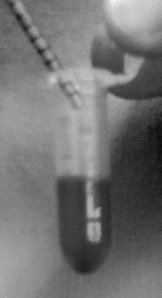
Test tube containing 1 mg/mL of iron-based microparticles (Fe-MP) with 2% solution of blood, the highest concentration of blood tested. Also shown is the 8F magnetic tool (upper left) used to extract the magnetized stone fragments.
Discussion
The goal of the surgical treatment for urinary tract calculi is to ensure complete stone removal. A stone-free state prevents potential long-term sequelae of residual fragments, such as continued stone growth, obstruction, and repeated surgical intervention. 2,9,10 Stone-free rates after minimally invasive treatment of upper tract calculi, however, vary from 50% to 95%, depending on the treatment modality and the imaging study used to define stone-free status. 3,4 The difficulty in achieving complete stone removal can be attributed to limited intraoperative visibility, inadequate endoscopic access to some calices, and lack of spontaneous fragment passage. 5,6,11
Despite advances in ureteroscopic technology and intstrumentation, 11 –13 surgeons still rely on repeated individual stone fragment retrieval to attain a stone-free state, often at a cost of longer operative time and higher cost, increased morbidity, and physician fatigue. Given the current challenges of complete stone removal with minimally invasive techniques, we developed Fe-MP that bind to human kidney stone fragments, rendering them paramagnetic. When compared with standard nitinol basket extraction in an inanimate model, this technology reduced extraction time by up to 53% and decreased extraction attempts for complete stone removal by nearly 40%. 7 To characterize safety and performance before in vivo assessment, we undertook assessing Fe-MP in vitro toxicity and magnetic binding performance when conducted in the presence of blood and urine.
Toxicity of foreign, therapeutic materials can be assessed in a variety of ways, and our in vitro methodology warrants discussion. Three established cell lines—murine fibroblasts, human urothelium, and human TCC—were chosen because of commercial availability, demonstrated robustness for in vitro culturing, and anatomic relevance. Anticipating use of a dilute Fe-MP irrigation solution for endoscopic surgery, Fe-MP would initially encounter the superficial uroepithelial layer of the renal pelvis and ureter. If the particles are able to permeate the epithelial layer, an underlying connective tissue layer containing numerous fibroblasts would be encountered. TCC was used to assess whether Fe-MP could affect viability of “sturdy and aggressive” cells that exhibited homology to urothelium. Indirect contact toxicity through an agarose overlay enabled gradual Fe-MP exposure to cells and sought to replicate the collecting system environment whereby urothelium is protected by mucus composed of proteoglycans rich in glycosoaminoglycans and glycoproteins. 14 –16
In our study, toxicity was limited to urothelium at a concentration of 2×10⁵ cells/dish (average absorbance of 0.268 vs 0.559 in PBS control) when exposed to 1 mg/mL Fe-MP for 48 hours. This 48% decrease in viability is concerning, because a 30% decrease in viability is considered cytotoxic according to the ISO. 8 While it is important to note that no toxicity occurred in the higher concentrations of urothelium, it is possible that lower concentrations of urothelium result in a higher “particle to cell” ratio, allowing any toxic effects of the particles to become additive or to perhaps disrupt cell-cell communications, which are important for urothelial maintenance. 17 There was no indication, however, of increasing toxicity with decreasing cell concentration in any of the other cell lines, specifically fibroblasts, which also rely heavily on autocrine and paracrine cell-cell regulation for growth and survival. 18 Finally, it is understood that in vitro toxicity serves solely as an initial screening tool to hypothesize potential outcomes for in vivo testing if it is perceived to be safe to pursue.
Urine and blood did not significantly affect binding performance or extraction time of any sizes of calcium oxalate stone fragments. Testing concentrations of urine (10%, 20%, 50%) and blood (0.5%, 1%, 2%) were chosen to exceed commonly occurring concentrations of these fluids in irrigant during endourologic stone surgery, given the constant inflow of irrigation solution through the endoscope. The results indicate that commonly occurring electrolytes in urine and blood, such as Ca2+, K+, and Na+, do not interfere with Fe-MP efficacy. All stone fragments were incubated with 1 mg/mL of Fe-MP for 10 minutes, which was previously determined to be an optimal concentration and time for in vitro extraction of both small and large stones. 7 Although 10 minutes was allotted for incubation, further refinement of this technology may possibly allow real-time magnetization of stone fragments with microparticle-infused irrigation during surgery.
Small (3–3.9 mg) or large (6–6.9 mg) fragments used for this study correspond to average weights of stone fragments with diameters of 1 to 2 mm and 3 to 4 mm, respectively. This size criterion was chosen to represent fragments commonly accepted as clinically insignificant residual fragments (<4 mm). While 2 to 4 mm residual fragments have been suggested to increase the risk of subsequent stone-related morbidity after percutaneous stone surgery, the cumulative stone burden of smaller residual fragments (<2 mm) also contributes to this risk. 19 Thus, it is imperative to develop means to extract all residual fragments, including those that are traditionally too small or difficult to extract with conventional retrieval devices.
This Fe-MP technology is still clearlyin its developmental infancy. Optimization of performance and issues of clinical implementation still need to be addressed. This study, however, represents the first step in establishing Fe-MP biologic compatibility and functionality. Of note, Fe-MP is currently only designed to work with calcium oxalate stones. Further modification of binding targets will be needed to allow noncalcium-containing calculi to be magnetically extracted. Likewise, our 8F magnetic extraction device might function in PCNL but will need to be significantly downsized to be accommodated by a ureteroscope. Efforts are currently under way to miniaturize magnetic tools while still preserving adequate magnetic force to replicate current performance.
Conclusion
Fe-MP do not appear to cause cellular toxicity and do function well in the presence of physiologically relevant concentrations of urine and blood that would likely be present if the microparticles are suspended in irrigant fluid for endourologic stone surgery. Further in vivo toxicity and performance testing in animal models is warranted and in progress.
Footnotes
Disclosure Statement
No competing financial interests exist.