
Abstract
A case of moving ileal Taenia saginata parasites is presented with demonstrative images. We came across the parasites surprisingly while performing robot-assisted laparoscopic radical cystoprostatectomy with intracorporeal Studer pouch urinary diversion. We recommend stool sample evaluation in the preoperative period for possible presence of intestinal parasitic diseases, particularly in patients with bladder cancer who are admitted from areas with an increased incidence of intestinal parasitic diseases, before opening the bowel segments during surgery to perform radical cystectomy and urinary diversion.
Introduction
Intestinal parasitic infections are still an important public health problem in our country, particularly in the southeastern region of Turkey. 2 We present a case of ileal Taenia saginata parasitic infection in which we saw the moving parasites in the ileum while performing robot-assisted laparoscopic intracorporeal Studer urinary diversion after performing radical cystoprostatectomy and extended bilateral lymph node dissection in a patient with invasive bladder cancer.
Case Report
A 52-year old man who lives in a small village that is located close to the Turkish-Syrian border underwent transurethral resection of the bladder for bladder tumor that revealed high-grade, muscle-invasive transitional-cell carcinoma. The patient was then referred to our institution for further treatment. No metastatic disease was detected on radiologic evaluation of the patient, including abdominopelvic ultrasonography, CT, and chest radiography. His preoperative International Index of Erectile Function (IIEF) score was 53.
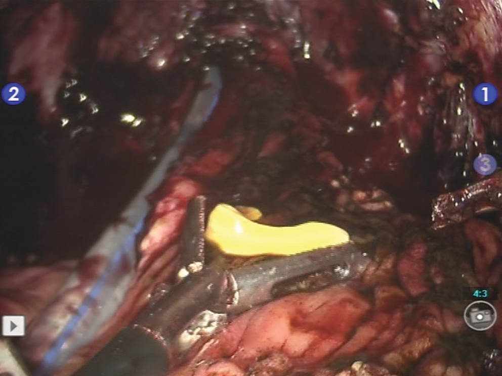
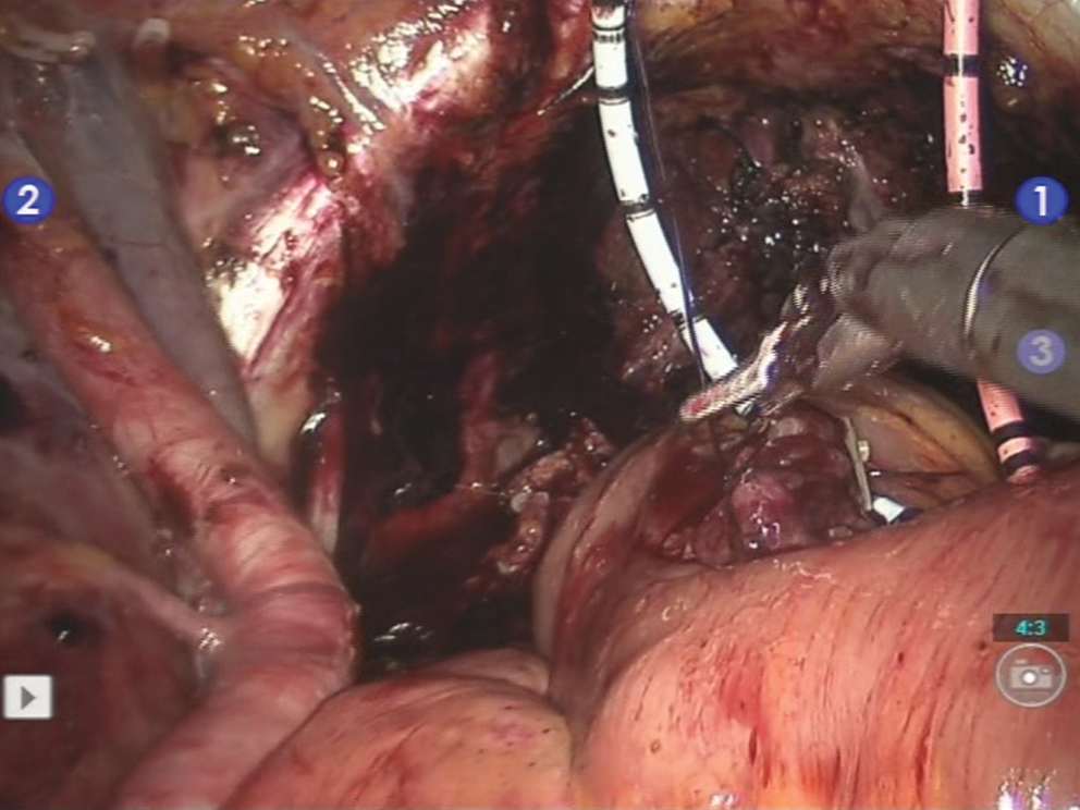
Bilateral nerve-sparing robot-assisted laparoscopic radical cystoprostatectomy, extended bilateral pelvic lymph node dissection, and intracorporeal Studer pouch formation was performed using the da Vinci-S surgical system (Intuitive Surgical, Sunnyvale, CA). Overall, six trochars were used for this procedure: A 12-mm port for robotic three-dimensional lens, three 8-mm sized robotic ports for robotic arms, a 12-mm port for assistant surgeon and a 15-mm port for introducing bowel stapler. To form a Studer pouch intracorporeally, a 45-cm length ileum segment was used following preservation of the 20 cm length distal part of the terminal ileum. When we opened the ileum we saw the moving parasites on the mucosal surface by great surprise (Figs. 1 –3). They were 5 mm sized and yellow-white colored (Figs. 1 –3). We removed all of them cautiously one by one with the laparoscopic grasper and sent them for pathologic and parasitologic evaluation. Both the pathology and the microbiology departments reported these parasites as Taenia saginata. Niclosamide (P.O.) was administered by the department of microbiology.

Appearance of a Taenia saginata parasite in the ileum during robot-assisted laparoscopic intracorporeal Studer pouch formation.
Removal of the Taenia saginata parasite by a laparoscopic grasper.
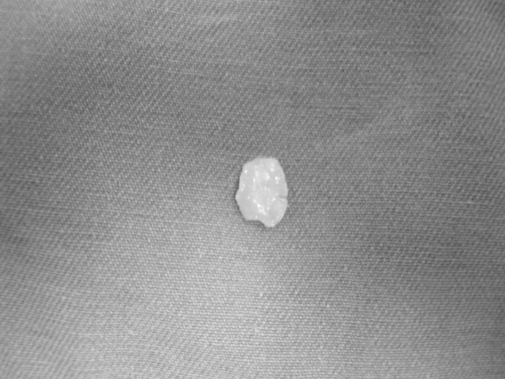
Appearance of the removed Taenia saginata parasite from the abdominal cavity.
Discussion
Parasitic infections are commonly seen in the developing countries including our country which constitute an important public health problem. The incidence of parasitic infections has been reported to be high particularly in the southeast of Turkey. 2 Social, cultural, ecologic, hygenic, sanitation and economic status are regarded as the risk factors for parasitic infections. 2 Contamination occurs commonly by fecal-oral route, generally by contamination of the foods. 2
This disease is more commonly seen in African region, Eastern European countries, Middle East region and Latin America. The length of Taenia saginata might be up to 20 m long in some cases and they have 4 powerful suckers. Human is the definitive host and cattle is the intermediate host for these parasites. They inhabit in the small intestines of humans. Although generally asymptomatic as in our case, they might cause abdominal pain, nausea, loss of appetite, diarrhea or constipation. The diagnosis is simple and requires stool samples to be investigated for parasite eggs. Anti-parasitic drugs are used for the treatment of this disease. 3
Advantages of performing robotic-assisted laparoscopic intracorporeal urinary diversion has been suggested as: i. causing less insensible losses due to a closed abdomen resulting in decreased fluid shifts and earlier bowel function, ii. having a smaller incision with less retractor strain to the abdominal musculature leading to decreased analgesic requirements and earlier postoperative ambulation and, iii. resulting in less complicated postoperative care with shorter hospital stays while continuing to provide equivalent oncologic results (2). In our case, the whole procedure took 8 hours without any complications (Fig. 4). Abdominal drain was removed on the 9th postoperative day and the patient was discharged on postoperative day 14. Currently, our patient is on the 4th-month follow-up without any complications. He does not have urinary incontinence in the day time however he sometimes experiences night time urinary incontinence. His IIEF score is 7. Microscopic and microbiologic stool sample evaluations are negative for parasitic eggs and parasites.
Appearance of the completed robot-assisted intracorporeal laparoscopic Studer pouch.
We recommend stool sample evaluation in the preoperative period for possible presence of intestinal parasitic diseases particularly in patients with bladder cancer admitting from areas with an increased incidence of intestinal parasitic diseases before opening the bowel segments during surgery to perform radical cystectomy and urinary diversion.
Footnotes
Disclosure Statement
No competing financial interests exist.