
Abstract
Purpose:
To evaluate a materials model for laparoscopic guided cryotherapy or radiofrequency tissue ablation (RFA) of kidney tumors through expert surgeon assessment.
Materials and Methods:
During the inaugural American Urological Association 2010 Tissue Ablative course content, validity testing of a renal tumor model was undertaken. Five expert faculty in cryotherapy and RFA techniques for renal tumors performed laparoscopic ultrasonography (US) examination of the tumor model. They performed US guided placement and activation of the treatment probe into the tumor of the model. They completed a questionnaire and rated the quality of the renal tumor model on a 5 point Likert scale.
Results:
All of the subjects assigned a score of 5 of 5 on the Likert scale regarding the ability to identify the tumor with US, were able to deploy the ablative probe into the model under US guidance, and would recommend the use of this teaching model to residents or fellows. They thought that this tumor model was appropriate for teaching laparoscopic US imaging of a renal tumor during ablative treatment procedures, teaching and practicing laparoscopic US-guided cryotherapy, and teaching and practicing laparoscopic US-guided RFA.
Conclusion:
We have developed a unique model that simulates small kidney tumors that can be used for training surgeons in ablative techniques.
Introduction
Minimally invasive energy-based ablative procedures, such as cryoablation and radiofrequency ablation (RFA), have become alternatives for the management of clinically localized small renal tumors 4 allowing a less invasive approach than open and even laparoscopic surgery; its indications have been expanding from renal compromised patients, elderly patients with comorbidities that make them poor surgical candidates, and patients with multicentric tumor disease to include younger, healthier patients with a small renal mass who request ablative therapy. 3 Advantages of the ablative techniques include reduction in blood loss, shorter length of hospital stay, decreased postprocedure pain, and fewer complications when compared with LPN. 5
These procedures necessitate percutaneous imaging with simultaneous accurate needle placement for biopsy and treatment ablation, which mandates accurate dexterity and understanding of the ablative modality.
To date, there has been no realistic, high fidelity model on which to practice these skills and techniques before moving to a more complex scenario, such as the animal model or clinical setting. The ideal model should have the image quality similar to that of the clinically normal kidney and tumor, the texture to provide realistic haptic feedback during needle placement and biopsy, and demonstrate a response to the therapy modality that is similar to the clinical surgical response. 6 –8
Organ mimics are most commonly made with agar or a gelatin base. 9 These, however, can be challenging to reliably create and replicate and do not always have the same characteristics as clinical renal tumors during needle placement and the ablative process. Polyvinyl alcohol cryogel (PVA-C) has been used as an imaging phantom for MRI. From previous laboratory studies, it has been shown to form into a gel with tissue-mimicking properties, can be modeled into anatomic structures, and can be tailored for very realistic imaging with ultrasonography (US), CT, and MRI by exposing the samples to repeated freeze-thaw cycles and adding radiographic contrast material to the PVA 9 –11 solution before implementing the freeze-thaw cycles.
Materials and Methods
PVA model preparation
Commercial PVA powder (Airvol Grade 165, Air Products and Chemicals, Inc, Allentown, PA) is used to prepare the PVA liquid. To make a 10% by mass PVA solution, 900 mL of distilled water and 100 g of PVA powder are mixed in a 1 L tempered glass kettle (Pyrex, Corning Inc, New York) and heated for 30 minutes to up to 75°C with a heating mantle (Glas-Col, Terre Haute, IN) on 80% power. Temperature is measured constantly with a digital thermometer (Traceable Calibration, Friendswood, TX) and stirred with a mechanical mixer (Fisher Scientific, Pittsburgh, PA) at 300 to 350 rpm (Fig. 1). The heater is then turned down to 5% to 15%, keeping the temperature no higher than 87°C, ideally between 83°C to 87°C, for 1 hour.
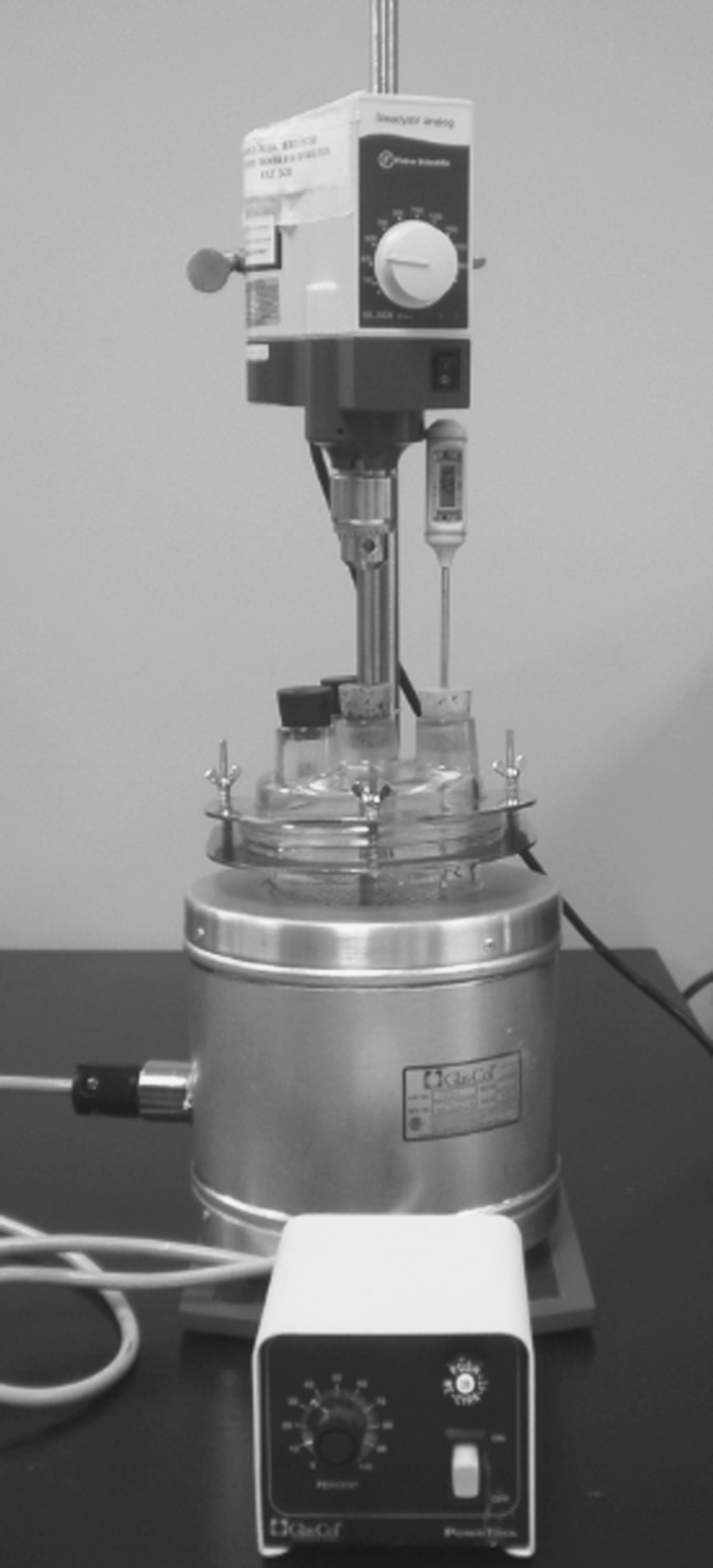
Glass kettle, digital thermometer, heating mantle, and mechanical mixer in polyvinyl alcohol cryogel preparation setting.
The resulting clear liquid is then poured into a plastic container, cooled to room temperature, and rested for 24 hours to allow air bubbles to rise to the surface and then be removed from the resultant slurry. If a dry skin forms on the surface, it can removed with a spoon. The solution is kept sealed and at room temperature for subsequent model creation. To form the PVA liquid solution into a solid tumor phantom, the PVA liquid is poured into an aluminum mold custom designed by the authors with five 1.5 cm spherical spaces and is cooled in a commercial chest freezer to −25°C for 12 hours. At the end of the 12 hours, the freezer is turned off, and its lid kept open 3 cm with a wedge; a small commercial fan circulates the air out of the freezer, allowing a gradual return to room temperature over 12 hours.
The tumor models are removed from the mold, and two tumor models are threaded with a regular cotton suture and straight needle, which is used to suspend the tumor model inside a kidney mold that is custom designed by the authors (Fig. 2). The kidney mold with the tumor models is sealed and filled with liquid PVA using a 60 mL syringe, and the freeze-thaw cycle described above is repeated. This process results in double freeze-thaw cycles for the tumors and a single freeze-thaw cycle for the kidney model. The final model (Fig. 3) is removed from the mold and stored in a container with clean tap or distilled water at room temperature, allowing the phantoms to be kept indefinitely. The model's echogenic characteristics are shown in Figure 4.

Aluminum kidney mold; note the suspended threaded tumor models.
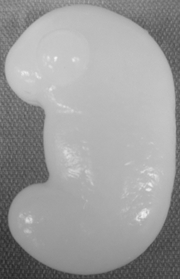
Final polyvinyl alcohol cryogel kidney model.

Final polyvinyl alcohol cryogel kidney model's echogenic appearance.
Evaluation of the PVA model
During the inaugural American Urological Association (AUA) 2010 Tissue Ablative course, content validity testing of the renal tumor model was undertaken. This course was designed to help urologists initiate a renal tumor ablation program and incorporate these technologies into their clinical repertoire. Five nationally recognized expert faculty in cryotherapy and RFA techniques for renal tumor disease and within the standards of the Helsinki Accord performed laparoscopic US examination of the tumor model and identified the various lesions in each of the five different models created for this study. Each model had a different number and location of the tumor model within the kidney model. They then performed US-guided (B&K, Peabody, MA; Aloka, Wallingford, CT) placement of the ablative treatment probe into the tumor of the model and activation of cryotherapy (Galil Medical, Yokneamor, Israel; HealthTronics, Augusta, GA) or RFA probe (Covidien, Dublin, Ireland; AngioDynamics, Latham, NY) to the clinically accepted parameters of the two treatment modalities.
After this, they were asked to complete a questionnaire regarding the ease of visualization of the tumor within the model on US and the ease and realism with which they were able to deploy the ablative therapy probe and perform the simulated ablative technique. They were also asked to rate the quality of the renal tumor model, on a 5 point Likert scale, as to its appropriateness for teaching cryotherapy and the RFA ablative procedure to residents, fellows, and practicing urologists. The model was also incised over the area of the ablative procedure to compare the model deformity of the treated area to what would be expected in clinical tissue using the same ablative technique.
Results
Five faculty experts, all practicing academic urologists reporting more than 50 renal surgeries performed as an assistant and more than 50 cases as the primary surgeon, participated in the study. They each performed at least one laparoscopic renal tumor identification by US and ablation technique with their preferred modality of cryotherapy or RFA. The entire faculty reported that they were able to identify the tumor within the models with US, declaring it to be “very easy” to image the tumor model with US (5 of 5 in the Likert scale). In addition, they were each able to deploy the ablative probe into the model under US guidance and perform the ablative procedure similar to the clinical scenario. They all indicated that they would recommend the use of this teaching model for residents or fellows learning to perform tumor imaging by US, to perform cryoablation and RFA of renal tumors, and to practice cryoablation and RFA of renal tumors in a simulated environment.
Three of the five faculty declared that placing the ablative probe into the tumor model was “very easy” (5 of 5 in the Likert scale), one found it “moderately easy” (3 of 5), and one did not answer this question. Three faculty assigned a 4 of 5 in the Likert scale as a score of the tumor's realism, while two thought that the model was “moderately realistic,” a 3 of 5 score on the Likert scale.
Four surgeons were able to monitor the iceball during cryotherapy ablation. During RFA, three faculty noted that they could monitor the bubbles during the heating process on US. One faculty indicated that cryoablation was “very easy” (5 of 5) to perform, two faculty thought it was “easy” (4 of 5), one faculty stated that cryoablation was “moderately easy” to perform (3 of 5), and one did not answer. Regarding RFA, one faculty member stated that it was “very easy” to perform (5 of 5), two assigned an “easy” (4 of 5) score, and two did not answer. Finally, four faculty members thought that this model was helpful in learning to perform the technique of cryoablation and RFA, and one did not provide an answer.
After the RFA treatment, one of the tumor models was incised, and the area liquefied was consistent with the anticipated treatment area effect in real renal tissue. Similarly, the iceball formation in the cryotherapy model was similar to the expected size of the lesion in clinical tissue.
The cryotherapy mock-treatment procedure did not significantly alter the tumor model when it thawed, suggesting this technique could be repeated without untoward effects on the model. Unanimously, the expert faculty thought that this tumor model was appropriate for teaching laparoscopic US imaging of renal tumor disease and for teaching and practicing laparoscopic US-guided cryotherapy and US-guided RFA techniques.
Discussion
Trainers and simulators play an important role in the acquisition of surgical and percutaneous skills, because they enable surgeons to hone their skills before advancing to the clinical setting in a variety of procedures. 12,13 Models may provide insufficient training, however, because many do not accurately resemble the clinical conditions that may be unique and specific to the urologic procedures being learned. 14 Tissue-mimicking phantoms have been shown to be an important component in medical US, CT, and MRI research, offering an effective alternative to real tissue, because they can replicate the imaging characteristics of the tissues being studied. 10
PVA-C is a useful phantom material with well recognized US, CT, and MRI characteristics. 9 Multiple anatomic phantoms have been successfully developed using PVA-C, and most of these phantoms have been used in published research, demonstrating that small, regularly or irregularly shaped objects that are frozen a different number of times than the surrounding model tissue can be easily targeted with US or MRI for segmentation or biopsy. 9,15 PVA-C vascular models have been produced with and without artificial plaques, even achieving pulsatile motion using different techniques, and its mechanical and MR properties in both T1 and T2 phases have been assessed. 16
Orr and associates 17 investigated the feasibility of creating a PVA phantom with an agarose additive to emulate the MRI's T1 and T2 phases of neonatal brain, finding that by adjusting temperature as well as PVA and agarose concentration, the phantom could be fashioned to accurately reproduce the clinical T1 and T2 phase images in human tissue. 17 There have also been reports of PVA prototypes for photoacoustic mammography, noting that the optical and acoustic properties of the PVA models are close to the normal properties of human breast tissue. 18,19 Interestingly, PVA-C is a recyclable material, so that once a model has been used in a teaching session, it can be melted, remodeled, and reused. This unique model can be used to create a variety of solid organs with corresponding tumor lesions, including a wide variety of teaching objectives including liver, pancreas, brain, breast, and kidney. These models have potential for use in the basic skills teaching of surgeons learning tissue imaging techniques, open surgery, laparoscopic surgery and percutaneous ablative therapies.
One of the observations by the faculty during this course was the inexperience that most of the course participants had with laparoscopic US examination of the kidney and tumors in general. They suggested that adding a dedicated US skills training portion to the program would be beneficial, and the renal tumor model could be used for this training, thereby reducing the number of animals needed for the course. With the addition of enhancing imaging agents, such as gadolinium and barium, this model may also provide effective imaging by CT and MRI. Studies of these additional agents are presently being undertaken by our researchers. Although this was not specifically evaluated in this study, these imaging modalities could be used for the tissue ablative skills training, thereby providing less expensive training strategies than animate or cadaveric models.
Although the expert faculty thought that this tumor model was appropriate for teaching laparoscopic US imaging of renal tumors during ablative treatment procedures and for teaching and practicing laparoscopic or percutaneous US-guided cryotherapy and RFA, the information obtained with this model study represents the preliminary step in designing a teaching model for renal ablative techniques. The model limitations identified during the study were related to storage and use. First, PVA-C undergoes desiccation when exposed to air, degrading its optical properties. For this reason, the gels have to be stored under water. Secondly, PVA-C models are susceptible to proliferation of fungal growth, a situation that can be prevented by storing the phantoms in a 0.01% solution of sodium azide; and finally, outer dimensions expand on freezing and shrink with subsequent freeze-thaw cycles. 16,18
Although it is not part of the study because only a laparoscopic US probe was used, in previously published reports, an external transducer from 3 to 8 MHz has been used. 9 Further assessment of this model is necessary to reproduce with more accurate fidelity the mechanical and ultrasonographic characteristics of the model and determine if it could be used in an animate model for even more realistic skills practice. In addition, concurrent validity testing of this model will be needed before widespread adoption of the model as a training tool occurs.
In this work, we investigated the feasibility of creating a phantom material to reproduce the ultrasonic characteristics of renal tumors and to create a teaching model for percutaneous cryoablation and RFA. Our results demonstrated that it is possible to recreate the environment encountered during US imaging of a renal tumor.
Conclusion
Preliminary study of a PVA-C renal tumor model for teaching US-guided cryotherapy and RFA of renal tumor disease has demonstrated it to be realistic and accurate for training purposes. The limitations of this study are that a small number of experts evaluated the models, and that PVA-C preparation and model manufacturing are labor intensive and time consuming. Further investigation is necessary to refine the model and determine its efficacy with other imaging modalities and as a potential adjunct or alternative to the animate and cadaveric training models for teaching these surgical techniques. The model should be properly validated by increasing the subject cohort and the number of procedures per subject.
Footnotes
Acknowledgments
We acknowledge the support of the American Urological Association Office of Education, Dr. Jaime Landman and Dr. Raymond Leveillee, the course codirectors, for allowing the PVA tumor to be presented and assessed at the 2010 Tissue Ablative Course at the University of Miami.
Disclosure Statement
No competing financial interests exist.