




Abstract
Background and Purpose:
Extended ureteral stricture is a rare complication of renal transplantation. Its management remains challenging. The aim of our study was to report our 15 years of experience with subcutaneous pyelovesical bypass graft (SPBG) in selected renal transplant patients who presented with extended ureteral stricture and who were not eligible for open ureteral reconstruction.
Patients and Methods:
Seven patients were operated on between 1992 and 2007—six men and one woman—with a mean age at surgery of 47 years (range 30–67 y).
Results:
We report no encrustation or dislodgment with a mean follow-up of 6 years (range 1–14 y). Postoperative mortality was null. During the follow-up, three patients died: Two from an extrarenal cause with a functional SPBG and a stable renal function; one from septic shock after fungic colonization of the prosthesis. Asymptomatic urinary tract infection developed in two other patients, with no renal function impairment.
Conclusion:
Despite a 47% rate of infection (3/7), SPBG can be a safe and efficient alternative to open surgery to save many years of graft function in renal transplant patients who present with extended ureteral stricture.
Introduction
Patients and Methods
Between February 1992 and December 2007, 1510 renal transplantations were performed in St. Louis Hospital, Paris. Ureteral complications necessitating surgical or endourologic management (4%) developed in 60 patients. Among them, seven patients underwent SPBG for extended ureteral stricture: Six men and one woman, with a mean age at surgery of 47 years (range 30–67 y). All were kidney transplant recipients, with a diagnosis of extended ureteral stricture that was responsible for rising serum creatinine levels and hydronephrosis on imaging. Ureteral stricture was confirmed by nephrostography at the time of nephrostomy tube insertion, which was first performed.
Ureteral stricture was located at the pyeloureteral junction and was associated with a urinary fistula in one case, secondary to pyeloureteral anastomosis because of the shortness of the graft ureter. The ureteral stricture affected the inferior third of the ureter in two cases and the total ureter in four cases. Because of the failure of the endourologic approach in all cases and no possibility of open surgery, all patients underwent SPBG placement as a salvage procedure for their kidney transplant.
Details of the artificial ureteral replacement technique have been described (Fig. 1). Briefly: The patient was placed in the supine position, and a nephrostomy tract was progressively dilated to insert a 30F Amplatz sheath into the graft cavities. A tunneler was used to create a subcutaneous tract starting in the suprapelvic region and ending at the level of the Amplatz sheath. The ureteral prosthesis was inserted into the graft pelvis through the Amplatz sheath. A composite structure ureteral tube was used (Detour, Coloplast Inc), with the external tube composed of e-PTFE (from 1992 to 2000) or woven polyester (from 2001 to 2007). The internal tube, in direct contact with urine, was always made of silicone. The tube was brought down through the subcutaneous tunnel to the suprapubic area and introduced into the bladder via a short incision and attached to the bladder wall with four absorbable sutures. Since 1992, we have reviewed retrospectively all the charts and office notes to assess patient clinical characteristics, the time to diagnosis of ureteral stricture, and its modalities of initial management. We also reported the kidney function before and after SPBG, the immediate complications of the technique, and its long-term outcome.
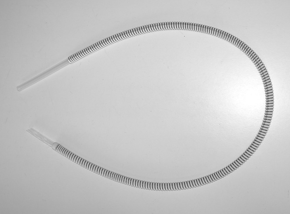
Photograph of the subcutaneous pyelovesical bypass graft. Reproduced with permission from Coloplast, Inc.
Results
Patient characteristics are summarized in Table 1. All transplanted kidneys were from deceased donors. Ureteral stricture management is described in Table 2. Mean diagnosis time of the ureteral extended stricture was 5.6 years, while mean delay to surgery was 1.7 years.
EMG=extramembranous glomerulonephritis; IL=ipsilateral; DM=diabètes mellitus; CL=contralateral; FSGS=focal segmental glomerulosclerosis; NA=not available; TMA=thrombotic microangiopathy; POD=postoperative day.
SPBG=subcutaneous pyelovesical bypass graft; NA=not available; UTI=urinary tract infection.
All patients had attempts of ureteral stent placement with a few months of delay between diagnosis and surgery. Patients #2, #3, and #7 benefited each from one retrograde ureteral stent placement, while patients #4 and #5 had one attempt of retrograde stent placement after balloon dilation of the stricture after two and three simple retrograde stent placements, respectively. Patient #6 was an exception: He received a diagnosis of ureteral stricture 12 years after renal transplantation, with successive retrograde ureteral stent placements during nearly 10 years, which has probably led to secondary fibrotic ureteral obstruction.
Short and long term complications
No immediate complication was observed, and postoperative mortality was null. During the follow-up, three patients died, and one death was directly related to the SPBG. This patient (#6) died 2 years after SPBG from septic shock from the infection of the prosthesis first by multiresistant microorganisms, then by Candida albicans. The initial management of the infection consisted of removing the prosthesis 1.5 years after surgery, and then removing the infected graft 6 months later. Unfortunately, this management did not prevent him from dying from septic shock consecutive with a pulmonary fungal infection that was resistant to all antifungal treatments.
Patient #1 died 11 years after SPBG from an aneurysm of the abdominal aorta with an intact and functioning prosthesis. He was operated on twice 4 and 5 years after surgery from an abdominal eventration.
Patient #4 died 1 year after SPBG from extensive pulmonary fibrosis associated with pneumocystis with an asymptomatic colonization of the prosthesis by multiresistant microorganisms: Acinetobacter and Enterobacter cloacae.
The last patient (#2) with long-term complication of the ureteral replacement was also the third one with occurrence of infection of the prosthesis, 14 years after SPBG placement, secondary to a sigmoid abscess associated with peritonitis and enteric perforation that was treated with ileostomy. Since digestive surgery, recurrent urinary tract infections (UTIs) have developed with multiresistant microorganisms: Pseudomonas aeruginosa, Enterobacter, and recently C albicans. He recovered his basic renal function (creatinine: 130 micromoles/L) after effective antimycotic treatment, without any hydronephrosis on imaging. No obstruction or displacement of the prosthesis was observed.
Creatinine levels before and after SPBG and at last follow-up are detailed in Table 3.
Cr=creatinine; SPBG=subcutaneous pyelovesical bypass graft; LFU=last follow-up; eGFR=estimated glomerular filtration rate using the sex variables Modification of Diet in Renal Disease method; FU=follow-up; PD=peritoneal dialysis; HD=hemodialysis.
All patients had an immediate benefit of ureteral replacement with a decreasing postoperative serum creatinine level, except for patient #3, who had a chronic vascular graft dysfunction 10 years after renal transplantation and 2 years before the diagnosis of ureteral stenosis; this patient is at present being treated with peritoneal dialysis and included on the waiting list for a second renal transplantation, with no obstruction or infection of the prosthesis.
Because mean follow-up was 6 years (range 1–14 y), the five remaining patients had a good and stable renal function at last follow-up, dead from an extrarenal cause (#1 and #4), or alive (#2, #5, and #7).
Discussion
Extended ureteral stricture is a rare complication of renal transplantation, and its management remains difficult. The standard ureteral reconstruction procedure consists of either direct ureteral reimplantation in the case of short distal stricture or pyeloureterostomy using the native ureter in the case of extended stricture. In the case of no native ureter available, a wide range of techniques has been described for ureteral reconstruction, including modified Double-J stent, 7,8 interposition of an intestinal conduit, 9 and pyelovesicostomy with or without a Boari flap. 10,11 Difficulties raised by these techniques are numerous: Double-J pigtail stents are associated with a high risk of obstruction by incrustation; the acidosis from reabsorption within the intestinal conduit can be dangerous in patients with renal function impairment; pyelovesicostomy is challenging in case of a small volume bladder with poor compliance.
Regarding all these arguments, SPBG was first successfully used in 1992 at St. Louis Hospital as an original treatment of extended ureteral stricture in selected patients. 3,4 In surgical experience, the success of a technique usually relies on acute and precise indications, the key to a low short- and long-term morbidity. The indication for SPBG according to our experience concerns patients for whom endourologic management has failed, with no available ureter. Reoperation on a transplanted kidney may be difficult and exposes the patient and the renal graft to potentially serious secondary complications. Open surgery was avoided in high surgical risk patients: Obese patient with multiple previous laparotomies (#2), location of the renal pelvis beside iliac vessels necessitating vascular surgery (#1), or proximal stricture needing extensive dissection of the renal pelvis (#3). The initial kidney disease explains in the other cases (#4, 5, 6, and 7) the lack of a native ureter available for open reconstruction: Bilateral reflux, bilateral nephroblastoma, adenocarcinoma located on a unique kidney, lack of length of the graft ureter necessitating a primary pyeloureteral anastomosis.
Thus, SPBG can be used as graft salvage therapy when open surgery is not possible on the preoperative evaluation. This technique may also provide a second treatment option as described in the Canadian experience for recipients who had failed ureteral reimplantation or pyeloureterostomy for extended ureteral stricture. 6
In our series, no immediate complication occurred, such as urine leak secondary to dislodged SPBG as described in the Canadian experience. 6 The learning curve associated with this technique is rather short, especially in a surgical team trained in bypass of extrinsic ureteral obstructions. 5 In this indication, Aminsharifi and associates 12 suggested in 2009 percutaneous access to the bladder using a split Amplatz sheath during placement of the SPBG to avoid open cystostomy incision and make the procedure even more minimally invasive. This modification of the technique may be applied to kidney transplant patients. After a follow-up of as long as 15 years, no incrustation, kinking, or obstruction was encountered, as suggested by our experience in SPBG for extrinsic ureteral obstruction. 13 No replacement of prosthesis was necessary because of stone formation in kidney transplant patients, with more than 10 years of follow-up in two cases. This is certainly because of a good nephrologic management of the patients, by maintaining a high fluid intake, and a good correction of secondary hyperparathyroidism and distal renal tubular acidosis. 3,4 It can also be linked with the large caliber (17F) and large dead space of the prosthesis, as suggested in the Canadian report. 6
No secondary skin erosion exposing the prosthesis was reported in our series: This complication happens mostly in patients with pelvic malignancies and concomitant radiochemotherapy. 5,13
The main problem in long-term treatment of immunosuppressed patients with transplanted kidneys remains infection of the prosthesis. Because the infection rate with SPBG in immunocompetent patients is 30%, 14 we observed a rate of 42% (3/7) in our series after a mean follow-up of 6 years, against 25% in the Canadian experience, 6 which displayed a mean follow-up of 19.4 months. Bacterial colonization of the graft was not severe in all cases: One case of asymptomatic UTI with no renal dysfunction, one case of multiresistant microorganisms UTI occurring 14 years after SPBG secondary to digestive surgery, one case of death after graft removal because of fungal colonization of the prosthesis. Long duration, high dose, and large spectrum antimicrobial treatment for multiresistant microorganisms provides a high risk of fungal infection of the SPBG, potentially lethal in an immunosuppressed patient. The Canadian series reported a case of graft loss from an infection by a microorganism that was resistant to continuous antimicrobial treatment. 6 This experience may lead us to advocate the early ablation of the prosthesis in the case of positive urine culture with C albicans.
No antimicrobial-coated ureteral stents have been designed yet, to the best of our knowledge, to reduce the incidence of UTI.
Conclusion
In selected patients who presented with extended ureteral stricture after renal transplantation with no native ureter available for reconstruction, we have successfully used the SPBG since 1992 with good immediate and intermediate results. Its safety and efficiency remained to be verified in the long term. Although the use of a prosthetic device is not recommended in immunosuppressed patients, our series showed an acceptable rate of infection, with no mechanical complications. Its ability to save many years of graft function makes the SPBG a good solution for selected patients with an extended ureteral stricture after renal transplantation.
Footnotes
Disclosure Statement
No competing financial interests exist.